Keywords
Trauma, vein of Labbé, outcome
Trauma, vein of Labbé, outcome
Dural venous sinus thrombosis after blunt head trauma has been reported in few case series1–6. Only a few studies have been done on outcome following traumatic vein of Labbé haemorrhagic infarction7. It is an important neurosurgical entity because of the involvement of the area that it drains in language comprehension and processing, as well as possessing a higher propensity for causing early uncal herniation with a sometimes fatal outcome8–10. As such, stringent monitoring of the patients and early surgical evacuation if required is the key to the successful management of this condition. One of the important differential diagnoses for vein of Labbé infarction is traumatic temporal artery damage wherein damage to the medial temporal region including the insular territory is also seen. Another entity to be excluded is transverse sinus thrombosis. This case highlights the importance of close observation of patients with petrous bone fracture and transverse sinus thrombosis for evolving vein of Labbé haemorrhagic infarction and early uncal herniation. There have only been a few studies highlighting this clinical entity and the cumulative advice from these is the suggestion of performing cerebral venographic studies in suspected cases so as to make a timely and correct surgical decision.
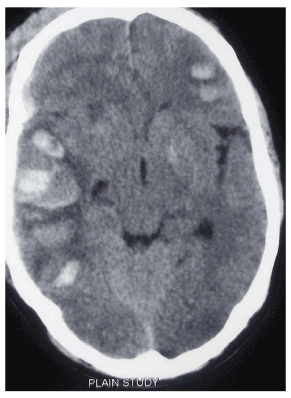
A 55-year-old Nepalese male from a remote village in Nawalparasi, Nepal was brought to the emergency room after being hit by a moving car. Medical history turned up no significant past medical illnesses or surgical interventions. At the time of arrival, his Glasgow coma scale (GCS) was E3M5V4. Vital parameters like blood pressure (130/80), pulse rate (76/min), respiratory rate (23/min) and oxygen saturation (99% at room air) were within normal range. Both of his pupils were equally sized and equally reactive to light. A primary and secondary injury survey did not reveal other systemic injuries. An urgent head CT scan revealed the presence of a hyperdense lesion in the right temporal region with mild effacement of the ipsilateral ambient cisterns and widening of the cerebello-pontine cisterns suggestive of early uncal herniation (Figure 1). We made the provisional diagnosis of traumatic vein of Labbé hemorrhagic infarction with traumatic contusions as the main differential diagnosis. Screening MR venography proved the findings of traumatic vein of Labbé haemorrhagic infarction through identification of the absence of vein of Labbé on the right side (Figure 2).

Due to the risk of imminent herniation, patient’s relatives were counseled regarding the benefits and risks involved in the surgical management. After verbal and written consent, the patient was taken up for surgery. Fronto-temporo-parietal flap craniotomy was performed. Durotomy was done and the posterior temporal corticostomy with evacuation of the hematoma was undertaken. Brain was lax and pulsatile at the end of the procedure. Patient was extubated the following morning after a repeat CT showed resolution in the herniation effect without any untoward post-operative events. Patient was started on the antiepileptic Sodium Valproate (300 mg intravenously, every 8 hours) for seizure prophylaxis and was advised to continue on this regiment for at least 6 months. Patient showed remarkable improvement, attaining a GCS score of E4M5V5 but with deficits in the prosody of his speech, attributable to the involvement of his right temporal lobe. The patient was early ambulated after the second post-operative day so as to prevent complications like chest infection and deep venous thrombosis due to prolonged immobilization. He was discharged home on the seventh post-operative day, after removal of his wound stitches, with the advice of taking the antiepileptic regularly. The patient was followed up in the outpatient clinic 3weeks later. Patient still had some deficits to prosody of his speech but language fluency and content were normal. Repeat CT showed complete resolution in hematoma and mass effects (Figure 3). Patient was advised to continue with the antiepileptic medication (Sodium Valproate 300mg via oral route three times daily) for 6 months.
Named after the French surgeon Charles Labbé, the vein of Labbé (also known as the inferior anastomotic vein) crosses the temporal lobe between the Sylvian fissure and the transverse sinus and connects the superficial middle cerebral vein and the transverse sinus.
Since there is higher propensity for early uncal herniation and concurrent rapid neurological deterioration, any traumatic temporal lobe lesion poses an enigma for neurosurgeons.
Impact injury and counterblow are the main causes of injuries to the vein of Labbé, which can consequently lead to serious traumatic cerebral infarction with its associated poor prognosis7. Temporal bone fracture was associated in 15 of all the 16 cases in a study by Long et al.7
In a study by Giannetti et al.11, CT scan findings such as mediolateral diameter of the lesion, location of the hematoma, status of the ambient cisterns and position of the midline structures were used as a criteria to decide which patients would benefit from early surgery. In this case, we used the location of the hematoma, its volume and features of obliteration of ambient cisterns to assess the need to surgically evacuate the hematoma.
In previous studies of patients with blunt head trauma who have skull fractures extending to a dural venous sinus or jugular bulb, multi-detector CT venography identified dural venous sinus thrombosis (DVST) in 40.7% of cases, and of these 55% were occlusive12. There is a high risk of evolution of vein of Labbé haemorrhagic infarction in the subsets of patients with petrous bone fracture. So proper monitoring is justified for any signs and symptoms of increased intracranial pressure.
Also given the nature of the area of brain that vein of Labbé drains (language processing and comprehension), there is a need for long-term follow up of these patients to determine any neurological sequelae.
A high index of suspicion needs to be kept in patients with petrous bone fractures for probable vein of Labbé hemorrhagic infarction following transverse sinus thrombosis. In those with traumatic venous infarction, stringent monitoring needs to be taken for evidence of early uncal herniation. In case of lesions more than 25ml, anisocoria, uncal herniation and asymmetric ambient cisterns, early surgical evacuation is justified.
Written consent for publication of clinical data and images was sought and received from the son of the patient.
SM wrote and formatted the paper. BMK revised and edited the final format. All authors have seen and agreed to the final content of the manuscript.
| Views | Downloads | |
|---|---|---|
| F1000Research | - | - |
|
PubMed Central
Data from PMC are received and updated monthly.
|
- | - |
Competing Interests: No competing interests were disclosed.
Competing Interests: No competing interests were disclosed.
Alongside their report, reviewers assign a status to the article:
| Invited Reviewers | ||
|---|---|---|
| 1 | 2 | |
|
Version 1 19 Aug 15 |
read | read |
Provide sufficient details of any financial or non-financial competing interests to enable users to assess whether your comments might lead a reasonable person to question your impartiality. Consider the following examples, but note that this is not an exhaustive list:
Sign up for content alerts and receive a weekly or monthly email with all newly published articles
Already registered? Sign in
The email address should be the one you originally registered with F1000.
You registered with F1000 via Google, so we cannot reset your password.
To sign in, please click here.
If you still need help with your Google account password, please click here.
You registered with F1000 via Facebook, so we cannot reset your password.
To sign in, please click here.
If you still need help with your Facebook account password, please click here.
If your email address is registered with us, we will email you instructions to reset your password.
If you think you should have received this email but it has not arrived, please check your spam filters and/or contact for further assistance.
Comments on this article Comments (0)