Keywords
bone fragment, brain surgery, compound-depressed fracture
bone fragment, brain surgery, compound-depressed fracture
We have revised and added the reason for opting to remove the depressed fracture despite its anatomical location.
To read any peer review reports and author responses for this article, follow the "read" links in the Open Peer Review table.
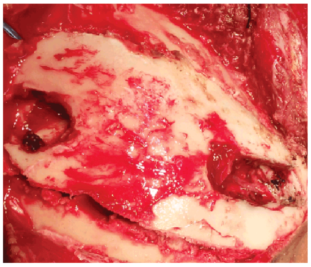
A 22 year-old female, with no significant past medical and surgical illnesses, was brought to the casualty room with a Glasgow coma scale of 6/15 following a collision between two bikes three hours earlier. Local examination revealed two compound depressed skull fractures in the frontal and the parietal region with egress of brain matter. Following primary resuscitation, computed tomography (CT) of the head confirmed the local findings along with the presence of one bone fragment in the third ventricle (Figure 1). The patient was taken for debridement of the wound and craniotomy circling the depressed sites. Since the patient was already extending and because there was already hemoventriculi, we opted for removal of the fragment despite its anatomical location so as to minimize further damage and chance of hydrocephalus. The bone fragment in the third ventricle was easily accessible following the hematoma track. An endoscope was also kept ready just in case the corridor to the fragment was difficult to access. Following retrieval of the bone fragment (Figure 2, Figure 3), intraventricular drain was placed and neurosurgical intensive care was provided. Repeated CT scans showed hypodensities around the third ventricle (Figure 4). On the second post-operative day, the patient was started on ionotropic support because of the refractory hypotension, and was also replaced with hydrocortisone, fludrocortisone and thyroid hormones. Wound dressing and the ventricular drain care was continued. Cerebrospinal fluid (CSF) culture from the drain resulted sterile. The patient died on the 8th post-operative day because of the traumatic severe hypothalamic insult.

As brain abscesses may result from driven bone fragments and other retained foreign bodies in the brain, the removal of readily accessible foreign bodies has received much attention3–6. Migration of foreign bodies can occur because of gravitational force. Other routes of migration can be subdural, parenchymal, transventricular or along streamlining along the white matter track7. The removal of foreign bodies is mostly done via craniotomy8,9, but other methods such as burr hole, stereotaxy10 and sometimes by ventriculoscopy11 have also been described.
The goals of modern treatments include removal of the foreign body under a controlled environment in the neurosurgical operation setting. Surgical principles include removal of bone fragments, intracerebral hematoma, control of hemorrhages and prevention of further loss of neural tissue. Patients should receive a broad spectrum intravenous antibiotic therapy along with tetanus prophylaxis. Monitoring and control of elevated intracranial pressure with maintenance of cerebral perfusion pressure plays a significant role in the patient’s survival and outcome. The follow-up of such patients is essential, considering known complications like cerebrospinal fluid fistula in the early post-operative period and brain abscesses and seizures which may occur years after injury. Outcome after a penetrating head injury is directly related to the Glasgow coma scale at the time of presentation, which is the reflection of the extent of brain tissue damage caused directly by the primary impact. Intensive post-operative monitoring of intracranial pressure, cardio-respiratory function and metabolic status are required for optimizing the outcome of victims of penetrating craniocerebral injuries12. Penetrating head injuries have a higher mortality and morbidity than blunt trauma even in a civilian set up13. Even after timely removal of the penetrating objects and intensive medical management, the outcome may remain poor.
Informed written consent for publication of images and clinical details was obtained from the patient’s husband.
Sunil Munakomi wrote and submitted the manuscript. Balaji Srinivas, Binod Bhattarai and Iype Cherian formatted and reviewed the paper.
| Views | Downloads | |
|---|---|---|
| F1000Research | - | - |
|
PubMed Central
Data from PMC are received and updated monthly.
|
- | - |
Competing Interests: No competing interests were disclosed.
Competing Interests: No competing interests were disclosed.
Competing Interests: No competing interests were disclosed.
Alongside their report, reviewers assign a status to the article:
| Invited Reviewers | |||
|---|---|---|---|
| 1 | 2 | 3 | |
|
Version 2 (revision) 31 Mar 15 |
read | read | |
|
Version 1 11 Mar 15 |
read | ||
Provide sufficient details of any financial or non-financial competing interests to enable users to assess whether your comments might lead a reasonable person to question your impartiality. Consider the following examples, but note that this is not an exhaustive list:
Sign up for content alerts and receive a weekly or monthly email with all newly published articles
Already registered? Sign in
The email address should be the one you originally registered with F1000.
You registered with F1000 via Google, so we cannot reset your password.
To sign in, please click here.
If you still need help with your Google account password, please click here.
You registered with F1000 via Facebook, so we cannot reset your password.
To sign in, please click here.
If you still need help with your Facebook account password, please click here.
If your email address is registered with us, we will email you instructions to reset your password.
If you think you should have received this email but it has not arrived, please check your spam filters and/or contact for further assistance.
Comments on this article Comments (0)