Keywords
LHCGR, insertions, exon-1, poor ovarian response
LHCGR, insertions, exon-1, poor ovarian response
Luteinizing hormone (LH) is a key regulator of the female menstrual cycle1 and is produced by the anterior pituitary gland of gonadotroph cells. During ovulation, the LH surge is not only essential for the release of an egg from the follicle, but also regulates the formation of the corpus luteum. The corpus luteum in turn produces progesterone, which helps in the maintenance of pregnancy2,3. Therefore, LH along with its receptor complex has a cardinal role during reproductive processes4,5. The heterodimeric glycoproteins LH and chorionic gonadotrophin (CG) are mediated by luteinizing hormone/choriogonadotrophin receptor (LHCGR), which is a member of the G-Coupled receptor protein family. This receptor is expressed by the LHCGR gene located on chromosome 2p21 that consists of 11 or 12 exons and codes for around 600 amino acids. The LH receptor is located on Leydig cells in males, and theca, granulosa cells of the ovaries20. The receptor activation and inactivation are brought about by the conformation of amino acids in their localized regions6.
Women, whose LH receptor is down regulated with GnRH agonists or antagonists, may have decreased concentrations of luteinizing hormone and follicle stimulating hormone. This results in poor outcome due to low endogenous LH levels. Results from our earlier study demonstrated more number of oocytes and clinical pregnancies after use of recombinant LH(rLH) combined with recombinant follicle stimulating hormone (rFSH) in assisted reproductive therapy (ART)7. Although supplementation of LH is beneficial in a selected category of patients, a number of studies have also demonstrated that LHCGR mutations at specific amino acid regions result in mild or complete loss of receptor function8.
LHCGR gene mutations were first studied in men with Leydig cell hypoplasia, later they were extended to female reproductive disorders11. Studies have shown that inactivating base pair insertions in exon-1 of LHCGR gene are associated with low oocyte yield and also negatively associated with intrauterine insemination (IUI) and in vitro fertilization (IVF) outcome in infertile population9–11. A recent investigation by Bentov et al., 2012 reported a 6bp insertion in exon-1 of LHCGR gene which was associated with recurrent IUI and IVF failures. Although many factors like age, low anti-mullerian hormone (AMH) etc. are indications for decreased oocyte yield, poor ovarian response and low ovarian reserves, recent publications have focused on FSHR and LHCGR mutations/polymorphisms. A study was initiated to delineate the possible role of LHCGR dysfunction in patients with poor ovarian response, low and moderate ovarian reserves11.
A total of 114 patients was screened for LH receptor gene polymorphisms/mutations with the following inclusion criteria: number of antral follicle count <8, normal testosterone levels, low antimullerian hormone (AMH) and poor response to previous infertility treatment. Out of 114 patients, 3 patients showed a 6bp homozygous insertion and 23 patients showed heterozygous 6bp insertion as described previously11,12. However, a 54bp insertion in exon-1 of LHCGR gene was detected in a patient with a clinical history of poor response to human menopausal gonadotrophin (hMG) stimulation and recurrent cyst formation.
The study was approved by the institutional ethics committee and informed consent was obtained from the participants.
A 28-year old Indian woman with a marital life of 5 years suffering with infertility referred to the Krishna IVF clinic for evaluation and treatment. Physical examination revealed normal breast development, normal external and internal female genitalia with irregular menstrual cycles. The patient has a sibling with two children.
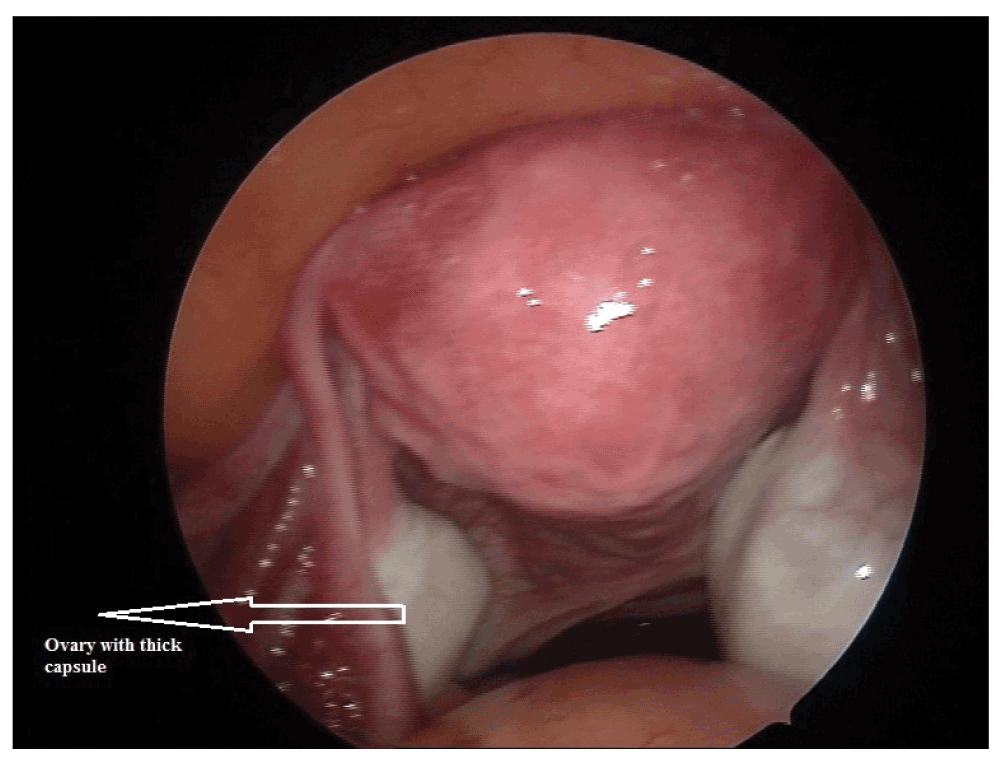
At day 2, basal ultrasound examination showed an anteverted uterus volume:48ml, a right side ovary volume of 14.43 ml with a growing follicle and a left side ovary volume of 6.32 ml. Antral follicle count was 6 combining both ovaries. The patient underwent diagnostic laparoscopy and hysteroscopy and the ovaries had a normal volume with thick capsules as shown (Figure 1). On patient’s request, gonadotrophin/IUI was planned three weeks after laparoscopy. Ultrasound examination detected a large functional cyst which was managed using oral contraceptive pill. In the first cycle, the patient was super ovulated with a dose of hMG of 150 units/day. No response was observed after 4 days and 6 days of stimulation. As the AMH (0.2 ng/dl) and the ovarian reserves were low and there was no other option available, in the next cycle patient was super ovulated with a hMG dose of 225 units/day for 10 days. On the 9th day of stimulation, one follicle with 18 to 20 mm diameter, 2 follicles with 1 to 9 mm and endometrial thickness of 10 mm were observed. A trigger dose of hCG 5000 IU was administered. Luteal support was provided with susten 200 mg vaginally. The patient had started bleeding p/v from 6 days after trigger dose of hCG. A day 2 scan in the subsequent cycle showed an ovarian cyst formation again.
Because of the discrepancy between AFC, AMH and the ovarian volume, and the poor response to IUI stimulated cycles and the recurrent cyst formation, hCG stimulation test and LHCGR gene sequencing were performed.
The hCG stimulation test was performed in a natural cycle. Baseline data of the patient hormonal profile were recorded. These showed serum FSH 10 mIU/ml, serum LH 8 mIU/ml, serum dehydroepiandrosterone (DHEA-S) 3.71 mcg/ml, serum testosterone 20 ng/dl, serum prolactin 14.1 ng/ml, indicating normal values, and serum AMH showed a value of <0.2 ng/ml. hCG increases the serum concentrations of androstendione and testosterone by acting through LHCGR. A dose of 10,000 units of hCG was administered subcutaneously. A blunted response to hCG was observed after 4 hours of administration as there was no significant change in the serum testosterone concentration (22 ng/dl) compared to baseline values. Based on the patient’s poor response to ovarian stimulation and hCG stimulation test, the patient was further referred to the molecular genetics department to investigate the possible LHCGR mutations/polymorphisms.
Genomic DNA from peripheral blood of patient’s was isolated using a modified salting out method as described previously13. All the coding exons of the gene were amplified by polymerase chain reaction using primers designed using primer express software from Applied Biosystems, the amplified product was observed on 2% agarose gel electrophoresis and purified with Exo-Sap enzyme from Thermo Scientific21. The purified product was sequenced using big dye cycle sequencing kit on 3500 genetic analyzer and analyzed using Seq Scape (Life Technologies, USA).
The sequencing results revealed a 54bp insertion (GCTGCTGAAGCTGCTGCTGCTGCTGCA)2 in exon-1 as shown (Figure 2). This insertion sequence gave rise to glutamine, lysine and leucine rich regions. Insertion in exon-1 of LHCGR gene, according to previous authors11,12,14, will have a profound effect on the LHCGR function as reflected by the patient history and hCG test.
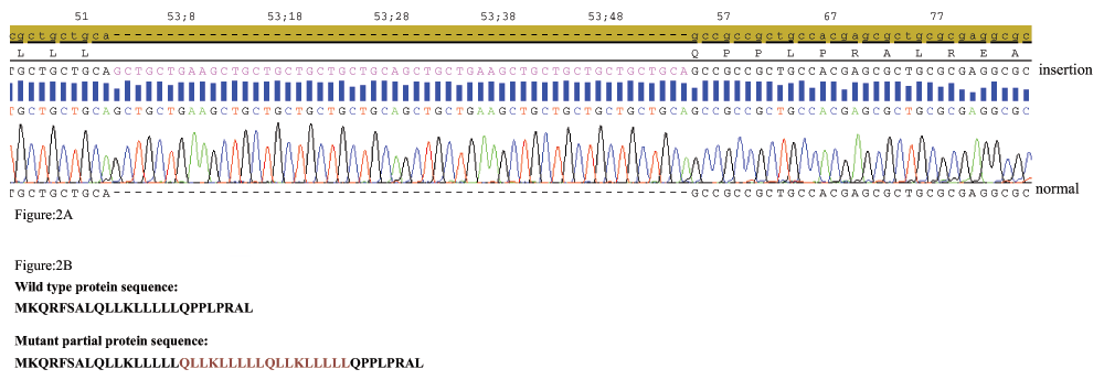
A) A dark yellow portion showing the reference sequence of LHCGR exon 1 from NCBI database and the insertion sequence shown in pink. B) Wild type protein sequence and the inserted aminoacids shown in red.
LH regulates a number of reproductive functions in males as well as in females. LH and CG hormones act through the LH/CG receptor which facilitates the activation of different cell groups in the ovaries. LH triggers follicular maturation, ovulation and formation of corpus luteum and increases progesterone secretion in luteal phase2,3,14. Different chronological, hormonal, functional biomarkers are used to assess the ovarian response during controlled ovarian stimulation (COS)15. Along with these biomarkers, the possible roles of LHCGR mutations associated with different female reproductive disorders like oligo amenorrhea, recurrent pregnancy loss, ovarian resistance, infertility and IVF outcome have been studied by previous authors10,11,16,17. A 6bp (CTGCAG, amino acids: LQ) exon-1 insertion was first described in LHCGR after nucleotide position 54 in Leydig cell hypoplasia9,18. Later, a homozygous 33bp and a 27bp insertions (CTGCTGAAGCTGCTGCTGCTGCTGCAG, amino acids: ) were described by two authors after nucleotide position 54 presenting Leydig cell hypoplasia in males explaining a possible role in signal transduction12,19.
Here we present a homozygous mutation with a 54bp insertion (GCTGCTGAAGCTGCTGCTGCTGCTGCAGCTGCTGAAGCTGCTGCTGCTGCTGCA) in the exon-1 of LHCGR of an infertile woman. Inactivating mutations of the hLHR are known to affect signaling pathways, decrease LH and its receptor binding. Improper folding of this receptor protein structure in the endoplasmic reticulum may result in decreased LHR expression which leads to reduced cell response14. One of the above stated mechanisms might have played a role in the poor ovarian response in the present case. The serum hCG stimulation test also showed a blunted response indicating reduced receptor function. The data from the present case are highly significant as the involvement of a possible genetic cause was delineated at an early stage of the infertility treatment. LHCGR mutations may lead to a reproductive catastrophe. Supplementation of LH in patients with LHCGR polymorphisms might offer a benefit for those who opt for IVF treatment. However the concept of LH supplementation may not yield a good response in such a type of homozygous LHCGR mutations. A blunted response might be observed at a very high doses with mature and immature oocytes. In such situations, in vitro maturation of oocytes, where in the dependence of hCG is less to cause the follicle maturation, can be an option for such patients. This study concludes that examination of LHCGR mutations/polymorphisms would be a useful investigation for classifying response, prognosis and management in infertile patients with low ovarian reserves.
Informed written consent to publish clinical data was obtained from the patient.
| Views | Downloads | |
|---|---|---|
| F1000Research | - | - |
|
PubMed Central
Data from PMC are received and updated monthly.
|
- | - |
Provide sufficient details of any financial or non-financial competing interests to enable users to assess whether your comments might lead a reasonable person to question your impartiality. Consider the following examples, but note that this is not an exhaustive list:
Sign up for content alerts and receive a weekly or monthly email with all newly published articles
Already registered? Sign in
The email address should be the one you originally registered with F1000.
You registered with F1000 via Google, so we cannot reset your password.
To sign in, please click here.
If you still need help with your Google account password, please click here.
You registered with F1000 via Facebook, so we cannot reset your password.
To sign in, please click here.
If you still need help with your Facebook account password, please click here.
If your email address is registered with us, we will email you instructions to reset your password.
If you think you should have received this email but it has not arrived, please check your spam filters and/or contact for further assistance.
Comments on this article Comments (0)