Keywords
Surgical Resection, Primary dural diffuse large B-cell lymphoma, Paediatric cancer, Herniation syndrome, Intracranial lymphoma
Surgical Resection, Primary dural diffuse large B-cell lymphoma, Paediatric cancer, Herniation syndrome, Intracranial lymphoma
Primary dural diffuse large B-cell lymphoma (DLBCL) is an extremely rare entity with only five cases reported so far1. The symptoms are nonspecific. The main differential diagnosis of the condition remains meningioma2. Currently there is no standard treatment due to a paucity of cases3. A high index of suspicion should be kept in order to diagnose the condition in a timely fashion and then plan for appropriate management since diffuse large cell lymphoma has a relatively benign clinical prognosis4. Here we report a case of a primary dural based DLBCL in a 14 year-old boy presenting with herniation syndrome, who improved after surgical excision and is currently on chemotherapy.
A 14 year-old Tharu boy, from Siraha (a remote village in Nepal) presented to our emergency department with a sudden onset altered sensorium which lasted for 1 day. The patient had a history of intermittent headaches and vomiting over the last 3 months. The patient’s parents also noticed significant weight loss and the presence of scalp swelling for the last 2 months. There was no remarkable family history. Previous treatment history revealed that the patient had been taken to India 1 month back, where fine needle aspiration cytology (FNAC) of the scalp lesion in the parietal region had revealed Non-Hodgkin’s lymphoma. The patient party was told the prognosis and advised for chemo- and radiation therapy but this was refused because of their poor financial status and so the family returned back to Nepal.
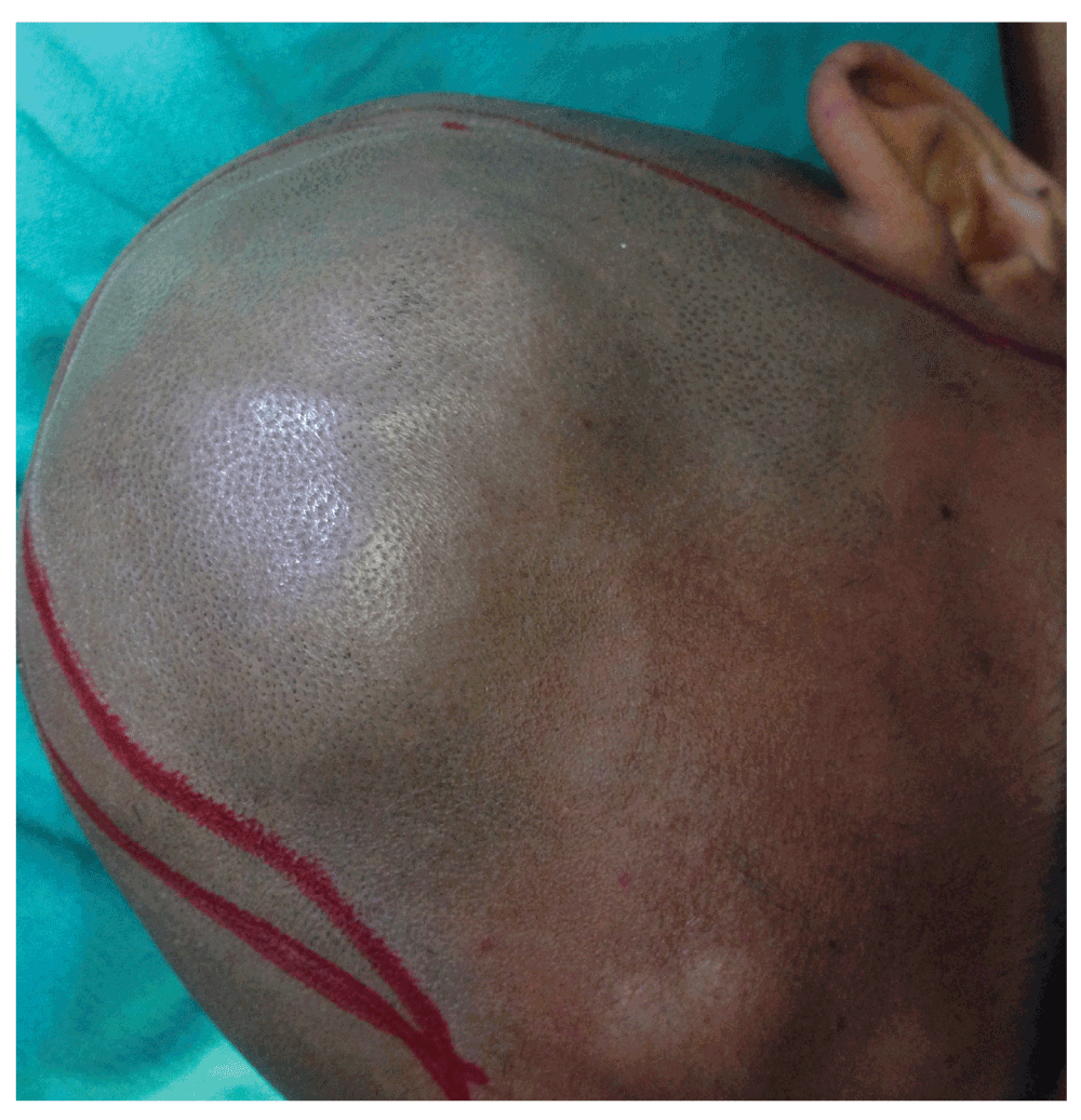
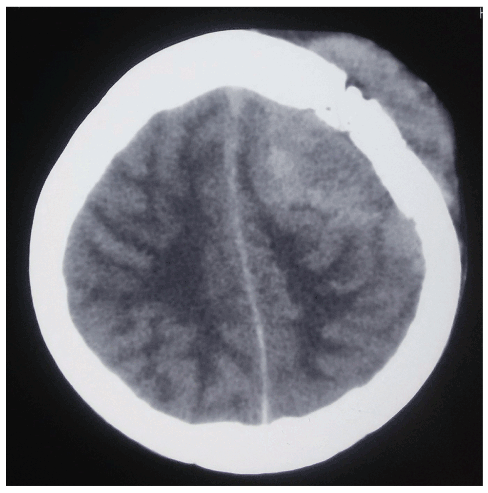
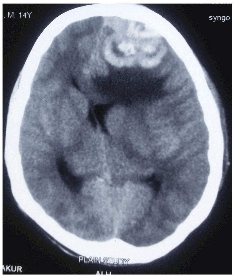
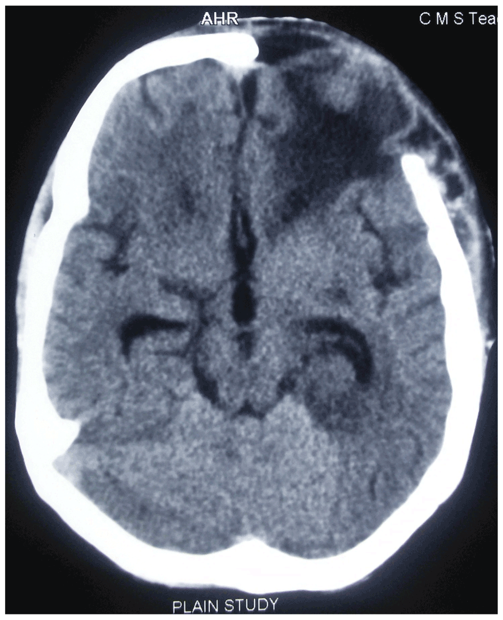
On initial examination at our ER room, the patient attained a Glasgow Coma Scale (GCS) of E2M4V2 with anisocoria on the left side. There were two scalp swellings on the left parietal and the frontal regions (Figure 1) which were soft and fluctuant. Serology performed was negative for human immuno-deficiency virus (HIV) and hepatitis B and C. Computed tomography (CT) scan of the head was performed, revealing a dural-based hyperintense lesion on the frontal and parietal region with subfalcine herniation (Figure 2 and Figure 3) and honeycomb appearance of the involved bone (Figure 4). Ultrasonography of the abdomen revealed no significant lymph nodes.
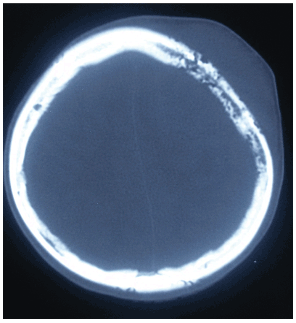
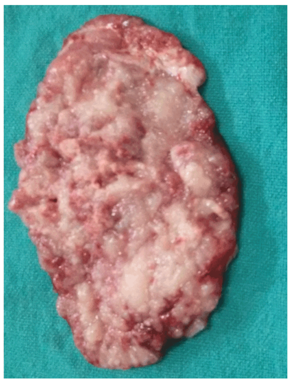
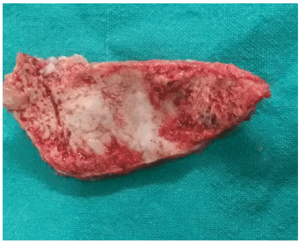
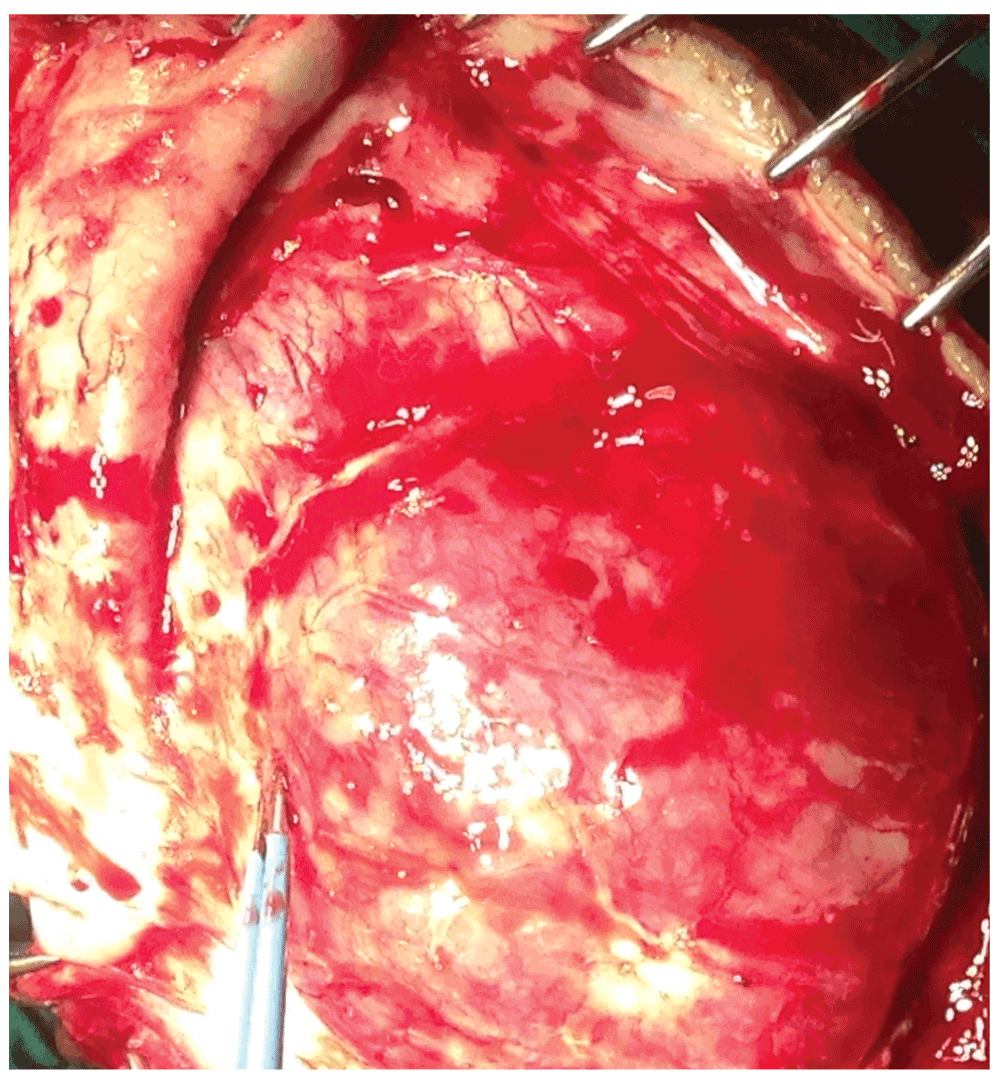
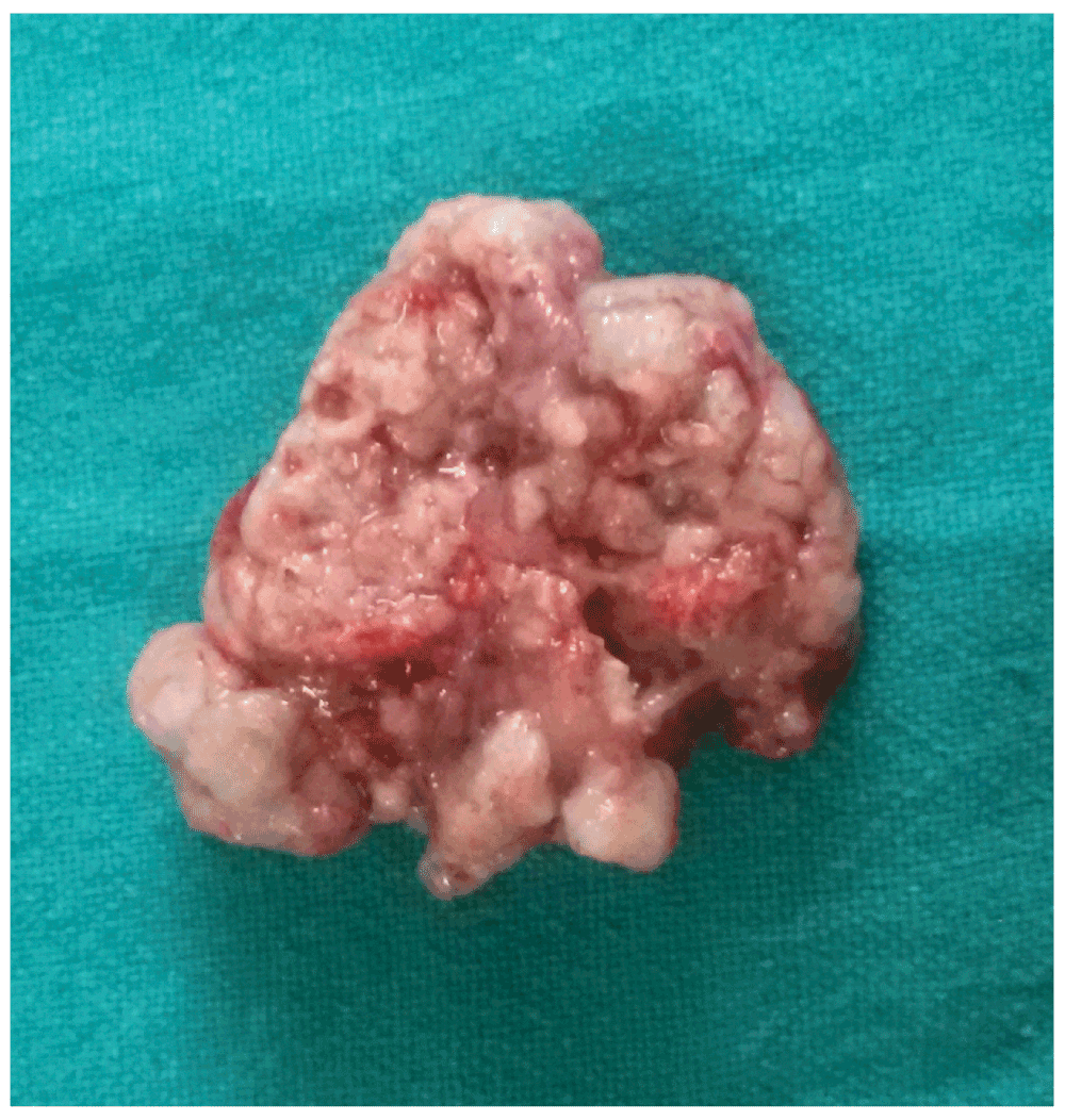
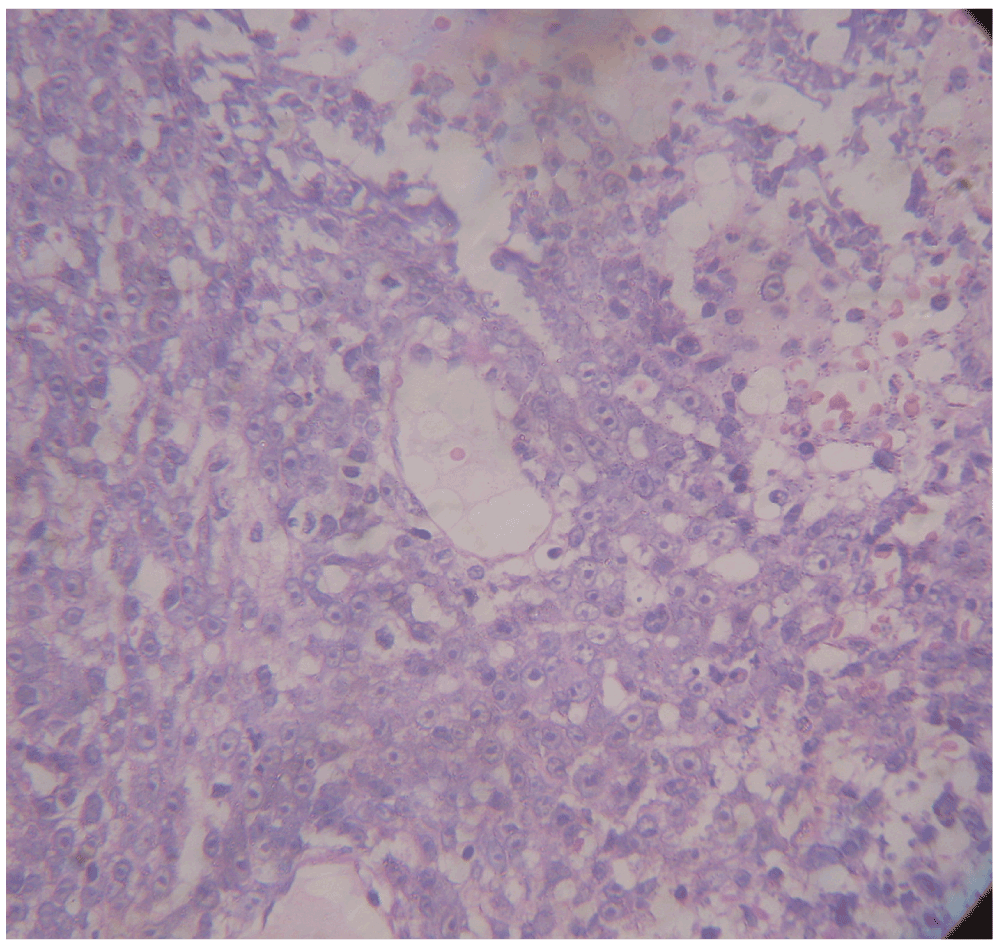
Because the child was already herniating, he was started on intravenous dexamethasone (4mg over 8 hours) and a single 100ml dose of 25% mannitol was given. Parents were counseled and written consent was taken for operative management. Surgery revealed a dural-based lesion (Figure 5) that was moderately vascular, soft and friable in consistency with involved bone showing a moth-eaten appearance (Figure 6). Both extra- and intra-dural extension (Figure 7 and Figure 8) of the lesion was seen. Scalp lesions, involved bone, and the dural and intradural component were all excised and sent for histopathological (HPE) study. A post-operative scan showed gross excision of the lesions and absence of mass effect (Figure 9). The HPE revealed an immunoblastic variant of diffuse large cell lymphoma (Figure 10).
Postoperatively, the patient improved to GCS 15. The steroids were slowly tapered off as the mass effect and edema were absent on repeat CT image and also prolonged usage would hamper healing of scalp surgical wound. The patient was thoroughly counseled and then referred for free chemo- and radiation therapy in a government oncology hospital.
Primary dural lymphoma, first described by Oberling5, is an exceedingly rare disease entity. Only five cases of primary dural diffuse large B-cell lymphoma have been described so far with a median age at diagnosis of around 50 years1. Trauma, inflammation and viral infection have been postulated as probable causes6. The symptoms of the disease are variable and non-specific. The radiological findings are indistinguishable from other dural-based lesions such as meningiomas and hemangiopericytomas2. Since the prognosis of intracranial DLBCL is favourable4, it is important to make a correct and timely diagnosis. Rapid progression of the symptoms, lytic lesions on the bone and restricted diffusion in magnetic resonance imaging (MRI) may provide additional clues to the diagnosis. In cases where there are no obvious neurological symptoms, it may be advisable to take a needle biopsy of the scalp tumor as described by Ochiai et al.7. There has been no consensus on the correct treatment protocol in the management of dural large-cell lymphoma due to a paucity of cases3. Previous cases have been treated with tumor resection followed by cyclophosphamide, hydroxy-doxorubicin, oncovin (vincristine) and prednisone (CHOP) with or without rituximab- or methotrexate-based chemo regimes. Additional radiation was also tried in some cases1. This case is the youngest age where the entity has been observed and showed good recovery despite initial presentation with herniation syndrome. Therefore, we suggest that maintaining a high index of suspicion and timely intervention is the key to better outcome in the patients.
Written informed consent for publication of their clinical details and images was obtained from the father of the patient.
| Views | Downloads | |
|---|---|---|
| F1000Research | - | - |
|
PubMed Central
Data from PMC are received and updated monthly.
|
- | - |
Provide sufficient details of any financial or non-financial competing interests to enable users to assess whether your comments might lead a reasonable person to question your impartiality. Consider the following examples, but note that this is not an exhaustive list:
Sign up for content alerts and receive a weekly or monthly email with all newly published articles
Already registered? Sign in
The email address should be the one you originally registered with F1000.
You registered with F1000 via Google, so we cannot reset your password.
To sign in, please click here.
If you still need help with your Google account password, please click here.
You registered with F1000 via Facebook, so we cannot reset your password.
To sign in, please click here.
If you still need help with your Facebook account password, please click here.
If your email address is registered with us, we will email you instructions to reset your password.
If you think you should have received this email but it has not arrived, please check your spam filters and/or contact for further assistance.
Comments on this article Comments (0)