Keywords
Urinary bladder cancer, Squamous cell carcinoma, Non-urothelial bladder cancer, Fungal ball
Urinary bladder cancer, Squamous cell carcinoma, Non-urothelial bladder cancer, Fungal ball
Squamous cell carcinoma (SCC) of the bladder is a rare malignancy in Western countries, accounting for only about 5% of all primary bladder cancers1. Chronic irritation is the predominant risk factor, with recurrent infections, bladder stones and long term catheterisation common precursors, and the highest incidence occurring in patients with spinal cord injuries who rely on indwelling or self-catherisation for bladder drainage2,3. In the Middle East and Northern Africa, where schistosomiasis is endemic, the incidence is much higher, with SCC being the predominant subtype of bladder cancer4. We report a case of primary SCC of the bladder secondary to a fungal ball located in the renal pelvis.
A 72 year-old Caucasian lady was referred to our unit for further investigation of recurrent polymicrobial urinary tract infections (UTI) complicated by sepsis and associated with intermittent left flank pain. An ultrasound performed by the treating team revealed a poorly defined, poorly vascularised mass in the lower pole calyx of her left kidney associated with moderate ipsilateral hydronephrosis.
The patient had extensive medical co-morbidities including chronic renal impairment, ischaemic heart disease with unstable angina, aortic stenosis and mitral regurgitation in addition to a previous cerebrovascular event (CVA) 8 years prior with residual left sided weakness and blindness, and a known but unclipped middle cerebral artery aneurysm.
We arranged further investigations including a mercaptoacetyltriglycine (MAG 3) renogram, computerised tomography (CT) of her renal tracts and urine cytology. The MAG3 renogram demonstrated obstructed drainage of urine from the left renal pelvis. CT again demonstrated left-sided hydro-nephrosis, with a poorly defined mass in the lower pole calyx, with no obvious contrast enhancement. Urine cytology was negative for malignancy with only non-specific inflammatory changes present.
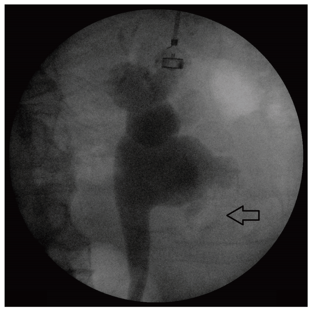
Cystoscopy, retrograde pyelogram (RGP) and insertion of ureteric stent were arranged to further investigate her renal mass and hydronephrosis. Cystoscopic examination of the bladder was unremarkable. RGP demonstrated a “moth-eaten” left lower pole filling defect (Figure 1). Washings from the renal pelvis were taken via ureteric catheter. A ureteric stent was placed and she was discharged on the same day.
The day following her procedure she presented to the emergency department with urosepsis. Her urine and blood failed to culture a causative organism and she was subsequently discharged on oral trimethoprim 300mg daily for seven days after clinical improvement following intravenous piperacillin/tazobactam, 4.1g three times daily.
Urine cytology from operative renal pelvis washings were again inconclusive, with non-specific inflammatory changes only, and flexible pyeloscopy was arranged two weeks later in order to better assess her renal mass. The bladder was again unremarkable on cystoscopic examination. Flexible pyeloscopy demonstrated a poorly defined mass in the lower calyx. Visualisation of the mass was poor due to debris in the renal pelvis and contact bleeding. Washings and tissue biopsies were retrieved and her ureteric stent was replaced. The patient was discharged the following day on oral norfloxacin 400mg twice daily for seven days.
Biopsies and washings following pyeloscopy demonstrated no evidence of malignancy, but fungal elements were identified on microscopy. Re-look pyeloscopy was arranged in two weeks to exclude urothelial malignancy as visualisation of the mass at the previous pyeloscopy had been poor.
At repeat flexible pyeloscopy we again saw no macroscopic evidence of a renal pelvis tumour. However, a white plaque was seen extending over the lower pole calyces suggestive of a fungal ball. After subtotal removal of the fungal plaque with an endoscopic basket, a temporary ureteric catheter was placed. Post-operatively, the patient became haemodynamically unstable and unresponsive to fluid resuscitation and was transferred to the ICU. Treatment with teicoplanin (800mg twice daily loading dose, following by 400mg once daily dosing) and piperacillin/tazobactam (4.1g three times daily) was initiated due to her history of polymicrobial UTI. Intravenous caspofungin (70mg once daily loading dose, 50mg once daily maintenance) was used in addition to cover for possible fungaemia. Vasopressor support necessitated ICU stay for three days prior to a further seven days of intravenous piperacillin/tazobactam on the surgical ward. She was discharged on oral fluconazole 200mg once daily for two weeks.
As a result of urosepsis repeatedly complicating any urological procedure, the patient remaining currently asymptomatic and her multiple medical comorbidities, we decided to manage her fungal ball conservatively and no further procedures were planned at this stage.
After several uneventful months, the patient represented with a further episode of urosepsis again unresponsive to fluid resuscitation. A further extended stay in ICU on vasopressor support was required. CT of the urinary tract at this time demonstrated recurrent mild left hydronephrosis and reappearance of debris in the lower pole calyces. A ureteric stent was placed in case upper tract obstruction had precipitated her sepsis. In view of her continued septic episodes and recurrence of her fungal ball, we decided in conjunction with the patient to prepare for an elective simple nephrectomy. Infectious diseases prescribed oral voriconazole 200mg twice daily to be continued prophylactically on discharge until her planned nephrectomy which was tentatively scheduled in 1 months’ time.
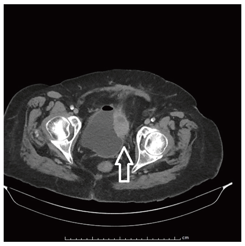
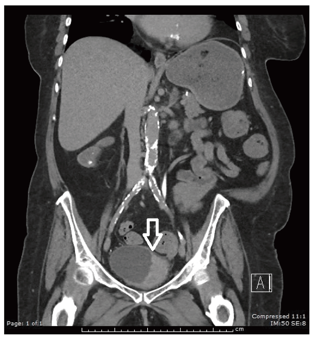
Prior to her planned nephrectomy she was re-admitted for a further episode of urosepsis, this time complicated by the new symptom of macroscopic haematuria with passage of multiple clots. After failed conservative management of her haematuria with bladder irrigations, flexible cystoscopy was performed to identify the source of ongoing bleeding. A large ulcerated erythematous mass was found, adjacent to her left ureteric orifice (Figure 2 and Figure 3). CT confirmed an enhancing bladder mass 5cm in diameter, arising from the left wall of her bladder (Figure 4). Resection of bladder tumour was performed and confirmed a pure squamous cell carcinoma of the bladder with invasion into muscularis propria (Figure 5, Figure 6, Figure 7 and Figure 8).
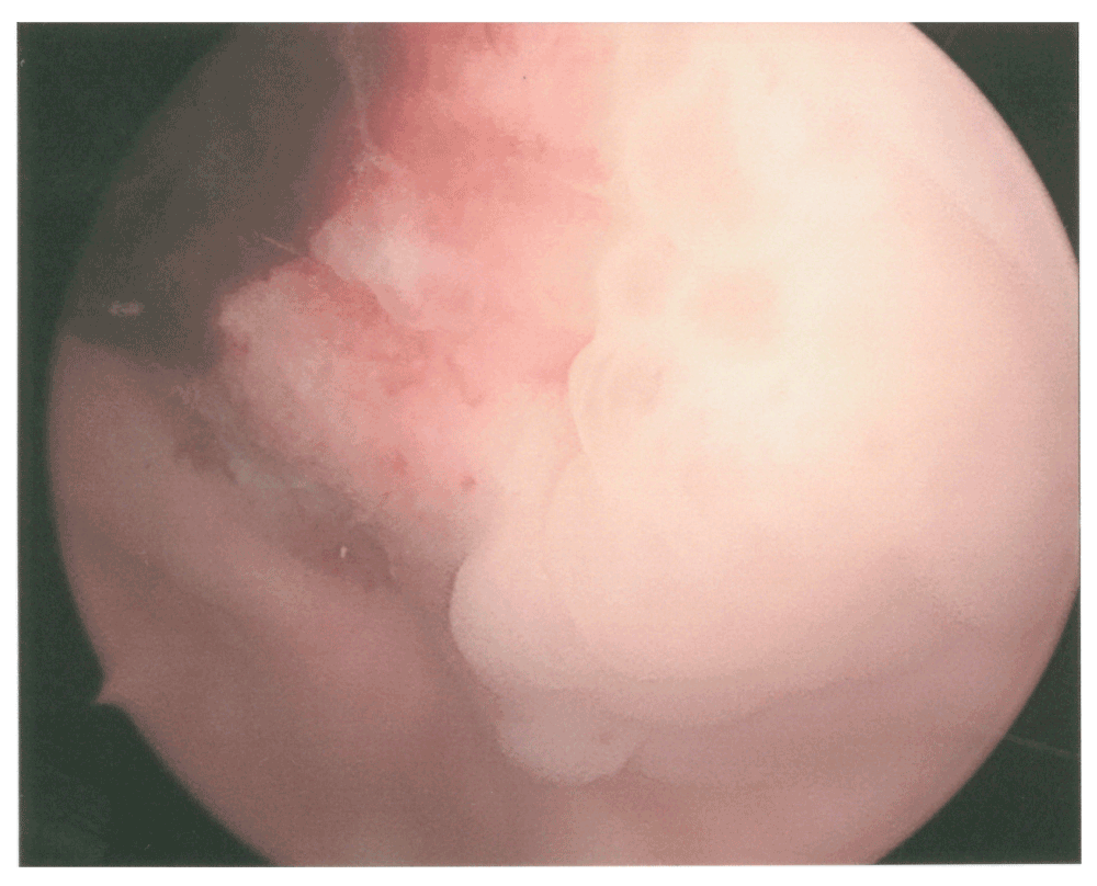
Cystoscopic examination revealed a large irregular ulcerated bladder mass with adherent blood clot.
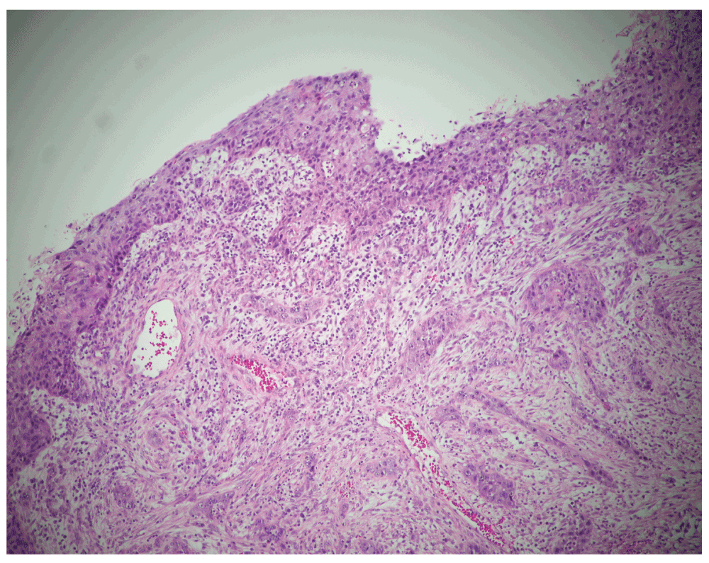
Squamous carcinoma in situ is seen overlying a focus of SCC which has invaded into the lamina propria.
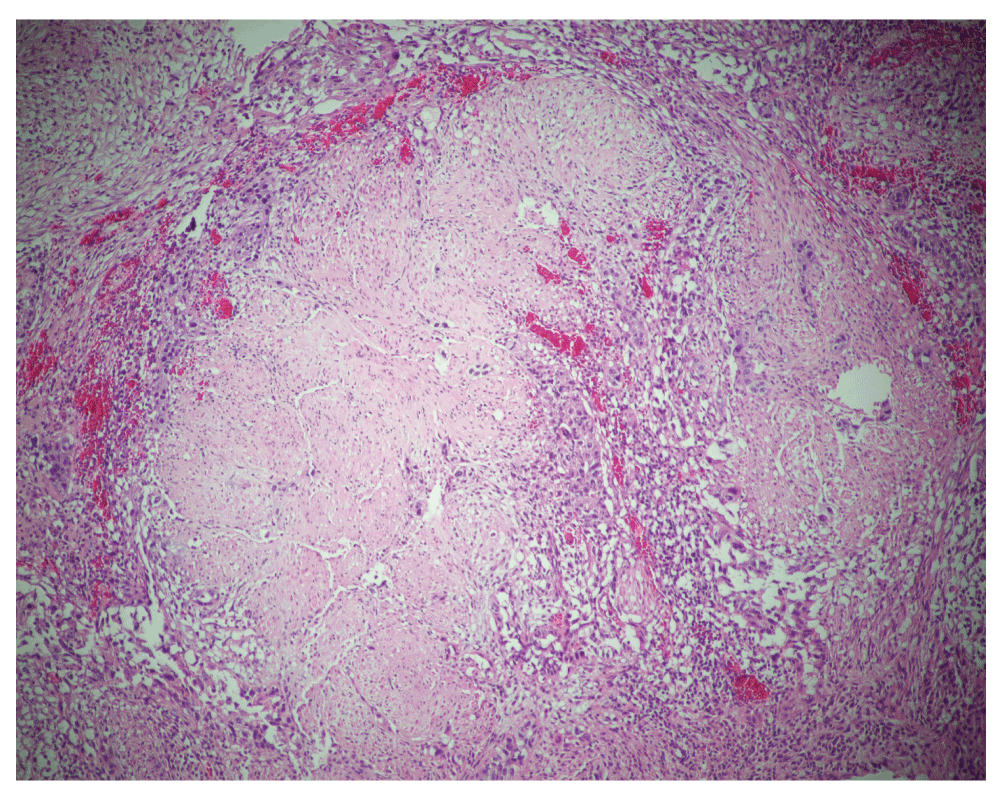
SCC is here seen invading into the muscularis propria.
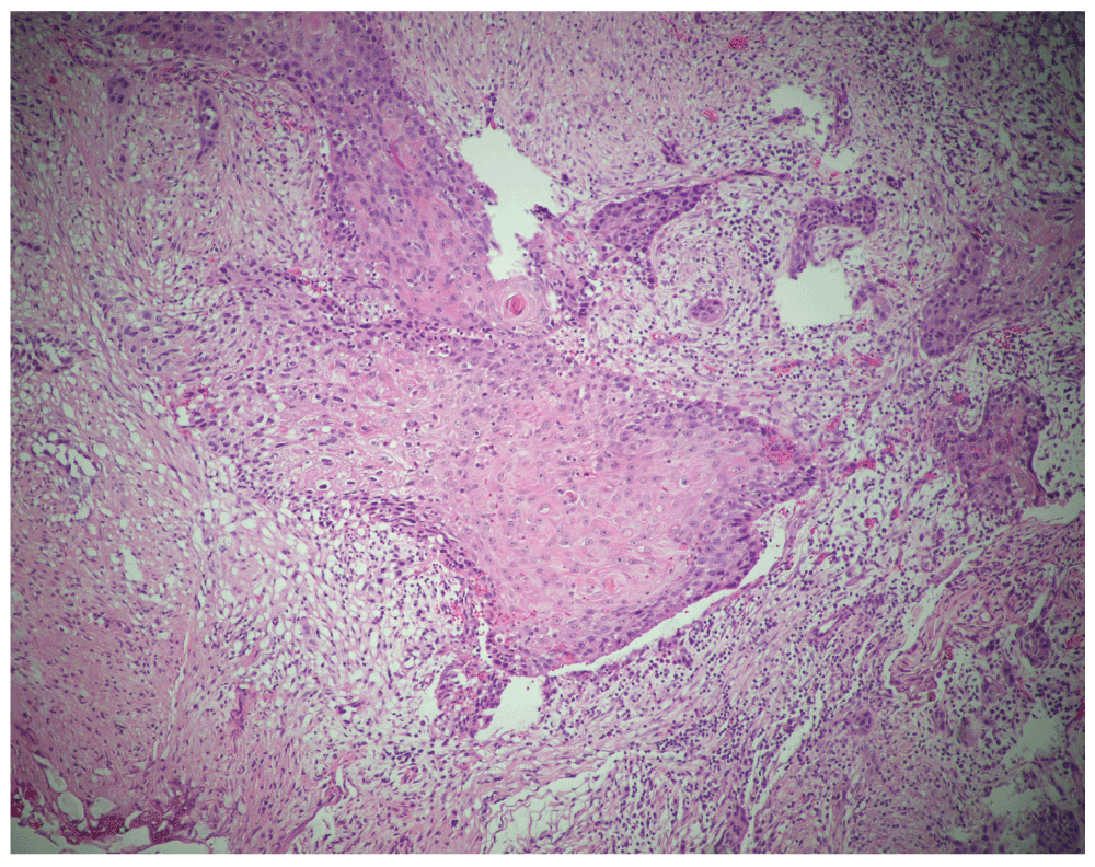
Invasive carcinoma with squamous differentiation with inclusion of a keratin pearl.
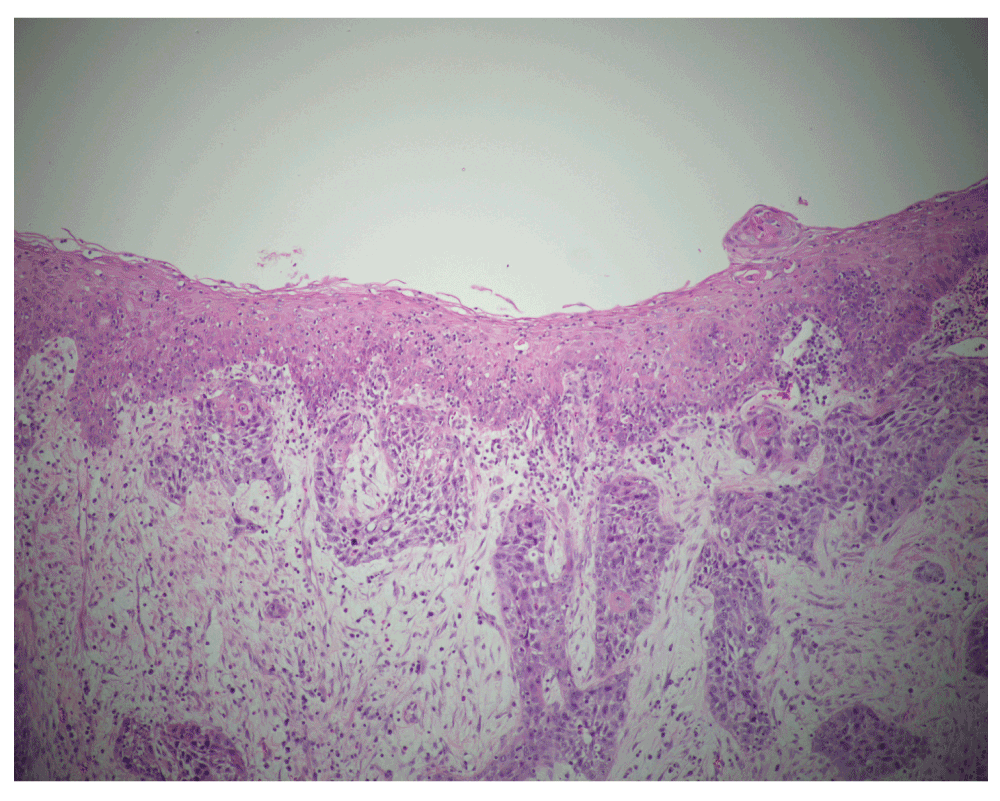
Superficial squamous metaplasia without atypia is seen overlying SCC which has invaded the lamina propria.
The patient was booked for a radical cystectomy and formation of an ileal conduit. At operation the tumour was found to have invaded pubic symphysis and was deemed unresectable. The planned cystectomy was aborted and instead a palliative urinary diversion was created. The patient was reviewed by medical and radiation oncology in regards to her suitability for adjuvant chemo-radiotherapy but the consensus was that she would not benefit from further aggressive treatment.
One month post-operatively she developed recurrent bleeds from her bladder with difficulty and much discomfort expelling the clots. She received two fractions of palliative radiotherapy to her bladder.
Prior to receiving further planned doses she was again admitted to hospital with another urinary tract infection and further bladder haemorrhage. Whilst in hospital she continued to deteriorate despite antibiotics and after discussion with the patient and her family, comfort cares were initiated. She passed away just over three months following her urinary diversion and less than four months following her diagnosis of squamous cell carcinoma.
SCC of the bladder is an uncommon primary malignancy of the bladder. Whilst chronic irritation, and chronic urinary tract infection are well known predisposing factors in Western populations, there is to date, no reported cases of bladder SCC secondary to fungal balls2,3.
SCC of the urinary tract typically presents at an advanced stage. In a large series of 1422 non-bilharzial SCC cases 85% were muscle invasive at diagnosis5. Additionally 56% of all bladder SCC were graded as American Joint Committee on Cancer (AJCC) stage T3 or T4 at diagnosis5.
SCC of the urinary tract has a significantly higher mortality than primary urothelial carcinoma, even after adjustment for higher tumour stage for SCC at diagnosis5. Scosyrev et al. found that for T4 tumours, even with cystectomy two year survival rates for SCC were low at 28%, where-as for stage matched urothelial carcinoma survival was significantly improved at 42%. Two year survival rates for T4 SCC without cystectomy is poor at only 5%5.
We present, we believe, the first documented case of primary SCC of the bladder secondary to an upper tract fungal ball. Despite the patient being under close urological surveillance for her fungal ball at the time of tumour development, the cancer demonstrated trans-mural invasion at initial diagnosis and was surgically unresectable. The disease was rapidly progressive and the patient passed away fewer than four months following diagnosis.
Written informed consent for publication of their clinical details and clinical images was obtained from the patient’s next of kin.
| Views | Downloads | |
|---|---|---|
| F1000Research | - | - |
|
PubMed Central
Data from PMC are received and updated monthly.
|
- | - |
Provide sufficient details of any financial or non-financial competing interests to enable users to assess whether your comments might lead a reasonable person to question your impartiality. Consider the following examples, but note that this is not an exhaustive list:
Sign up for content alerts and receive a weekly or monthly email with all newly published articles
Already registered? Sign in
The email address should be the one you originally registered with F1000.
You registered with F1000 via Google, so we cannot reset your password.
To sign in, please click here.
If you still need help with your Google account password, please click here.
You registered with F1000 via Facebook, so we cannot reset your password.
To sign in, please click here.
If you still need help with your Facebook account password, please click here.
If your email address is registered with us, we will email you instructions to reset your password.
If you think you should have received this email but it has not arrived, please check your spam filters and/or contact for further assistance.
Comments on this article Comments (0)