Keywords
Computed tomography, Conventional angiography, laparoscopic donor nephrectomy, renal artery, renal vein, Vascular anatomy
Computed tomography, Conventional angiography, laparoscopic donor nephrectomy, renal artery, renal vein, Vascular anatomy
Renal transplantation is the treatment of choice for patients with end stage renal disease. With less morbidity and early recovery laparoscopy has become the standard of care for donor nephrectomies1–3. Laparoscopic donor nephrectomy has a steep learning curve and the most limiting factor of the surgery is the vascular anatomy of the kidney. There are different ways of imaging the kidney before donor nephrectomy. These include conventional angiography (CA), digital subtraction angiography (DSA), computed tomography (CT) and magnetic resonance imaging (MRI).
The most preferred imaging modality worldwide is CT angiography. Traditionally conventional angiography was used for assessing the vascular anatomy in case of donor nephrectomies. The advantages of conventional angiography is that it uses less dye than CT angiography and it gives information regarding differential contribution in case of multiple arteries. The drawback of conventional angiography is that it does not delineate the venous anatomy. It does not give an idea about the surrounding anatomy and their relations. Conventional angiography is not useful in diagnosing associated findings such as gall stones and adnexal pathologies. The disadvantage of CT angiography is the larger amount of contrast used and the radiation exposure.
There is paucity of literature describing the accuracy of CT angiography in detecting the vascular anatomy. The reported accuracy of CT angiography in assessing the vascular anatomy is around 85 to 100%4,5. We did a retrospective study to assess the diagnostic accuracy of CT angiography in evaluation of vascular anatomy in comparison with intraoperative findings in 392 patients who underwent laparoscopic donor nephrectomy in our hospital from January 2010 to December 2012.
392 consecutive patients who underwent laparoscopic live donor nephrectomy from January 2010 to December 2012 were included in the study. Their CT scan findings were noted from the stored archives of our hospital. The data of patients who did not consent to participate in the study were excluded. All CT data were obtained using a 4-MDCT scanner. The parameters noted in the CT angiography are the artery and vein number on both the sides, any prehilar branching, the number of lumbar veins, relationship of the artery to the vein and other morphological abnormalities or any other incidental findings. Ethical approval was obtained from the institutional ethics committee (EC/317/2015).
The patients were accepted for donor nephrectomy after approval by the transplant board of the hospital and after due clearance from the regulatory authorities. Donor nephrectomy was done using a laparoscopic approach and recipient surgery was done using an open approach. One patient required conversion to open surgery. The intraoperative findings noted were the artery and vein number, prehilar branching, and lumbar vein anatomy. The intraoperative findings were recorded by viewing all the operative videos of patients who underwent laparoscopic donor nephrectomy. All the data obtained were analysed. The preoperative findings in terms of number of artery, vein, and lumbar vein were compared with the intraoperative findings. The specificity, sensitivity, and positive and negative predictive values were calculated. Any incidental finding present during the surgery was also noted.
Of the 392 patients who underwent laparoscopic live donor nephrectomy, 353 patients underwent left donor nephrectomy and 39 patients underwent right donor nephrectomy. Of the 392 patients, 263 were females and 129 were males. In 353 patients who underwent left donor nephrectomy, 26 patients had double renal artery and 17 had double renal vein. Two had both double artery and vein. Triple renal artery was found in 4 patients and triple renal vein was found in 1 patient and the rest had single renal artery and vein. Two patients had double ureter. In 39 patients who underwent right donor nephrectomy, 5 had double vein and 1 had double artery and the rest had single renal artery and vein. The CT scan was able to identify prehilar branching on both sides in 84 patients, on the left side in 40 patients and on the right side in 12 patients. The CT angiogram showed retroaortic left vein in 13 patients and circumaortic renal vein in 7 patients and 1 patient had IVC duplication. The additional findings identified incidentally in CT angiogram are renal calculus in 5 patients bladder mass in one patient, uterine fibroid in 2 patients, renal mass in 1 patient, gall bladder calculi in 2 patients, ovarian cyst in 2 patients, hemangioma of liver in 2 patients, adrenal myolipoma in 1 patient, and utero vesical fistula in 1 patient (Table 1).
The patient with an utero vesical fistula underwent hysterectomy followed by laparoscopic donor nephrectomy.
CT interpreted a case of double renal vein as single and a case of circumaortic vein reported on CT was not detected intraoperatively. A case of right side early branching was not detected on CT. A case of a retroaortic branch of renal vein was missed on CT scan. Rest of the intraoperative findings were correlating with that of the CT scan.
The intraoperative data was available in the case of 147 patients for lumbar vein and correlated with the CT angiogram in all cases (Table 2).
Single renal artery and vein was found in 86% of patients, 14% of patients had multiple vessels. The incidence of double renal artery was 7.3% on the left side and 12% on right side. The incidence of double renal vein was 4.8% on left side and 2.5% on the right side (Table 3).
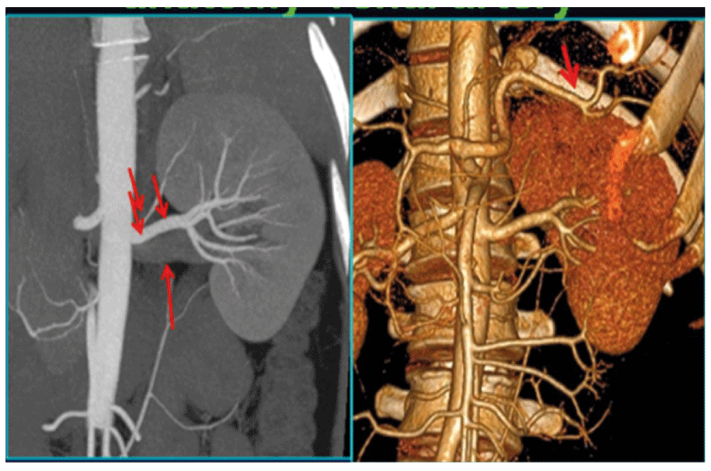
Precise knowledge of vascular anatomy before laparoscopic donor nephrectomy is vital in avoiding complications as it helps the surgeon to be better prepared in case of an emergency. The identification of accessory vessels on CT scans helps in avoiding inadvertent injury and reduces complications. When an accessory vessel is identified on CT preoperatively appropriate management of the vessel can be planned i.e bench surgery or separate anastamosis. In our institute it is mandatory for the operating surgeon to study the detailed vascular anatomy of the donor in the CT console before surgery. The surgeon sees the 3D reconstruction of the kidney along with the vascular anatomy. The surgeon discusses the relationship of the artery and vein and the number of vessels with the radiologist and gets the approximate idea about the lie of the artery and vein beforehand which helps in the dissection. Apart from predicting the number of vessels, the CT angiography study helps in strategic planning for laparoscopic donor nephrectomy. For instance in Figure 1, it can be noted that there is a small subcapsular branch from the origin of the artery, the artery lies at the upper border of the vein posteriorly. It shows that the arterial stump is of adequate size. In addition the images show that the splenic vessels are close to the upper pole.

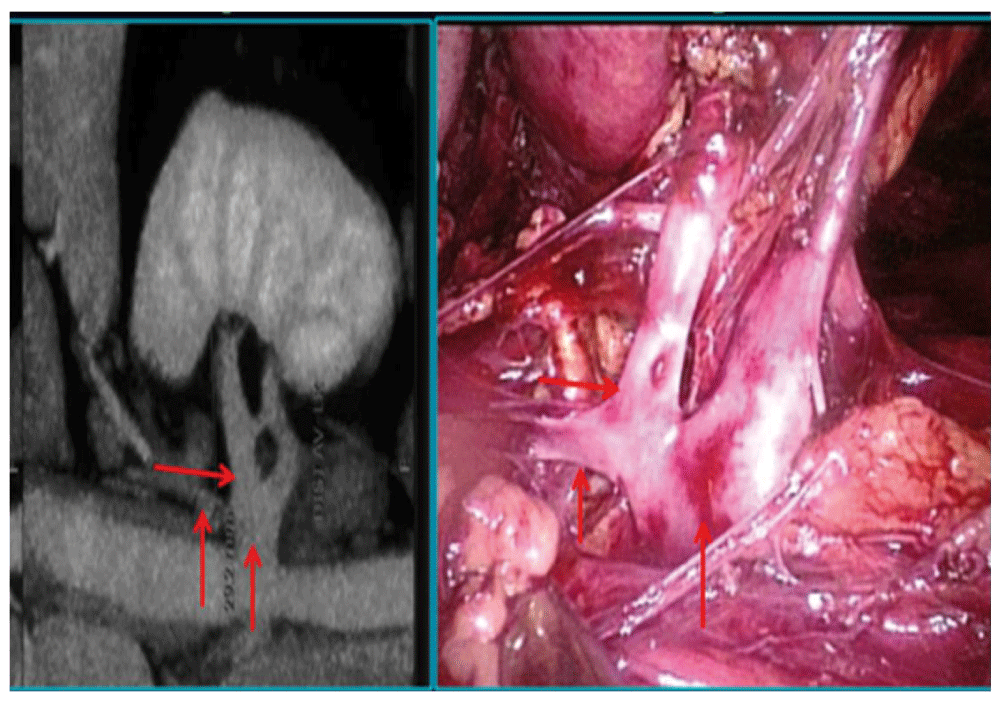
Apart from arterial anatomy, CT angiography also gives us the detailed venous anatomy. For example in Figure 2, it can be seen that the vein branches in two and the adrenal vein is close to the upper branch. This gives an idea about the length of the common stump of the vein.

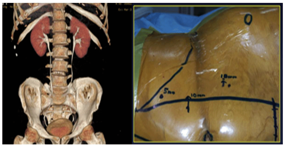
The location of hilum and the relationship of the kidney with the ribs on CT also helps in deciding the port placement. For example if the hilum of the kidney is above the 12th rib, the ports will be placed cranially and vice versa (Figure 3, Figure 4).


In our study the incidence of single renal artery and vein was 86% which was higher than the literature7. The incidence of double renal artery was 7.3% on the left side and 4.8% on the right side in our study, which was lower in comparison to the literature7. The incidental findings detected by CT scan such as calculi, mass and hemangioma or fibroid can be of help in managing the patient after surgery. In our case where we incidentally detected a case of uterovesical fistula, the patient underwent a hysterectomy followed by laparoscopic donor nephrectomy (Figure 5).
The incidence of multiple arteries was 14.2% in our study. The incidence of multiple renal arteries was 9% on the left side which was lesser compared to the study by Shetty et al.7 who reported the incidence rate of 17% on the left side. We had 2% incidence of multiple renal arteries on the right side whereas Shetty et al.7 did not have any multiple vessels on the right side. The incidence of multiple renal veins was 5.6% on the left side and 12.8% on the right side in our study whereas Shetty et al.7 reported an incidence of 1% on the left side and 8% on the right side.
The positive and negative predictive value in our study for single artery and vein and double artery was 100 for both the left and right side. In case of double vein on the right side, the positive and negative predictive values were 100 whereas on the left side the positive predictive value was 99.7 and the negative predictive value was 100. In a study by Shetty et al.7 the positive predictive value and negative predictive value for venous anatomy in left side correlation was 99.5. Similarly for arterial anatomy in left side it is was 96.8. It was 100 for arterial and venous anatomy in the right side7. Pozniak et al.8 mentioned that sensitivity and specificity for identifying specific vessels was 99.6% and 99.6% for main renal arteries, 76.9% and 89.9% for polar arteries, and 98.7% and 95.5% for main renal veins, respectively. Satomi et al.9 found that CT and surgical findings were accurate in 96% of cases for arteries and 99% of cases for veins.
In comparison with traditional angiography which was used in the past for evaluation of donors, CT angiography has the advantage of visualisation of venous anatomy and better delineation of extravascular anatomy.
Laparoscopic donor nephrectomy requires vital information regarding the vascular anatomy of the kidney. CT scan helps in accurately mapping the vascular anatomy. CT angiography helps in strategic planning of the surgery in terms of port placement and vascular dissection and helps in avoiding vascular complications. The incidental findings detected by CT scan can also help in holistic management of the patient in donor nephrectomy because it is a zero error procedure.
Written informed consent for the publication of clinical data and images was obtained from each patient.
MV: substantial contributions to conception and design, acquisition of data, and analysis and interpretation of data and drafting the article or revising it critically for important intellectual content; SG: substantial contributions to conception and design, acquisition of data, and analysis and interpretation of data and drafting the article or revising it critically for important intellectual content; AG: substantial contributions to conception and design, acquisition of data, and analysis and interpretation of data and drafting the article or revising it critically for important intellectual content; VM: Contributions in acquisition and analysis and interpretation of data and drafting and revising the article; SM: contributions in drafting the article or revising it critically for important intellectual content, and final approval of the version to be published; RS: contributions in drafting the article or revising it critically for important intellectual content; and final approval of the version to be published; MD: contributions in drafting the article or revising it critically for important intellectual content, and final approval of the version to be published.
| Views | Downloads | |
|---|---|---|
| F1000Research | - | - |
|
PubMed Central
Data from PMC are received and updated monthly.
|
- | - |
Peer review at F1000Research is author-driven. Currently no reviewers are being invited.
Provide sufficient details of any financial or non-financial competing interests to enable users to assess whether your comments might lead a reasonable person to question your impartiality. Consider the following examples, but note that this is not an exhaustive list:
Sign up for content alerts and receive a weekly or monthly email with all newly published articles
Already registered? Sign in
The email address should be the one you originally registered with F1000.
You registered with F1000 via Google, so we cannot reset your password.
To sign in, please click here.
If you still need help with your Google account password, please click here.
You registered with F1000 via Facebook, so we cannot reset your password.
To sign in, please click here.
If you still need help with your Facebook account password, please click here.
If your email address is registered with us, we will email you instructions to reset your password.
If you think you should have received this email but it has not arrived, please check your spam filters and/or contact for further assistance.
Comments on this article Comments (0)