Keywords
DNA isotope labeling, PCR, DNA extraction, silicon chip, surface coating, isotope recombination
DNA isotope labeling, PCR, DNA extraction, silicon chip, surface coating, isotope recombination
The CIS method (for combing-imaging by SIMS) combines DNA combing on a silicon surface, flooding the surface with cesium, and the sensitive imaging technique of dynamic secondary ion mass spectrometry (D-SIMS) as provided by a Cameca NanoSIMS 50, a machine that allows five or seven different masses to be detected simultaneously, depending on the version1,2. This method may be of interest for those who wish to obtain fine-scale, quantitative information on DNA replication and protein-DNA interactions at the level of single molecules without the need to modify these molecules. Previously, we have used the CIS method and a NanoSIMS 50 to obtain quantitative images of single DNA fibers combed on modified, SIMS-compatible, silicon surfaces with lengths similar to those expected for the B-conformation and with a resolution of 50 nm, i.e., 150 bp3.
Variations in the rates and/or directions of DNA synthesis are implicated in many processes, including gene expression4, nutrient sensing involving the alarmone (p)ppgpp5, R-loop-mediated replication6 and DNA repair7. Variations between replisomes within the same chromosome have been observed using BrdU and light microscopy8. To study such variations on the 50 nm scale, CIS might usefully be extended to image not only the 12C15N- and 13C15N- recombinant ions but also other ions. Here, we describe in detail the protocols needed for CIS, we report using CIS to image the 13C-, we suggest ways in which 13C-imaging could be improved, and we mention possible applications.
In principle, DNA could be extracted from any organism to perform CIS. DNA generated in vitro can also be used. For the experiment reported here, for example, we extracted DNA from the 168 (trpC2) strain of Bacillus subtilis and a derivative (MT119, trpC2 leuB6 r- m-) harboring a 21.1 kb long, chloramphenicol-resistant plasmid (pHV1431 plus insert of 10.3 kb)3. On modified silicon surfaces at concentrations of 0.2 µg/mL, most of the DNA is combed as single fibers. Higher concentrations can lead to fibers running together whilst lower concentrations result in an insufficient number of fibers on the images. Combing works well with buffers containing NaCl at ionic strengths of around 0.2M.
Bacterial culture medium: 14g/L of K2HPO4, 6g/L of KH2PO4, 1g/L of sodium citrate, 10mM MgSO4, 0.01% (w/v) of tryptophan, 0.005% (w/v) of leucine, 0.2%(w/v) of 13C-glucose (Isotec) and 0.8g/L of 15NH4Cl (Isotec). Lysis buffer: 50mM of Tris-HCl (pH8.0), 10mM of EDTA (pH8.0), 150mM of NaCl and 5mg/ml of lysozyme. Proteinase K: Final concentration of 0.2mg/mL (Roche). Sarcosyl Buffer: Final concentration of 1.2%. Phenol/chloroform: Solution PCI: phenol-chloroform-isoamylalcohol (25:24:1 v/v) (the phenol is first saturated in NaCl 150mM and buffered at pH of about 7); Solution CI: chloroform-isoamylalcohol (24:1 v/v). RNase: Final concentration of 20µg/mL (Roche).
Other materials required are: 100% cold ethanol; 70% (v/v) ethanol; NaCl solution: 0.2M to 1M; PureYieldTM plasmid midiprep system (Promega); restriction enzyme; 0.3M Potassium acetate pH7.0; TaKaRa EX TaqTM system (TaKaRa); Expand Long Template PCR System (Roche Applied Science); QIAquick PCR Purification kit (QIAGEN).
DNA preparation from bacterial cultures. DNA can be prepared from any cells using a procedure that removes contaminating peptide and RNA molecules. In the case of the labeling and extraction of B. subtilis DNA, B. subtilis cells were cultivated at 30–37°C for ~20 generations in bacterial culture medium containing stable isotopes (here, 13C and 15N, see above) to saturation. Then, to prepare chromosomal DNA free of peptides and RNA, one milliliter of a freshly saturated culture was centrifuged at 15000 g for 2 min at 4°C. The supernatant was discarded. Pelleted cells were resuspended in 0.5 mL of lysis buffer. The cell resuspension was incubated for 20 minutes at 37°C. Proteinase K was added to a final concentration of 0.2 mg/mL in addition to 20µL of sarcosyl buffer 30% (1.2%, final concentration) and incubated for 20 min at 65°C. To remove peptides and cell fragments by a phenol/chloroform treatment, we lowered the temperature of the sample on ice for 3 min and then added 500µL of solution PCI (see Preparation of DNA) at 4°C and vortexed strongly for 30 s. The mixture was centrifuged at 15000 g for 15 min at room temperature in a benchtop centrifuge. The aqueous solution was recovered and 500 µL of solution CI was added to it (see Preparation of DNA). This mixture was vortexed strongly and centrifuged as above. RNase was added to a final concentration of 20 µg/mL to the aqueous, nucleic acid-containing phase and incubated for 10 min at 37°C. DNA was purified by a second phenol/chloroform extraction as above. We added 2.2 volume of 100% -20°C ethanol to the aqueous solution and centrifuged at 15000 g for 20 min at 4°C in a benchtop centrifuge. The supernatant was discarded and the DNA pellet was washed in 70% cold ethanol. This mixture was centrifuged again for 10 min at 4°C at 15000 g and the supernatant was discarded. The DNA pellet was dried for 10 min under vacuum and resuspended carefully in pure water for at least 12 h at room temperature (note that incomplete solubilization of ethanol-precipitated DNA severely perturbs combing). DNA concentration was measured by absorbance at 260/280 nm using a Nanodrop 2000 (Thermo Scientific). Depending on the fragment length, DNA concentration should be adjusted to within the range 0.2 to 2 µg/mL by dilution in NaCl solutions that may range up to to 1M; here, we used DNA at 0.2µg/mL in 0.2M NaCl.
In this paper, we only report CIS in the case of chromosomal DNA. CIS can also be used to analyse plasmid DNA (not shown). In the case, for example, of a 20 kb plasmid from B. subtilis, plasmid DNA may be extracted using the PureYieldTM plasmid midiprep system (Promega Corporation, Madison, WI, USA), followed by linearization of the plasmid by a single cutter restriction enzyme according to suppliers’ instructions (e.g. New England BioLabs, Inc., Hitchin, UK) to give fragments of the appropriate size; this linearization is needed before combing because of the difficulty of combing supercoiled circular molecules. After linearization, the DNA can be purified by the phenol/chloroform procedure and recovered by ethanol precipitation in the presence of 0.3 M potassium acetate pH 7.
DNA preparation by PCR (protocol used to prepare PCR-generated DNA fragments 1-20 kb long). DNA fragments generated by PCR can be analyzed by CIS3. We routinely produce fragments <5 kb using the TaKaRa Ex TaqTM system (Takara Shuzo Co., Ltd, Shiga, Japan). For products of 5-20 kb, we use the Expand Long Template PCR System from Roche Applied Science (Mannheim, Germany). The PCR products can be purified with the QIAquick PCR Purification kit (QIAGEN GmbH, Hilden, Germany). PCR products can then be diluted for CIS as described above.
This experiment required an efficient fume hood. The materials used were: P-type 100 crystalline silicon wafers (Siltronix) with a resistivity > 1 Ω cm (typically 10 mm × 10 mm × 380 μm); Piranha solution obtained by mixing 3 volumes of 96% of sulfuric acid [VLSI (very large-scale integration)-grade] and 1 volume of 30% hydrogen peroxide (VLSI-grade), to be used immediately; 50% hydrofluoric acid (VLSI-grade); acetone; isopropyl alcohol (iPrOH); chloroform (CHCl3); ethanol (EtOH); 1-tetradecene; a photochemical reactor for UV irradiation (λ=312 nm) (our home-made reactor has 8 tubes in a circle around a Schlenk tube with a fan at the bottom to limit any increase in temperature); a goniometer system (DIGIDROP, GBX, France).
Preparation of hydrogenated-terminated silicon surfaces. In the following preparation, it should be noted that the piranha solution is a powerful oxidant that reacts violently with organic materials; it can cause serious skin burns and must be handled with great care in a well-ventilated fume hood while wearing appropriate chemical safety protection. Moreover, HF is a hazardous acid that can cause severe damage to tissues if burns are not treated properly. Etching of silicon should therefore be carried out in a fume hood with the right safety measures, which include a face shield and double-layered nitrile gloves.
The silicon surface was cleaned in an ultrasonic bath for 5 min periods in acetone and isopropylic alcohol. It was then rinsed extensively with ultrapure water and immersed for 20 min in a piranha solution to remove organic contaminants on the surface. This was followed by the immersion of the clean surface in an aqueous solution of HF (50%, as provided by the supplier) to generate a hydrogen-terminated surface (Si-H)9. This surface was washed extensively with water and then dried by blowing with nitrogen. The resulting surface typically had a contact angle of 85° for a 1 µL water droplet. Note that freshly prepared Si-H surfaces were used immediately for DNA combing or for grafting hydrocarbon chains (see below). Before combing, coated silicon surfaces were protected from dust and dried because a thin film of water greatly reduces DNA adsorption.
Formation of organic monolayers on hydrogen-terminated silicon surfaces. To obtain highly hydrophobic surfaces, alkenes with C14 alkyl chains were grafted onto freshly prepared Si-H surfaces using the hydrosilylation reaction of 1-tetradecene with the hydrogen-terminated silicon surface9. The freshly hydrogen-terminated silicon surface (see above) was immersed in a Schlenk tube containing 10 ml of previously deoxygenated, neat 1-tetradecene under nitrogen bubbling. This was then irradiated at 312 nm in a photochemical reactor for 3 h. We removed unreacted and physisorbed 1-tetradecene by rinsing with chloroform and ethanol at room temperature. The silicon substrate was dried under a stream of nitrogen. We verified that water contact angles (using deionized water in the ambient atmosphere at room temperature) are around 104° as measured to an accuracy of ± 2° with a remote, computer-controlled, goniometer system (N.B., protect the surface from dust, see above).
Combing on silicon. The 'drop' and 'lift' methods10,11 can both be used to comb DNA on the Si-C14 surface. Both 'drop' and 'lift' methods entail the DNA becoming attached to the surface and then drawn out and aligned perpendicular to the triple line or the meniscus, respectively. The ‘drop’ method entails depositing 10 µL of prepared DNA solution on the silicon surface, incubating it for 10 min on the bench, tilting the surface with tweezers to 45° to cause the drop to roll off the surface, and washing the surface by immersion in water and rapidly air-drying; the quality of combing via this method can be adversely affected by vibrations occurring during the movement of the drop. Here, we used the ‘lift’ method, which entails immersing a part of the silicon surface in a DNA solution (see above); after 5 min incubation, the silicon surface was pulled out of the solution at a constant speed (600 µm/min) and rapidly air-dried. In both ‘drop’ and ‘lift’ methods, washing the surfaces after combing helps avoid perturbation of D-SIMS analysis by saline crystals. The tightly controlled, motor-driven ‘lift’ method gives more reproducible, better quality results but takes longer and requires more DNA than the manual ‘drop’ method. The ‘lift’ method is better than the drop method for fragments less than 5 kb; the ‘drop’ method is satisfactory with fragments equal to or greater than 5 kb.
Cs flooding method. To avoid a premature destruction of thin samples before SIMS detection12, a cesium flooding system is recommended to deposit neutral cesium on samples13,14. We used a UHV Cs evaporation system which is available for purchase from the Luxembourg Institute of Science and Technology (LIST) . It comprises a neutral cesium evaporator and an independent stand-alone UHV chamber. A suitcase under UHV (10-8 to 10-10 Torr) was used to transfer samples to the NanoSIMS50 thereby avoiding an immediate reaction between the neutral cesium deposit and air. To analyze combed DNA (as performed here), we flooded the surface with Cs0 at 1.5 Å/s for 1800 s before SIMS imaging. An alternative to cesium flooding is to coat samples with Au; a Cressington Sputter Coater can be used to deposit a layer of Au, approximately 60 nm thick, on wafers with combed DNA but the quality of the images subsequently obtained is often poor.
NanoSIMS 50 analyses. In SIMS analyses of samples labeled with carbon and nitrogen, there are several factors that should be borne in mind. First, carbon can be detected as C- and in multi-clusters that include CN- and C2- (N.B. CN is easier to ionize and therefore detect than C and C2 because the electron affinity is 3.8eV for CN-, 1.26eV for C- and 3.27eV for C2-). Second, nitrogen can be detected in the multi-cluster CN (note that nitrogen forms fewer types of multi-clusters thant carbon even though it can form to a very limited degree multi-clusters that include NO, N2H, and NS). Third, the probability of formation of the CN multi-cluster in the mixing-recombination under the primary beam is related to the distance between the carbon and nitrogen atoms15; this means that differentially labeled macromolecules can be colocalized if they are within 2 nm of one another; it also means that the carbon and nitrogen in the DNA are more likely to recombine with one another than with carbon and nitrogen that are further away. Note that even a trace amount of contaminants is an important problem for the highly sensitive CIS method and strenuous efforts should therefore be taken to protect samples from contamination by, for example, an atmosphere containing C and N. Silicon surfaces with combed DNA were analyzed in the multi-collection image mode of the NanoSIMS 50 (Cameca, Gennevilliers, France). The NanoSIMS 50 was used in the negative secondary-ion mode with the Cs+ primary ion beam. In this case high spatial resolution images were obtained using a primary beam around 0.5–1.0 pA in intensity and an impact energy of 16 keV. The surface was rastered by the cesium beam on a surface from 5 × 5 to 15 × 15 µm2. The instrument was tuned to limit the dispersion in aperture and energy in order to obtain a minimal mass resolution of M/ΔM = 5000 (10% height peak measurement).
Aphelion 3.2 was used to analyze the results pixel by pixel or line by line, whilst ImageJ plus the OPEN MIMS plug-in (which opens the .im format16 and is available from http://www.nrims.harvard.edu) was used to analyze the data in an ROI (Region Of Interest). The data were summed and the images colored using WinImage 2. The images of the 12C (m = 12.000000), and/or 13C (m = 13.00335484), 12C14N (m = 26.003074), 12C15N (m = 27.00010898) or 13C14N (m = 27.00642885), 13C15N (m = 28.00346382) were acquired simultaneously in 256 × 256 pixels with a dwell time of 10 ms per pixel. Note that 12C15N and 13C14N cannot be detected simultaneously in the same ‘sputter section’. The sulfur 32S (m = 31.9720718) distribution can be also acquired to control the quality of the preparation.
The CIS technique reveals individual DNA fibers of a wide variety of lengths lying in parallel (Figure 1). These fibers are particularly clear in the 13C15N image of the DNA (Figure 1a) but, importantly, they can also be discerned in the 13C14N image (Figure 1b). The images differ because (1) the coating on the silicon surface contains a large amount of the natural isotopes of carbon of which 1% is 13C; this 1% can then recombine with contaminant nitrogen to generate a high background of 13C14N that partially masks the signal from the labeled DNA (even though this DNA is 99% 13C) and (2) the background of 13C15N is low since this ion arises at a frequency of 1:25000 in combinations of naturally occurring carbon and nitrogen).
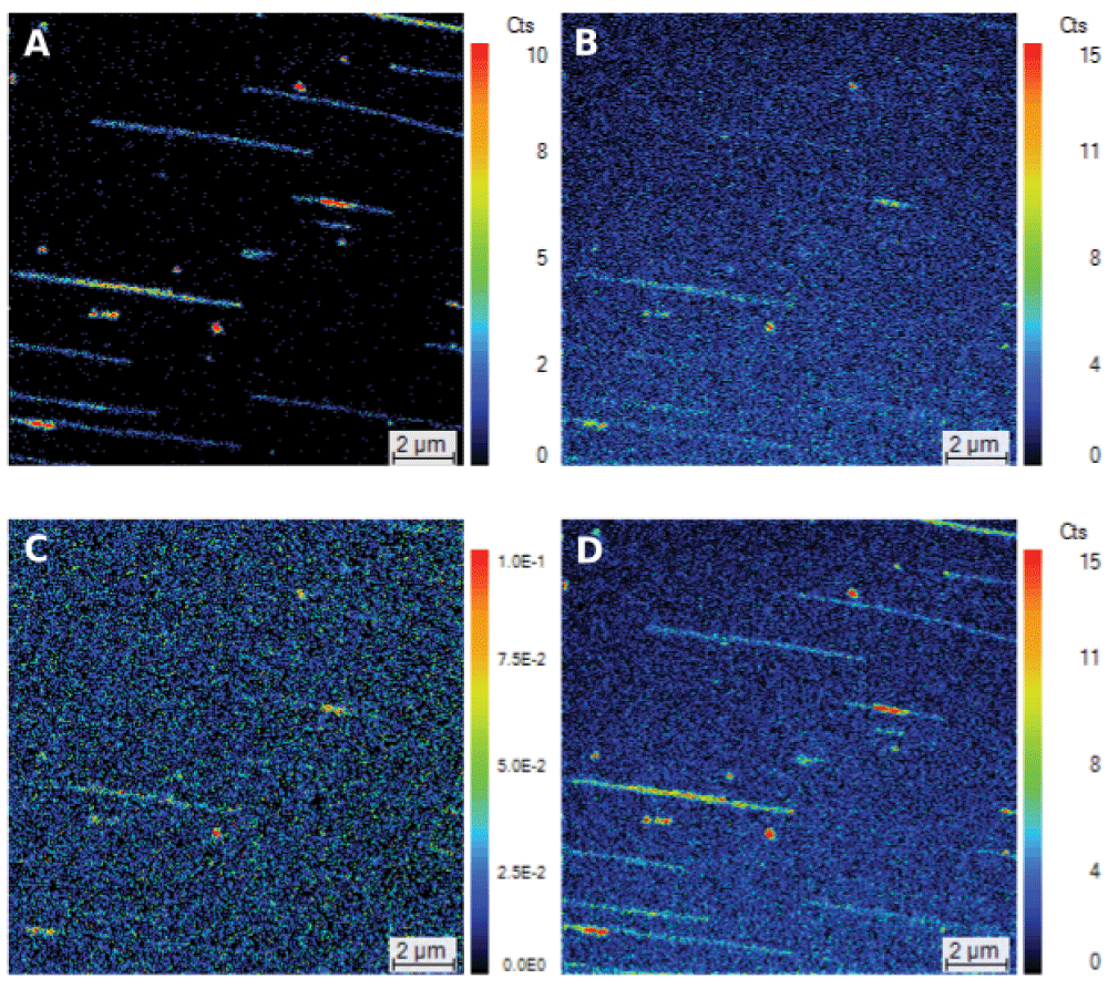
The DNA was combed at 0.2 μg/mL on a Si_C14 wafer using the lift method, the wafer surface was covered with cesium and analyzed with a NanoSIMS 50. Primary beam intensity, 1 pA; dwell time, 30 ms; field of view, 15 μm × 15 μm; 256 × 256 pixels; scale bar = 1 μm. (a) 13C15N, (b) 13C14N, (c) Isotope ratio 13C/12C, and (d) 13C15N+13C14N. (a) and (b) are the results of adding the counts from three successive sputter sections, (c) is the ratio between the two sets (13C and 12C) of three successive sputter sections, and (d) is the three sputter sections of (a) plus the three of (b). The count numbers of (a), (b) and (d), and the count ratio of (c) are given on the linear color scales.
Most of the fibers have similar counts throughout their lengths but, in a couple of cases, two fibers overlap and this results in more 13C15N- recombinant ions being detected (the red stretches in Figure 1a). The relative constancy of the counts and the similarity between the calculated lengths of the DNA and the lengths estimated by SIMS support the claim that CIS does not stretch or condense DNA3. However, an exception to this may occur when the DNA is so short that it cannot be combed properly, as is probably the case of the red spots of a few nm (Figure 1a).
The fibers in the image of the isotope ratio 13C/12C image (Figure 1c) are difficult to detect in their entirety. One reason for this difficulty is that the natural carbon in the coating on the surface contains 13C, as mentioned above. A second reason is that the yield of carbon in the C- form is relatively low and therefore the ratio can fluctuate (see Materials and methods). A third reason is that the carbon in the DNA reacts with neighboring atoms in the DNA (and elsewhere) such as nitrogen – and indeed carbon itself – to give multi-clusters (see Materials and methods); this means that much of the 13C is being distributed into multi-clusters that include 13C15N, 13C14N, 12C13C and 13C13C. It is therefore significant that, even in these unfavorable conditions for detecting 13C via the 13C:12C ratio, 13C-labeled fibers can just about be distinguished (Figure 1c).
In the experiment reported here, 98% of the nitrogen in the DNA is 15N. This means that the counts of 13C14N coming from recombination between the atoms within the DNA are nearly 98% lower than they would be if all the nitrogen in the DNA were present as the 14N isotope. To answer the question of whether labeling with 13C alone (i.e., without 15N-labeling) is sufficient to allow DNA to be detected, we added the counts of 13C15N (Figure 1a) to those of 13C14N (Figure 1b) to obtain an image of 13CN (Figure 1d). The fibers are again clear, showing that 13C labeling is indeed sufficient to allow DNA to be detected.
Previously, we have suggested that CIS could be used in conjunction with pulse-labeling with different isotopes to identify origins of replication or to study local variations in the rate of DNA elongation resulting from signals generated inside or outside cells or from addition of drugs3. Such pulse-labeling can be readily based on 15N since this is relatively rare naturally (0.36%) and enriching in 15N therefore makes fibers readily detectable. Here, we ask whether 13C labeling with 13C alone might suffice for detection via CIS. We try to answer this question in the context of having labeled the fibers with both 13C and 15N (Figure 1a). Even in these conditions, the image of the 13C14N- recombinant ion does allow fibers to be discerned, albeit with difficulty (Figure 1b). This difficulty is due not only to reduction of the ratio by surface contamination by carbon (which contains 1% 13C) but also to the recombination that generates multi-clusters (see Materials and methods); the latter includes recombination between the 13C and 15N in the fibers to give 13C15N (it is therefore not surprising that the fibers are hard to detect in Figure 1c using the ratio of the 13C- and 12C- ions). Another way to decide whether 13C14N can be used for imaging when DNA is labeled with just 13C is to add the counts from both 13C14N and 13C15N by combining the results in Figure 1a & 1b so as to increase the counts of 13C (Figure 1d). This shows that the fibers can indeed be discerned (although it should be noted that here both strands are labeled rather than just one strand being labeled as would occur after a short pulse). This result is significant because it means that, in the same experiment, one could label the DNA successively: first with 13C in 14N medium to detect 13C14N and then with 15N addition to detect 13C15N. One might even succeed in detecting three consecutive sequences, for example, by (1) labeling with 13C (via U-13C-Glc) to detect the 12C13C- or 13C13C- recombinant ion (or indeed simply the 12C- and 13C- ions), then by (2) labeling with 15N (via 15NH4Cl) to detect 13C15N, and finally by (3) replacing either the 13C with 12C (via U-12C-Glc) to detect 12C15N or the 15N with 14N (via 14NH4Cl) to detect 13C14N. Such consecutive labeling would allow investigation of local variations in the velocities of replication.
In principle, CIS could permit testing of several hypotheses. These include (1) the idea that the elongation step of DNA replication is coupled to central carbon metabolism17–19 and that this changes the local structure of the DNA20,21 or changes the relative copy numbers of genes4 and thereby affects the phenotype, (2) the idea that the strand separation resulting from ion decondensation is responsible for the initiation of DNA replication22, (3) the idea that the time to rereplicate the chromosomes in a cell population growing in steady state is highly variable since these populations are phenotypically diverse23,24, (4) the idea that the replication of a particular species of bacteriophages or viruses is very diverse. CIS could also be used to study the fine-scale interaction between DNA sequences and RNA, proteins and polyamines. Finally, CIS might eventually be used to study the modification of individual DNA fibers due to the covalent addition of methyl and acetyl groups, sugars, and other molecules, always providing that these molecules could either be labeled specifically by very rare isotopes such as 14C or bound specifically by antibodies or aptamers that were themselves labeled.
The value of such potential applications of CIS depends on the number of different ions that can be detected and on the quality of this detection (which depends on factors such as the signal:background ratios, the dwell time, the intensity of the primary beam, and the use of serial sputter sections). With the latest NanoSIMS 50L version of the NanoSIMS range, seven different masses can be detected simultaneously in the acquisition of a single ‘sputter scan’25, which increases the numbers of carbon multi-clusters that can be analyzed. Finally, the quality of detection of 13C- and the recombinant 13CN- ions could be improved by coating the silicon surface with carbon depleted in 13C. In principle, therefore, CIS could distinguish between DNA labeled so as to give consecutive stretches enriched in (1) 13C (detectable as 13C13C), then (2) 13C and 15N (detectable as 13C15N) and, finally, (3) 12C (detectable as 12C15N) (note there are other possible combinations). CIS could, of course, be extended further still if the DNA were labeled with Br or I or even 14C26.
CIS combines the advantages of DNA combing and SIMS in providing high sensitivity and high resolution since DNA fragments down to 1500 nm can be imaged at a resolution of 50 nm. Moreover, the labeling with stable isotopes does not significantly perturb cells. The fact that CIS can detect 13C14N increases the number of separate labels available to the technique, thereby making CIS particularly valuable for studying phenotypically important variations in the elongation of DNA replication, as well as the processes of initiation of replication, recombination and repair. The increase in the repertoire of labels available to CIS also supports the case for it being adapted to study the interaction between DNA and other molecules, including proteins and drugs, and perhaps even to the precise localization of covalent modifications to DNA.
F1000Research: Dataset 1. Raw data for Combing-Imaging by SIMS of bacterial DNA, 10.5256/f1000research.8361.d12027827
| Views | Downloads | |
|---|---|---|
| F1000Research | - | - |
|
PubMed Central
Data from PMC are received and updated monthly.
|
- | - |
Click here to access the data.
Spreadsheet data files may not format correctly if your computer is using different default delimiters (symbols used to separate values into separate cells) - a spreadsheet created in one region is sometimes misinterpreted by computers in other regions. You can change the regional settings on your computer so that the spreadsheet can be interpreted correctly.
Provide sufficient details of any financial or non-financial competing interests to enable users to assess whether your comments might lead a reasonable person to question your impartiality. Consider the following examples, but note that this is not an exhaustive list:
Sign up for content alerts and receive a weekly or monthly email with all newly published articles
Already registered? Sign in
The email address should be the one you originally registered with F1000.
You registered with F1000 via Google, so we cannot reset your password.
To sign in, please click here.
If you still need help with your Google account password, please click here.
You registered with F1000 via Facebook, so we cannot reset your password.
To sign in, please click here.
If you still need help with your Facebook account password, please click here.
If your email address is registered with us, we will email you instructions to reset your password.
If you think you should have received this email but it has not arrived, please check your spam filters and/or contact for further assistance.
Comments on this article Comments (0)