Keywords
Aortic stenosis, transcatheter aortic valve replacement, complications, intraoperative transesophageal echocardiography, coronary occlusion
Aortic stenosis, transcatheter aortic valve replacement, complications, intraoperative transesophageal echocardiography, coronary occlusion
Transcatheter aortic valve replacement (TAVR) is an increasingly popular approach for high-risk patients with severe aortic stenosis. Coronary ostium occlusion after valve implantation is a life-threatening complication that occurs in up to 0.4% of TAVR procedures despite the growing experience in performing this procedure and the constant improvements in TAVR devices1,2. We report the partial occlusion of the ostium of the left coronary artery (LCA) by the skirt of the core valve that was converted into a total occlusion following the initiation of heparin reversal with protamine and the value of multimodal imaging in the management of this case.
A 73-year-old woman with severe aortic stenosis (aortic valve area 0.7 cm2 and a mean gradient of 60 mmHg), preserved left ventricular (LV) systolic function, left ventricular hypertrophy, no significant coronary artery disease, and an Society of Thoracic Surgeons (STS) score of 3 presented to our institution for an aortic valve replacement (http://tools.acc.org/TAVRRisk/). The patient had compromised pulmonary function (FEV1 46% and DLCO 46%), and a catheter-based valve replacement was performed. Preoperative computer tomographic derived measurements demonstrated: aortic valve annulus diameter of 25 mm (major) and 20 mm (minor); coronary artery ostium distance of 18 mm (right) and 11.6 mm (left); coronary sinus diameter/height of 26/18 mm (right coronary), 28/19 mm (left coronary), 28/19 mm (non-coronary); and a moderate severe calcified valve with moderate-severe impaired leaflet excursion (Figure 1). A 26 mm Medtronic CoreValve (Medtronic, Minneapolis, MN, USA) was implanted via a right ileo-femoral approach. The patient had an uneventful valve deployment. A preoperative transesophageal echocardiography (TEE) verified a calcific, severely stenotic aortic valve with a normal left ventricular systolic function (ejection fraction 60% (Video 1 & Video 2). Deployment was facilitated using brief, temporary rapid pacing at 120 beats per minute; the patient was hemodynamically stable after valve deployment. A subsequent TEE examination revealed mild aortic paravalvular regurgitation (Video 3). A post-deployment angiography demonstrated a cephaled migration of the valve (from position 2 to -2), but coronary artery perfusion was maintained, as shown in a right anterior oblique (RAO) image (Figure 2B & Figure 3). Based on the images provided and the hemodynamic stability of the patient, the operative team was satisfied with the position of the valve and focused on repairing the femoral access site. The access site was repaired with normal flow twenty minutes after deployment as demonstrated by angiography. The decision was then made to reverse the anticoagulation with protamine. Shortly after the slow administration of protamine, the patient became hypotensive and unresponsive to vasopressor or inotropic therapy.
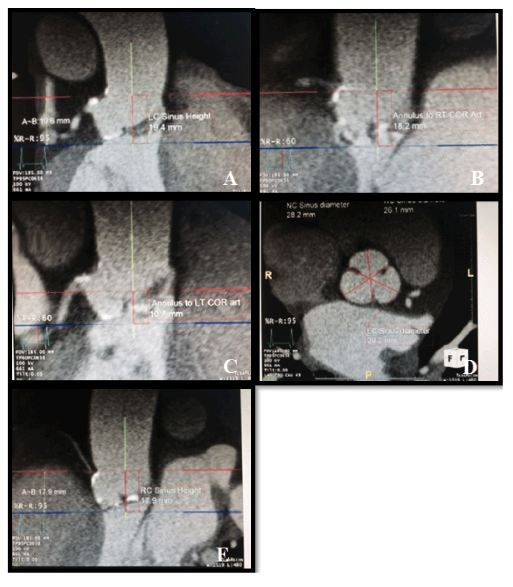
Preoperative measurements of the aortic valve.
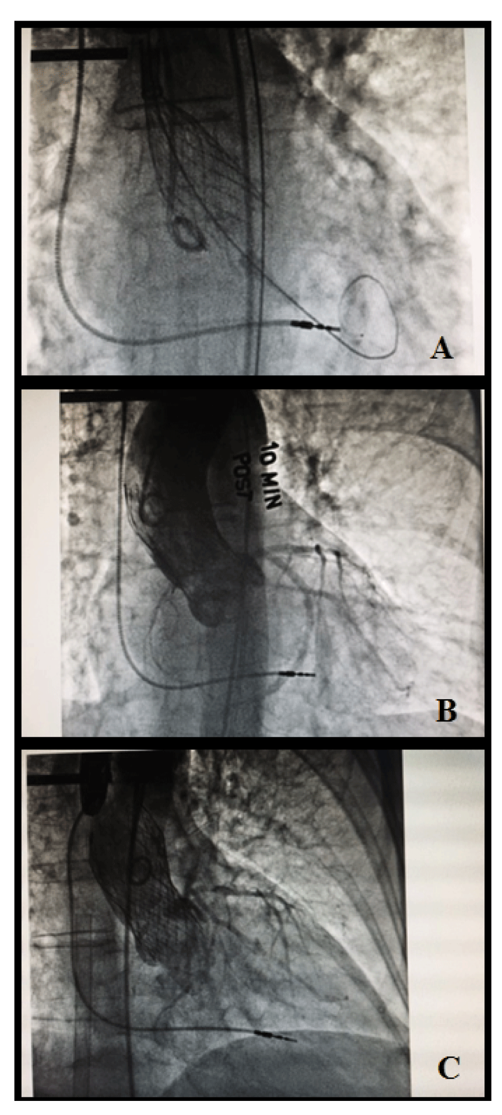
A. Details post-deployment of core valve. B. RAO view of the deployed valve, detailing flow. C. LAO view depicting lack of flow.
A TEE probe was emergently placed for examination and revealed normal right ventricular size and function, with global hypokinesis of the left ventricle (Video 4). These findings were consistent with a left main coronary artery occlusion in a left dominant system patient. Cardiopulmonary bypass (CPB) was instituted as a result of hemodynamic instability, and the diagnosis of coronary occlusion was confirmed with angiography (Figure 4). Due to the position of the valve, the decision was made to refrain from coronary stent placement and to convert to an open surgical replacement of the aortic valve. After the Medtronic CoreValve was removed, the left coronary ostium was examined and determined to be patent. After the placement of a 21 mm Perimount Magna valve (Edwards Lifesciences, Irvine, CA, USA), the patient was weaned from CPB without any problems. Further TEE was performed which revealed a normal left ventricular function. The patient was extubated on postoperative day one and remained hemodynamically stable without further concerns regarding her aortic valve or coronary ischemia. On the 7th postoperative day, the patient developed complete heart block that necessitated the placement of a dual chamber permanent pacemaker. She was discharged on postoperative day eight.
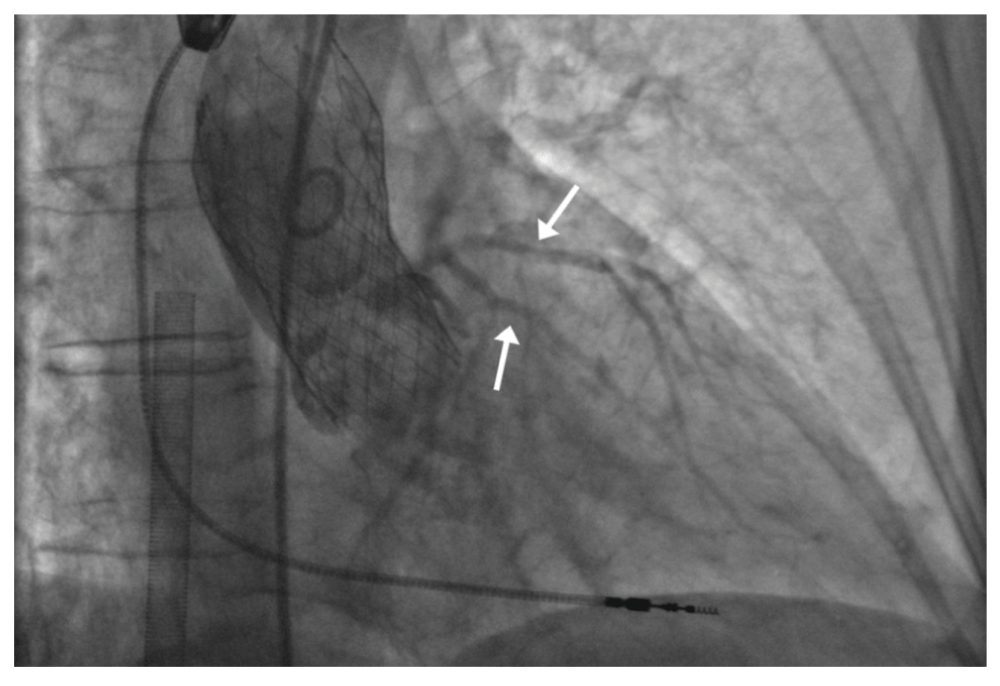
RAO coronary angiography showing flow through the left main coronary artery and branches.
TAVR is a novel approach for the treatment of high-risk patients with severe aortic stenosis, especially those deemed inoperable. Complications associated with TAVR include: (1) access related problems, including bleeding and occlusion (dissection or vascular); and (2) procedure-related problems such as cardiac tamponade, root rupture (annular or aortic), apical tears, right ventricular perforation, aortic regurgitation, coronary artery occlusion, renal failure, stroke, and conduction system disturbances requiring permanent pacemaker implantation.
Left main coronary artery occlusion after TAVR is a rare but potentially fatal complication. A complete occlusion of the left main coronary artery will manifest itself immediately; however, a partial compromise can be initially silent, such as the occlusion in our case. A partial LCA occlusion can result if calcific particles from the native aortic leaflet become displaced over the ostium while the aortic valve device is being expanded3,4. Other proposed mechanisms include incorrect positioning of the valve frame (obstructive portion) directly over the coronary ostium, hematoma formation or leaflet distortion, and apposition on the LCA ostium5,6. Valve design is an additional factor related to coronary obstruction7. Higher probabilities of occlusions occur with the balloon-expandable SAPIEN XT valve (Edwards Lifesciences, Irvine, CA, USA) than the Medtronic CoreValve4. This can be attributed to the hourglass-like design of the Medtronic CoreValve, which is less prone to native cusps displacement over the ostia4. However, a previous case has been reported using the Medtronic CoreValve that resulted in a left main coronary occlusion from calcium nodules3.
In our case, hemodynamic instability developed shortly after the administration of protamine. The initial working diagnosis suggested this instability resulted from an adverse reaction to the drug. Cardiovascular reactions to protamine vary from vasodilation-related hypotension to pulmonary hypertension, with subsequent right heart failure and cardiovascular collapse8. While treating our patient’s hypotension, which was unresponsive to vasopressors and inotropes, a TEE probe was placed and demonstrated global LV dysfunction. This observation was consistent with a LCA occlusion. Since neither right ventricular (RV) dysfunction nor indirect evidence of pulmonary hypertension were present, causality related to a severe protamine reaction was not likely.
In our case, a post-deployment coronary angiogram was done in the RAO projection and showed adequate coronary blood flow (Figure 3). Following the emergent implementation of CPB, another coronary angiography was performed in the left anterior oblique (LAO) projection and demonstrated complete cessation of flow through the left coronary ostium (Figure 4). Due to the high position (-2) of the Medtronic CoreValve, it was deemed impossible to place a stent across the occluded ostium and restore coronary blood flow in a timely fashion without lasting sequelae. Thus, the decision was made to surgically replace the calcific native valve after removing the prosthesis in place. After visualization of the valve in situ, it was noted that the skirt of the valve partially covered the left main coronary ostium. Due to the slightly high position of the prosthetic valve and reduced flow in the native “sinotubular reservoir”, protamine administration may have facilitated the formation of small micro thrombi aggregates adherent to the prosthetic material and led to a complete coronary occlusion (Figure 5). Even though a post-deployment cephalad migration of the valve (from 2 to -2) was immediately noted and raised concern regarding a coronary occlusion, an angiogram in the ROA view demonstrated good coronary flow. The assumption was made that the valve was situated in a fashion where adequate blood flow was maintained through the cage portion of the valve and was unobstructed by the overlying curtain (Figure 3).
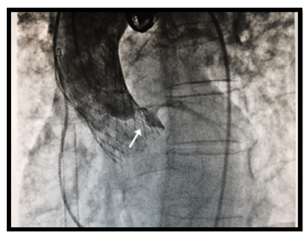
LAO coronary angiography showing lack of perfusion and stasis of blood within the left sinus of Valsalva.

This is the image of the core valve depicting point of obstruction.
In retrospect, an LAO view during the coronary angiography would have been helpful to visualize the ostium of the LCA. At the time that the RAO view was performed, filling of the coronary arteries was satisfactory and the ostium itself was not clearly visualized. This angiography most likely demonstrated adequate flow through a partial obstruction. After hemodynamic deterioration, a subsequent angiography in the LAO view did visualize the ostium of the LCA (Figure 2C and Figure 4). The Medtronic CoreValve was then removed and the left coronary ostium was noted to be completely patent, leading to no further LCA intervention. As a result of this case, the routine use of LAO coronary angiography has been instituted in our facility.
Several anatomic risk factors for coronary occlusion have been described in patients undergoing TAVR procedures. These include short distances to either coronary ostium (<10mm), shallow coronary sinuses, and narrow roots4,6. Our patient’s anatomy was appropriate for the placement of a Medtronic CoreValve; however, the distance to the LCA was just above 10 mm. This made the possibility of an occlusion after Medtronic CoreValve migration more likely.
Here we report a case of a coronary artery osteal occlusion from the skirt of the core valve that converted into a total occlusion possibly from micro thrombi after protamine administration. This case highlights the vital role of echocardiography in differentiating between a protamine reaction and a coronary occlusion. It also emphasizes use of proper views during coronary angiography to diagnose coronary ostium occlusion, in the context of TAVR. We recommend vigilance and detailed reexamination of any hemodynamic instability after TAVR in order to expeditiously search for and identify impaired coronary blood flow, as well as any additional causes.
Written informed consent was obtained from the patient for publication of this case report and any accompanying images and/or other details that could potentially reveal the patient’s identity.
Figshare: TEE short axis transgastric video. doi: 10.6084/m9.figshare.3507419.v19
Figshare: TEE aortic valve long axis color video. doi: 10.6084/m9.figshare.3507428.v110
Figshare: TEE four chamber video. doi: 10.6084/m9.figshare.3507431.v111
Figshare: TEE Post Deployment Aortic valve long axis color. doi: 10.6084/m9.figshare.357120612
| Views | Downloads | |
|---|---|---|
| F1000Research | - | - |
|
PubMed Central
Data from PMC are received and updated monthly.
|
- | - |
Provide sufficient details of any financial or non-financial competing interests to enable users to assess whether your comments might lead a reasonable person to question your impartiality. Consider the following examples, but note that this is not an exhaustive list:
Sign up for content alerts and receive a weekly or monthly email with all newly published articles
Already registered? Sign in
The email address should be the one you originally registered with F1000.
You registered with F1000 via Google, so we cannot reset your password.
To sign in, please click here.
If you still need help with your Google account password, please click here.
You registered with F1000 via Facebook, so we cannot reset your password.
To sign in, please click here.
If you still need help with your Facebook account password, please click here.
If your email address is registered with us, we will email you instructions to reset your password.
If you think you should have received this email but it has not arrived, please check your spam filters and/or contact for further assistance.
Comments on this article Comments (0)