Keywords
Radiation exposure, neuro intensive care, CT scans, Dose estimation, ALARA
Radiation exposure, neuro intensive care, CT scans, Dose estimation, ALARA
Computed tomography (CT) scans have become an integral part of the evaluation and management of patients with neurologic injury. The use of CT imaging has doubled every 2 years since the early 1980s to nearly 70 million scans worldwide in 20071,2. Widespread availability and ease of obtaining CT scans has tremendously aided diagnostics in hospitalized neurologic patients worldwide. This comes with an insidious risk of potential exposure to excessive quanta of ionizing radiation. The last decade has a seen an exponential increase in the number of publications related to all aspects of quality and patient safety in the intensive care unit (ICU), including ionizing radiation exposure3. However in spite of the recent publications highlighting the various risks, radiation exposure to patients seems to be in a blind spot of clinical decision-making, especially in the ICU. We seek to explore the patterns of radiation exposure to patients admitted to the neuro-intensive care unit (Neuro ICU) to better understand and review the methods and systems to implement to minimize radiation exposure in the ICU.
To describe the patterns of use of diagnostic CT imaging in a cohort of neuro intensive care patients during a single ICU admission.
The institutional review board approved our retrospective, quality improvement study (IRB # 141320 A QI project to determine pattern of CT scan usage and information to reduce the exposure of patients to ionizing radiation in the NCU). We included all adult patients who were admitted to our medical center Neuroscience intensive care unit between January 1, 2013 and December 31, 2013.
We retrospectively analyzed data on all CT scans that were completed on patients during their intensive care unit stay. The patient-specific dose of radiation delivered was not universally available on all patients at this time. We used previously published estimates of the doses of radiation based on individual types of diagnostic studies to calculate the arithmetic sum of estimated radiation exposure during the patient’s ICU stay. The radiation doses for CT scans used in our analysis were from previously published data from Mettler et al.4.
Radiation dose is measured by a number of units, including Sieverts (Sv) and Grays (gy). The Sievert reflects the effective dose and represents the stochastic biologic effects of ionizing radiation. The effective dose refers to the radiation risk averaged over the entire body because different tissues have different sensitivities to radiation. The Gray is the radiation measure used to reflect the absorbed dose. In our analysis we used the reported doses in Sievert units.
In the calendar year 2013, we had 2353 ICU admissions. A majority of the patients were admitted from the Neurology - Stroke service, general neurology service and the Neurosurgery service.
A total of 2028 CT scans were completed in this time period. 59.5% or 1208 studies were CT Head without contrast studies. The majority of these studies were done using a portable CT scanner (Model NL 3000, Neurologica Corp. Danvers MA, USA) in the N-ICU itself. A profile of the types and number of CT scans done in 2013 are shown in Table 2 (profile of types of CT scans).
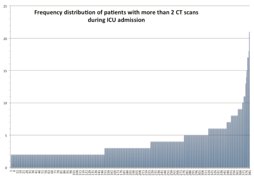
The frequency distribution of patients with two or more CT scans in the ICU is shown in Figure 1. 16.1% (379 of 2352) of patients had more than two CT scans during their ICU stay with the mean number of scans in this cohort being 3.8 ± 2 scans. 285 patients out of 2353 (12.11%) had ≥ 3 CT scans during their ICU course and the maximum number of scans done on a single patient was 21. In the multiple scan cohort, 148 patients were admitted by the Neurosurgical service and 111 patients were admitted by the Neurology and Stroke services. 123 (43.85%) were female patients in the high CT exposure cohort.
We believe that the pattern of CT scan usage and radiation exposure is not unique to our institution. Advances in neuroimaging have been instrumental in improving patient care and reducing both morbidity and mortality for perioperative neurocritical care patients. A brief list is shown in Table 2 (Imaging modalities radiation vs no radiation). In our practice, CT of the head without contrast was the most frequently completed study.
Currently, CT scans represents about 7% of all radiologic procedures in the world but accounts for more than 40% of the collective effective dose of radiation. Many factors have been suggested as explanations for the sharp increase in CT use, including increased scanner availability; favorable financial reimbursements for imaging procedures; and shifts in the practice of medicine including more time constraints and promotion of defensive medicine5.
The per-capita annual effective dose from medical procedures in the United States is among the highest in the world and is estimated to have increased six fold in the last 25 years6–8. Our analysis shows that a sizable percentage of patients hospitalized in Neuro ICU’s are exposed to radiation primarily from CT scans. These total estimated doses would be greater if exposure from other imaging modalities including X-rays and angiographic procedures were included.
Interpreting the risk and effect of exposure to radiation is complex and often cannot be explained just by the arithmetic sum of exposures from standard procedures. For example if a patient has 3 CT scans of the brain and if each CT scan of the brain without contrast has a reported exposure of 1mSV (per published reports), the cumulative radiation exposure is not always the product of exposures times the number of studies.
Cumulative dose by definition refers to the total dose resulting from repeated exposures of ionizing radiation to an occupationally exposed worker to the same portion of the body, or to the whole body (if the whole body is exposed at once during the procedure), over a period of time. To further confound the picture, the effective radiation dose is not intended to be a measure of deterministic health effects.
The topic of radiation exposure risks is controversial because the effects are uncertain. It has been known for several years that survivors of focused exposure to high doses of ionizing radiation (e.g. nuclear power plant accidents, nuclear explosion survivors, etc.) experience a higher-than-normal risk of malignancy several years after the initial exposure. Higher incidence of hematologic malignancies and certain solid tumors (esophagus, stomach, colon, liver, lung, non-melanoma skin, female breast, urinary bladder, brain and central nervous system and thyroid) is the most widely reported long term risks of radiation9. Controversy exists about the long-term effect of exposure to smaller doses of radiation from clinical imaging modalities. There is no consensus currently about the long-term effects of radiation exposure in ICU’s when patients are critically ill. This is in part because of the perception that the reason for ICU admission (e.g. septic shock, ARDS etc.) carries a higher mortality risk than the long-term effects of radiation exposure. This however does not negate us from identifying and addressing methods and processes of reducing the risk of radiation exposure in hospitalized patients.
Delivery and absorption of radiation doses are reported in variety of measures including air kerma, entrance surface dose, dose-area product, dose-length product, and administered activity. The effective organ absorbed doses are estimated by using validated anthropomorphic phantoms with internal dosimeters or specialized software such as the Monte Carlo system10. The Biological Effects of Radiation model (or BEIR), specifically the BEIR VII method is a dominant model used to estimate this risk.
National Research Council Biologic Effects of Ionizing Radiation report a projected risk of 4,100 cases of lung cancer per year could be related to CT scans alone11. The Life Span Study (LSS) cohort of atomic bomb survivors continues to be a cornerstone study that evaluates quantitative risk estimates that underlie radiation protection12. The mean radiation dose of atomic bomb survivors was estimated at 40 mSv13. Recent publications that have compared radiation effects from Hiroshima survivors and those exposed to radiation from medical procedures14. Extrapolating broadly based on this type of exposure is risky, primarily because of the differences in the type of radiation exposure. Atomic bomb survivors received radiation to the whole body over a very short period of time versus patients undergoing diagnostic studies.
There have been institutional efforts across the USA to influence practice patterns to decrease the exposure to ionizing radiation. One approach is to eliminating unnecessary ordering of routine CT scans especially when they do not change treatment decisions. An alternate approach to limit radiation exposure in general is to substituting imaging that does not use radiation, when possible.
With the advent of the patient safety monitoring systems in hospitals across the USA, there is a push towards implementing radiation dose tracking programs and modifications of radiology protocols to operate under the ALARA principle (As Low as Reasonably Achievable)15. As recent as April 2014, the USA Congress approved a bill, which includes verbiage about reporting the dosage of radiation especially from CT scans and about implementing decision support mechanisms to reduce exposure16.
Comprehensive dose tracking is valuable to a dose-reduction program in several ways. First, dose tracking allows parsing of dose information by examination type, examination protocol, scanner type and location, time of acquisition, and even the performing technologist or radiologist17. The State of California and the European Union have implemented a dose tracking mechanism in which CT studies must have their dose tracked in the radiologist’s report, including the dose-length product (DLP) and volume CT dose index (CTDIv). The dose report provides both a CT dose index (volume) (CTDIvol) in mGy, a measure of the energy output administered to a single axial “slice” of a patient, and a DLP in mGy-cm, an estimate of the total dose administered over the entire scan range (z-axis)18. It is important to note however that the CTDI values that are currently reported are not, individual patient dose estimates. The more widespread adoption of radiation exposure reporting is anticipated in the future.
Several commercial dose-tracking systems are available, including DoseMetrix (Primordial Design Inc, San Mateo, CA, USA), DoseWatch (GE Healthcare, Little Chalfont, UK) DoseMonitor (PACSHealth, Scottsdale, AZ, USA), and other platforms. The open-source, Radiance (www.radiancedose.com) is the most well known freeware dose-tracking software, which was developed at the University of Pennsylvania18. Other efforts include, the International Atomic Energy Agency’s transportable tracking system of an individual’s medical radiation exposure Smart Card/SmartRadTrack (previously Smart Card) (http://rpop.iaea.org/RPOP/RPoP/Content/News/smart-card-project.htm). The Society of Pediatric Radiology in collaboration with the American Association of Physicists in Medicine, American College of Radiology and the American Society of Radiologic Technologists initiated the educational campaign ‘Image Gently’ with a goal to reduce radiation exposure in pediatric patients (www.imagegently.org). The Image Gently campaign and its adult patient counterpart Image Wisely (www.imagewisely.org) seek to raise awareness of the methods to lower radiation dose in medically necessary imaging.
At our medical center multiple systems are in place to reduce the radiation exposure to patients. Protocols are crafted to optimize imaging while minimizing radiation. Iterative reconstruction on two of our scanners automatically reduces the radiation dose for head CT 20% and temporal bone CT greater than 20%. Low dose protocols are used with screening studies including chest CT for lung nodules and sinus CT. A dose report including DLP is now available with most CT studies at our institution. This report can be used to compare to diagnostic reference levels and ensure that any single study does not exceed recommended radiation levels. However in reality, the reduction in radiation exposure that can be achieved by the judicious selection of patients for CT has the potential to achieve a greater overall reduction than can be achieved through attempts at dose reduction through modulation of the peak kilovoltage and tube current alone19.
Currently there are no uniform standards to report radiation exposures for individual patients that can be easily interpreted by clinicians and are visible in the medical records.
Most non-radiologists have limited expertise and background to interpret the radiation exposure data from imaging modalities. Patient safety reports beyond incident reporting systems that specifically track radiation exposure events such as skin changes, hair loss etc., similar to transfusion reaction tracking, tracking patient falls, hospital acquired infection tracking are also not widely available. Above all, there is a tremendous need to increase the awareness of the non-trivial risks of radiation exposure in non-radiology specialties including amongst intensivists.
The immediate future needs include:
Implementing a visible standardized method of tracking cumulative doses of radiation in inpatients and in electronic medical records (similar to tracking allergies to medications and transfusion reactions).
A universally accepted and standardized format to report dose information that can be easily understood and interpreted by non-radiologists.
Education of clinicians and patients regarding the potential risks of radiation exposure and the pattern of cumulative risks.
More studies about the role of radiation exposure in the ICU and the future risk of malignancy.
Patients in Neuro ICUs commonly undergo multiple imaging studies, which require exposure to ionizing radiation. With increased utilization, improving the benefit-to-risk ratio has become a major challenge in medical imaging. A concerted push to educate the non-radiology medical practitioners will likely be a key step moving forward. Effective and standardized dose tracking and reporting mechanisms are currently not widely available but are eagerly anticipated in the near future.
All data were provided within the manuscript.
| Views | Downloads | |
|---|---|---|
| F1000Research | - | - |
|
PubMed Central
Data from PMC are received and updated monthly.
|
- | - |
Provide sufficient details of any financial or non-financial competing interests to enable users to assess whether your comments might lead a reasonable person to question your impartiality. Consider the following examples, but note that this is not an exhaustive list:
Sign up for content alerts and receive a weekly or monthly email with all newly published articles
Already registered? Sign in
The email address should be the one you originally registered with F1000.
You registered with F1000 via Google, so we cannot reset your password.
To sign in, please click here.
If you still need help with your Google account password, please click here.
You registered with F1000 via Facebook, so we cannot reset your password.
To sign in, please click here.
If you still need help with your Facebook account password, please click here.
If your email address is registered with us, we will email you instructions to reset your password.
If you think you should have received this email but it has not arrived, please check your spam filters and/or contact for further assistance.
Comments on this article Comments (0)