Keywords
CT angiography, MRI, left ventriculum, echocardiography
CT angiography, MRI, left ventriculum, echocardiography
Congenital left ventricular diverticulum is a rare cardiac abnormality, consisting of a localized out pouching from the free wall of the cardiac chamber. Commonly, this is from the left ventricular apex; however, non-apical diverticula may also occur1. There are two types of ventricular diverticulum: muscular or fibrotic2.
Ventricular diverticulum is usually associated with a thoracoabdominal wall defect as seen in the spectrum of Cantrell’s pentalogy1,2. Cantrell’s syndrome is a very rare congenital disease, described by Cantrell, Haller, and Ravitch in 1958, associating a lower sternal defect, a supraumbilical abdominal wall defect, a deficiency of the anterior portion of the diaphragm, a deficiency in the diaphragmatic portion of the pericardium, and cardiac malformations3.
This study reports a rare case of a left ventricular diverticulum on a new born infant with Cantrell’s syndrome.
A 3-day-new born African female, resulting from an irregularly monitored pregnancy, was referred to the Department of Pediatric Surgery of la Rabta Hospital of Tunisia for the investigation of an umbilical mass measuring 3cm in diameter. The baby was the first child of parents with no history of familial disease, and there was no significant antenatal history. Clinical examination showed a well-looking infant, presenting with a pulsatile mass with a palpable thrill in concordance with cardiac contractions. An electrocardiogram showed a normal sinus rhythm (150bpm) with a right deviation of the QRS axis and right ventricle hypertrophy signs.
Echocardiography showed a normal left ventricle with conserved contractility. Important dilatation of the right ventricle and a ventricular septum defect were seen. Both the aorta and pulmonary artery arose from the right ventricle, and the pulmonary artery was posterior to the aorta (Figure 1). Abnormal flow was seen in the cardiac apex.
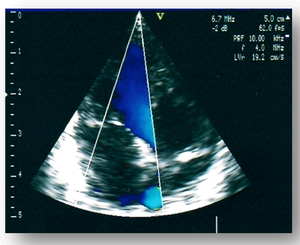
Echocardiography showing abnormal flow in the left ventricle apex.
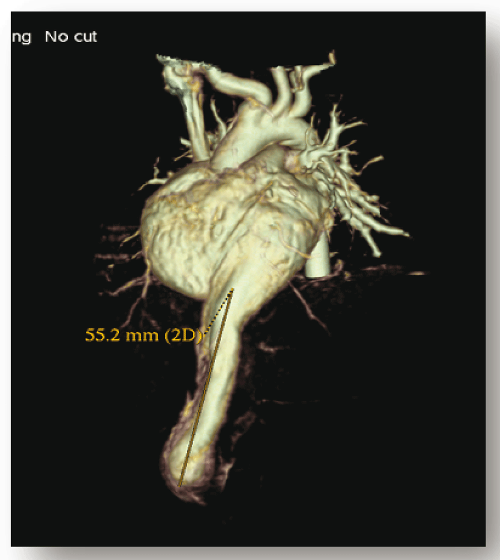
A 64-channel multi-detector CT (GE LightSpeed VCT) was performed for additional characterization. Sedation of the infant was not necessary. Thoracoabdominal helicoidal acquisition after the injection of non-ionic contrast agent was realized in a cranio caudal direction. The contrast enhanced multi-slice CT showed a thin walled channel extending up from the left ventricular apex to the anterior abdominal wall (Figure 2). This diverticulum was 6cm long following the abdominal midline through a defect of the anterior diaphragm and extending up to the umbilical region (Figure 3).
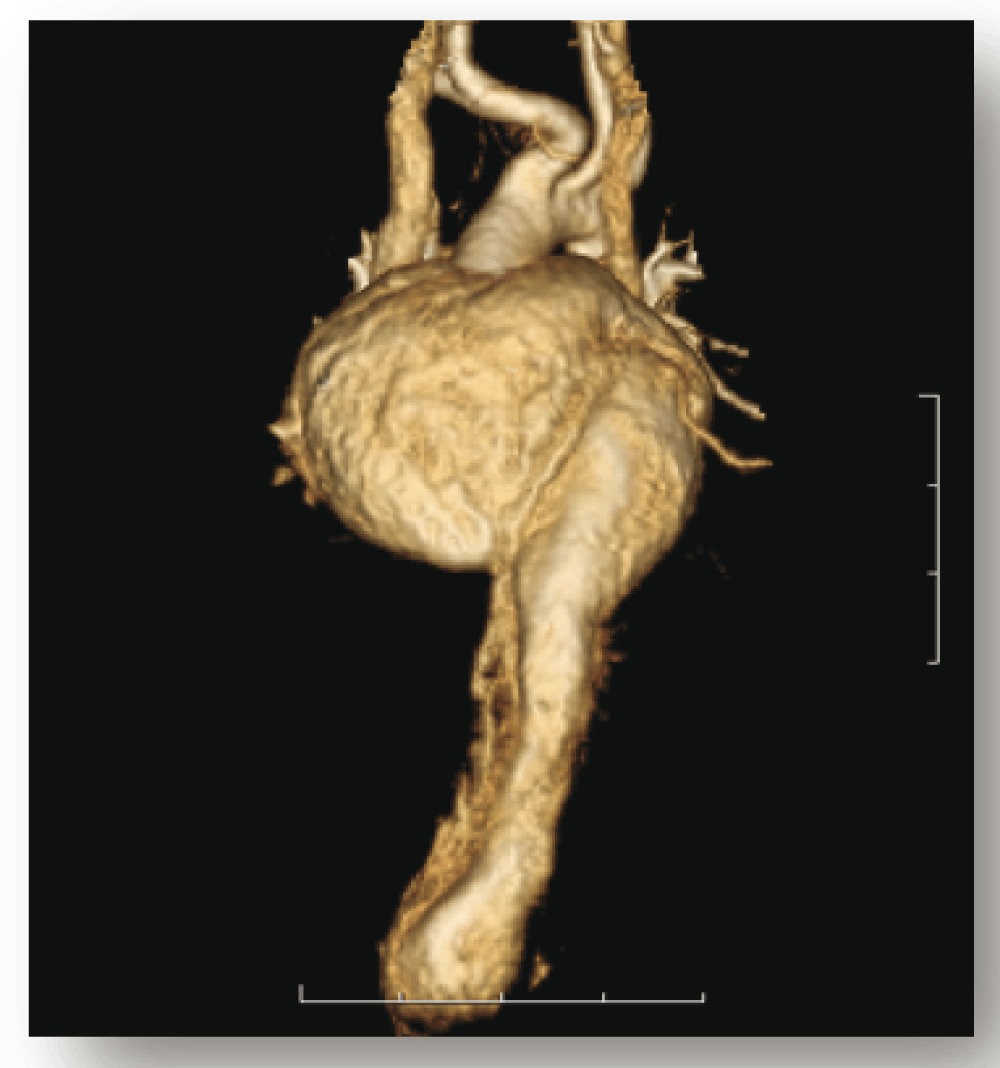
Volume rendered 3D CT image showing a diverticulum originating from the left ventricle free wall.
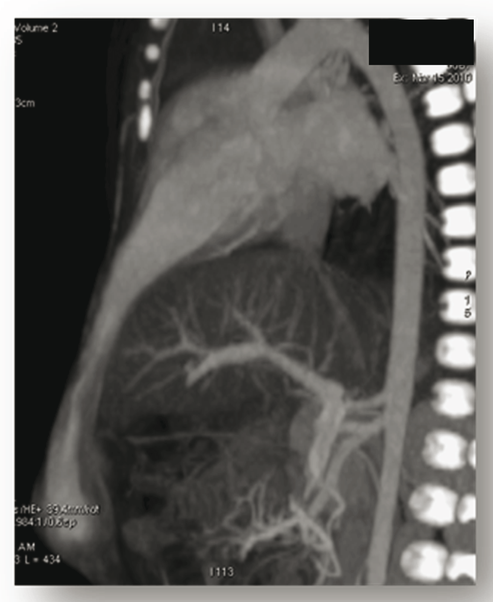
Mid-sagittal maximum intensity projection (MIP) thin CT image shows the diverticulum extending up to the umbilical region.
Myocardial thickness of the outpouching was 3mm (Figure 4A). No herniating bowels were seen (Figure 4B). The examination did not show any other abnormality of intra abdominal organs.
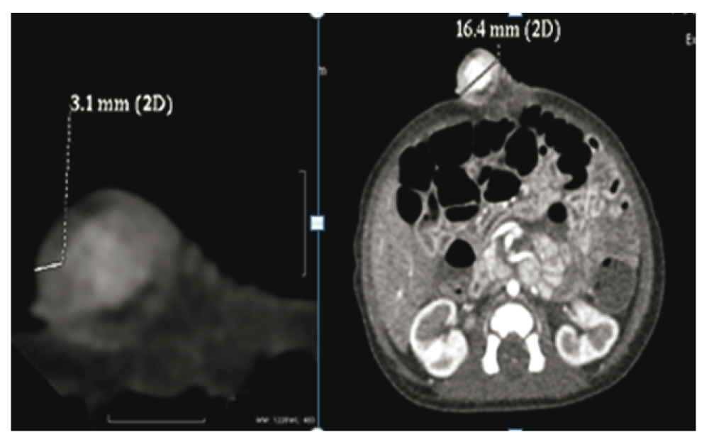
(A) Axial maximum intensity projection (MIP) CTscan showing the diverticulum wall consisting of a 3mm thick myocardium. (B) Axial enhanced multiple detector CT shows the defect of the anterior abdominal wall without herniating bowels.
The multi detector CT scan also confirmed the dextro transposition of the aorta and pulmonary artery (Figure 5) and the ventricular septum defect.
Surgical treatment was decided upon. The patient was connected to cardiopulmonary bypass and the diverticulum was opened and resected. The entry site was obliterated with a polytetrafluoroethylene patch. Further inspection revealed a normal-sized left ventricle and normal-sized coronary arteries, with no coronary aneurysms. Overlapping reconstruction of the abdominal anterior wall and diaphragm defects were also performed without using any prosthetic material.
Volume rendered 3D CT image showing the dextro transposition of the great vessels.
The postoperative period was uneventful, and the child was discharged from the hospital on the sixth postoperative day. We proposed medical management for our patient, comprising aspirin at a dosage of 5mg/kg/day to prevent any thromboembolic situation.
At a 6 month follow-up examination, the infant had a good clinical condition and a normal cardiac function on echocardiography.
Congenital left ventricular diverticulum is a rare cardiac malformation. Its incidence has been reported to be approximately 0.04% in the general population and approximately 0.02% in a consecutive pediatric autopsy series2,4. Although ventricular diverticulum may exist alone, it can also be associated with cardiac, vascular, or thoracoabdominal abnormalities4–6. In fact, cardiovsacular desease is a component of Cantrell’s pentalogy in some patients3. Cantrell’s pentalogy consists of a defect in the lower sternum, a supra umbilical abdominal wall defect, a deficiency of the anterior portion of the diaphragm, a deficiency in the diaphragmatic portion of the pericardium, and a congenital heart defect2.
Patients with isolated cardiac diverticulum are usually asymptomatic; however, there are reports associated with arrhythmias, embolic events, and even death due to diverticulum rupture2. Spontaneous rupture occurs very frequently, and can be explained by an increase in pressure inside the diverticulum as a result of a difference in the phase of contraction between the left ventricle and the diverticulum7.
Patients with a diverticulum sometimes present with an abnormal electrocardiogram8,9. In the case of our patient, a right deviation of the QRS axis and right ventricle hypertrophy signs were noted. Accurate diagnosis cans be made with ultra sonography or echocardiography2–10, and prenatal diagnostics have been reported in the literature5. CT angiography, MRI and invasive ventriculography give a clearer picture of the problem1,11. In the case of our patient, the diagnosis was suspected on echocardiography, and the CT angiography allowed a complete study of the pathology and confirmed the association with other cardiac, diaphragmatic and abdominal abnormalities. Surgical treatment is usually recommended when left ventricular diverticulum is associated with other cardiac or abdominal abnormalities. Perioperative management requests a multidisciplinary experienced team, due to the complexity of cardiac and thoraco abdominal abnormalities associated in Cantrell’s syndrome7. Recently, the field of percutaneous correction for congenital left ventricular diverticulum has witnessed tremendous development and a percutaneous transcatheter device treatment was reported12.
The strength of our study is the completeness of the observation with a 6 month follow-up. However, the limitation of our case is the absence of the full perioperative findings.
In conclusion, congenital left ventricular diverticulum is a rare cardiac malformation. The prognosis of this malformation is poor if not diagnosed in the perinatal period. Complications, such as embolism, infective endocarditis, arrhythmia and, rarely, rupture can occur. Although it may exist alone, it can also be associated with cardiac, vascular, or thoracoabdominal abnormalities (e.g., Cantrell’s syndrome). A diagnosis can be suspected with echocardiography. CT angiography allow a complete study of the problem. The treatment is always surgical with good postoperative prognosis.
Written informed consent for publication of their clinical details and/or clinical images was obtained from the parent of the patient.
All authors were involved in the revision of the draft manuscript and have agreed to the final content.
| Views | Downloads | |
|---|---|---|
| F1000Research | - | - |
|
PubMed Central
Data from PMC are received and updated monthly.
|
- | - |
Competing Interests: No competing interests were disclosed.
Competing Interests: No competing interests were disclosed.
Alongside their report, reviewers assign a status to the article:
| Invited Reviewers | ||
|---|---|---|
| 1 | 2 | |
|
Version 1 03 Nov 16 |
read | read |
Provide sufficient details of any financial or non-financial competing interests to enable users to assess whether your comments might lead a reasonable person to question your impartiality. Consider the following examples, but note that this is not an exhaustive list:
Sign up for content alerts and receive a weekly or monthly email with all newly published articles
Already registered? Sign in
The email address should be the one you originally registered with F1000.
You registered with F1000 via Google, so we cannot reset your password.
To sign in, please click here.
If you still need help with your Google account password, please click here.
You registered with F1000 via Facebook, so we cannot reset your password.
To sign in, please click here.
If you still need help with your Facebook account password, please click here.
If your email address is registered with us, we will email you instructions to reset your password.
If you think you should have received this email but it has not arrived, please check your spam filters and/or contact for further assistance.
Comments on this article Comments (0)