Keywords
Pelvic Congestion Syndrome, venous reflux, pelvic reflux, chronic venous insufficiency, PCS
Pelvic Congestion Syndrome, venous reflux, pelvic reflux, chronic venous insufficiency, PCS
Pelvic congestion syndrome (PCS) presents as a constellation of symptoms all caused by the development of varicosities in veins typically drained by either the ovarian or internal iliac veins. Its typical presentation is one of noncyclical dull, aching pelvic pain, typically unilateral, and persisting for more than 6 months. It was present in all patients in our series. Other symptoms that are commonly associated with the condition include dysmenorrhea and dyspareunia, seen respectively in case 1 and 4, and to some degree in all patients in our series.
Our experience has shown us that many authentic pelvic congestion syndrome diagnoses are being lost in a newfound reliance on cross sectional imaging as a diagnostic measure. We have found that to truly diagnose active pelvic congestion syndrome and treat the patient’s correlating symptomatic disease, the gold standard diagnostic imaging modality is fluoroscopic venography. In the following four cases you will read about patients who presented at our practice with historically atypical symptoms for pelvic congestion. After our suspicions were confirmed with duplex sonography revealing ovarian vein reflux and/or pelvic varicosities each patient underwent a fluoroscopic venogram and the appropriate image guided treatment. Although all of these patients presented with symptoms that are not indicative of pelvic congestion, all patients did indeed have advanced pelvic congestion. When each patient was treated for their specific disease state in the pelvic congestion syndrome treatment spectrum all symptoms resolved. Our intention is that the documentation of this experience will help other clinicians in their vigilance to rule out pelvic congestion syndrome with fluoroscopic venography before starting patients on a long road to a differential diagnosis.
A 61-year-old female with two children presented to our clinic with a chief complaint of left hip pain for 5 years, as well as pain in her lower back and pelvis, dysmenorrhea, and urinary frequency present for 2 years. Her hip pain was dull and aching in quality, though severe enough to interfere with exercise. She denied paresthesia radiating from her back down into either thigh, and she denied any history of prior trauma to the area. The patient had been seen both by orthopedists and pain management specialists, and both an MRI and a bone scan failed to pinpoint an etiology. She had taken non-steroidal anti-inflammatory drug (NSAID) regimens, systemic corticosteroids, and ultrasound-guided steroid injections without any improvement in her pain.
The patient’s past medical history was also notable for chronic venous insufficiency refractory to conservative and operative management, with endovenous thermal ablation therapy for the great saphenous veins in both legs. Three months after operative treatment, the patient reported continued pain, heaviness, fatigue, throbbing, cramping, and edema in both legs, aggravated by prolonged standing, and consistent with persistent chronic venous insufficiency.
On physical examination, the lower extremities were remarkable for spider veins, reticular veins, varicose veins, and mild pigmentation in her ankles and calves. Infragluteal varices were present. There was some mild pitting edema present, around the ankle and calf bilaterally.
A duplex sonogram was performed on both the pelvis and lower extremities. On sonographic examination of her pelvis, reflux was noted in her pelvic veins. In the legs, persistent reflux was noted in superficial veins in both legs.
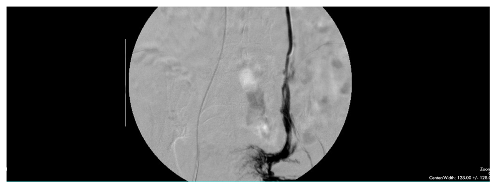
A fluoroscopic venogram was performed on the patient, with access obtained at the right common femoral vein. The study noted significant reflux in both internal iliac veins and reflux and stenosis in the left common iliac vein. Delayed washout of contrast was seen in the cross-pelvic collaterals of the tributaries off the internal iliac veins. Most notably, significant varicosities with delayed contrast washout were seen extending off the left ovarian vein (Figure 1) and communicating with the internal iliac veins. The varicose circulation form the refluxing left gonadal vein included prominent lateral extension towards the left side of the patient. Embolization of the left ovarian vein and collaterals from both internal iliac veins was performed. Post-embolization venography performed immediately after demonstrated successful closure of these refluxing venous branches.
The patient’s post-procedure course was uneventful. When seen again in our clinic 8 days later, her symptoms were significantly improved. Her hip pain had almost completely resolved, and at her 4 week post-operative checkup the patient reports she is again able to exercise without any impediment.
A 34-year-old female with 3 children by Cesarean section presented with an 8 year history of back pain. The pain was described as a dull ache, which increased throughout the day and improved after long periods in a recumbent position. She denied any radiation of her pain down her thighs. She denied any history of trauma. She did however, note some increase in the severity of her pain with increases in temperature and around the time of her menses. She had been on NSAIDs and had multiple spinal manipulations with a chiropractor with no improvement in her symptoms. An MRI of the lumbar spine showed only a mild L4 disc protrusion, and a nerve conduction study (NCS) showed no evidence of lumbosacral radiculopathy.
Upon further questioning, the patient noted a dull ache on the left side of her pelvis, and pain, cramping, and edema in both legs. As with her back pain, these symptoms would worsen with both daily activity and prolonged standing or sitting, and were worse on the left side. Her leg symptoms would improve slightly with leg elevation.
On physical examination, the lower extremities were remarkable for varicose veins and mild pitting edema noted around the ankle and calf bilaterally.
A duplex ultrasound examination was performed on the pelvis. A dilated left ovarian vein was visualized with 4.2 seconds of reflux.
An initial fluoroscopic venogram was performed that demonstrated severe reflux in her internal iliac veins bilaterally, with cross-pelvic collateral circulation lying near the sacrum, and also showed significant dilatation and reflux in both a left primary and accessory ovarian vein, both terminating into large and dilated pelvic varicosities that communicated with the left internal iliac vein and the right internal iliac vein via cross-pelvic collaterals. An intravascular ultrasound (IVUS) catheter noted dilation of the right ovarian vein as well. Embolization of both left ovarian veins was performed using a polidocanol and gelfoam slurry. The patient was brought back to the radiology suite 2 weeks later for a repeat venogram. During this procedure, the pelvic varicosities extending off branches of the left internal iliac vein approaching the sacrum, were embolized with gelfoam slurry and detachable coils. Closure of the left ovarian vein from the prior procedure was confirmed with a left renal venogram, and a post-embolization venogram confirmed closure of the internal iliac varicosities.
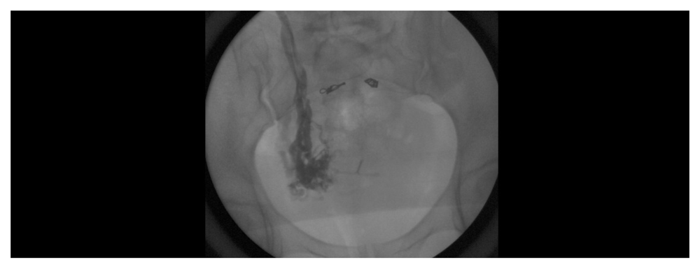
Roughly seven weeks after her two embolizations, the patient reported vast improvement in her symptoms on the left, both in her pelvis and in her back. She did, however, have persisting symptoms on her right side, and in addition noted post-coital tenderness that she was now noticing as some of her other discomfort was resolving. A follow-up sonogram demonstrated resolution of the left ovarian vein reflux, but significant reflux and varicosities on the right side of her pelvis consistent with right-sided pelvic congestion. At the 10 week mark from her initial treatment she has returned to the procedure suite for a third venogram with embolization of the right gonadal vein (Figure 2) and at the 12 week post-operative point has full resolution of her symptoms.
A 42-year-old multiparous woman presented to our clinic with 8 years tenesmus, fecal incontinence, and uncontrollable flatulence. These symptoms began shortly after the birth of her first child. The pregnancy was notable for a significant amount of pain and swelling in her legs and pelvis, and the vaginal delivery was assisted with an episiotomy. Although she had some improvement in her symptoms in the months after her pregnancy, she has since had 4 more pregnancies. Despite having Cesarean sections for her deliveries, each pregnancy has worsened her incontinence and overall bowel control, as well as her baseline control. She has been evaluated by gastroenterologists for the above problems, as well as for gas and bloating in her abdomen. She has undergone endoscopies and multiple imaging studies, and was diagnosed with Irritable Bowel Syndrome. Lastly, the patient had a history of hemorrhoids, which had bled on at least one occasion.
In addition to her bowel symptoms, the patient reported problems with urinary frequency, dyspareunia, and pelvic heaviness, and ovarian pain. Her pain would worsen around the time of her menses. She has been followed by a gynecologist for these issues, which were attributed to endometriosis. Her obstetrician has also attributed her incontinence issues to the episiotomy and postpartum pelvic floor dysfunction, and had suggested a potential sacral nerve stimulator implant to help her.
The patient also noted some cramping, heaviness, and swelling in both legs. These symptoms would worsen with daily activity, as well as with prolonged standing or sitting. She has had hemorrhoids and varicose veins over her perineum, and has had intermittent bleeding from both.
For all these symptoms, and despite her extensive workup by both gastroenterologists and gynecologists, the patient had been unable to find any relief for her numerous problems.
On physical examination, the lower extremities were remarkable for spider veins, varicose veins, prominent perineal veins, and vulvar varicosities. Mild pitting edema was noted as well. The abdomen was soft, with some ovarian tenderness on palpation. Duplex examination of the pelvis was performed. A collection of pelvic varicosities was seen surrounding the rectum.
A diagnostic fluoroscopic venogram was performed using a right common femoral vein approach. Moderate reflux was noted in the common iliac veins and significant reflux noted in the internal iliac veins, with delayed contrast washout noted in several varices and cross-pelvic collaterals. The exam, however, was most notable for an immensely dilated left ovarian and accessory left ovarian vein. They were roughly 28–30 mm in diameter, showing delayed contrast washout and collateralization with the internal iliac veins. A large and aneurysmal terminal branch of the vessels was noted to lie on the sigmoid colon. The left ovarian vein, its accessory, and its branches were embolized with a combination of gelfoam slurry and a 20mm detachable coil. Embolization of the internal iliac branches bilaterally was performed with gelfoam. Post-embolization venography performed immediately after embolization, demonstrated successful closure of all concerning branches.
At 1 week out from her embolization, the patient has had almost complete resolution of both her flatulence and her tenesmus. At the 4 week post-operative mark she had improvement with her bladder control and dyspareunia as well. She has some residual pelvic pain, and will be returning for another diagnostic venogram with an intravascular ultrasound (IVUS) to assess the reflux in her common iliac veins.
A 36-year-old multiparous woman presented to our clinic for pain, cramping, and edema in both legs for roughly 5 years. Of note, she was having significant back pain as well, which she chose to disclose after experiencing relief following venogram with successful embolization of the left gonadal vein and collaterals from the refluxing internal iliac veins.
In regard to her presenting complaint, her symptoms worsened over the course of the day and improved with leg elevation. They had shown no improvement with compression therapy and daily NSAIDs. Duplex sonography showed superficial venous reflux, which was ablated. Postoperative sonograms showed successful closure of each vein.
Four months later, the patient experienced a relapse of her leg pain and cramping. A repeat sonogram was performed, which showed reopening of all previously ablated veins. A sonogram was performed on the pelvis, which showed 2.9 seconds of reflux in the left ovarian vein. On further questioning and examination, the patient was found to have pelvic pain and varicosities in her inguinal area, extending to the vulva, most prominently on the left. These varicosities had been a cause of dyspareunia for the patient for several years, beginning shortly after the birth of her third child.
A diagnostic fluoroscopic venogram was performed from a right common femoral vein approach. Severe reflux was seen in the left internal iliac system, with cross-pelvic flow to the right side as well as retrograde contrast filling of the left ovarian vein. The left ovarian vein was almost as large as an iliac vein- roughly 14 to 16 mm in diameter. The left vein was embolized with a combination of polidocanol and gelfoam slurry. A post-procedure venogram was performed immediately after embolization and showed only moderate closure of the vein however, and intervention in the internal iliac system was thwarted due to vasospasm.
A repeat venogram was performed 10 days later, again with a right common femoral vein approach. Reflux was still seen in the branches off both internal iliac veins. The left ovarian vein remained open and dilated, with delayed contrast washout. Embolization was again performed, this time on the left ovarian vein and on the collaterals extending from both internal iliac veins, using a combination of gelfoam slurry and a 13 mm detachable coil. Post-embolization venography immediately after coil placement showed successful closure of the refluxing veins. Visualization of the right iliac veins was performed using an IVUS catheter and showed no evidence of stenosis.
At 8 weeks out from our first intervention, the patient has had significant reduction in her pelvic pain. She also mentions finding reduction in back pain which she reported upon experiencing relief status-post venogram interventions with embolization.
Pelvic congestion syndrome (PCS) presents as a constellation of symptoms all caused by the development of varicosities in veins typically drained by either the ovarian or internal iliac veins. Its typical presentation is one of noncyclical dull, aching pelvic pain, typically unilateral, and persisting for more than 6 months. It was present in all patients in our series. Other symptoms that are commonly associated with the condition include dysmenorrhea and dyspareunia, seen respectively in case 1 and 4, and to some degree in all patients in our series.
PCS is also seen in association with lower extremity varicose veins. As many as 15–20% of patients with lower limb varicosities have demonstrated pelvic venous reflux on venogram or duplex ultrasound, with a 30% incidence among those patients with varicosities that have recurred after prior treatment1. On physical examination, these patients may often have varicosities on the vulva, on or just beneath the buttocks, and on the upper thighs, particularly near the groin, owing to reflux from the pelvic veins communicating to the legs via the inferior gluteal and internal/external pudental veins2. In our series, cases 1 and 4 had reopening of veins previously closed by endovenous thermal ablations. Cases 1, 3, and 4 presented with either vulvar or infragluteal varices.
It is important to remember that PCS affects its patients due to the local compression and consequent irritation and inflammation of organs in proximity to the engorged and swollen plexus of pelvic veins. Since these veins drain the bladder, vagina, uterus, rectum, and sacrum, PCS can be responsible for several atypical symptoms, depending on the organ compromised. Some form of bladder instability is not uncommon, and was seen in cases 1 and 3. Irritation of the lumbosacral nerves, although uncommon, can result in either back or hip pain. In our series, back pain was seen in cases 1 and 2. Hip pain was seen in case 1. The finding of hip pain is particularly rare, with only 2 other cases being reported in the literature3. Pressure of these veins on the rectum, or perhaps reflux through collaterals feeding off a refluxing ovarian vein, can result in hemorrhoids, a finding seen in case 3. The finding of a dilated pelvic varicosity compressing the sigmoid colon and causing both tenesmus and uncontrollable flatulence in case 3 has never before been reported.
As shown in our first three cases, this atypical presentation of PCS can be the predominant issue impacting the patient’s quality of life. These atypical presentations are typically underdiagnosed, as seen by the extensive and ultimately unfruitful workups experienced by 3 of the 4 patients in our series. It is therefore important to maintain a low threshold for suspicion of PCS in any patient with symptoms that can be attributed to an organ in the pelvis.
Our workup for suspected PCS begins with pelvic sonography. Although there has been literature championing the efficacy of either the CT venography (CTV) or more often the magnetic resonance venography (MRV), in our experience, both studies have had a lower diagnostic yield4–6. Both imaging modalities require proper timing to catch the contrast in the pelvis during the venous phase, and are therefore very operator dependent. The studies also require the subject to be supine, a position that can compress ordinarily dilated pelvic veins. They are also expensive. In contrast, pelvic sonography is inexpensive, and can be performed with the patient upright, as the examiner looks for both dilated pelvic veins and flow reversal during Valsalva maneuvers7. The operator-dependent nature inherent to sonography mandates valuable sonogram technicians with high levels of training and experience in finding and reporting pelvic reflux and varicosities.
As with the CTV and MRV, the sonogram is also capable of ruling out other pelvic etiologies. Findings suggestive of PCS on ultrasound include ovarian veins >6 mm in diameter, the presence of dilated (>5 mm) arcuate veins crossing the uterine myometrium, and slow (<3 sm/sec) or reversed retrograde blood flow2,6. Polycystic changes in the ovaries have also been seen in about 50%4,6 of cases.
Diagnostic venograms remain the gold standard in identifying PCS, and are indicated for all suspicious presentations, even, in the appropriate clinical setting, if the other studies are negative. It is a low risk procedure, and permits treatment and potential curing of the patient’s symptoms at the same time. Evaluation of the left renal, bilateral ovarian, iliac, and internal iliac veins is performed, looking for dilatation and reflux. Venography findings include an ovarian vein diameter >5 mm, ovarian vein reflux, stagnation of contrast in the pelvic veins, contralateral reflux across the midline, and filling of vulvoperineal or thigh varices2,4. If there is a question of a stenotic lesion in either the renal or iliac vein, pressures across the area in question can be measured, or an intravascular ultrasound can be placed. In all of our patients, pelvic venograms successfully identified the condition.
In our clinic, we perform diagnostic venography with the patient awake. We begin our assessment at the most common site of pelvic reflux, the left ovarian vein. If reflux is present, we attempt to reproduce the patient’s symptoms using high pressure contrast flushes in a 5 french sheath. If the venous distension created by the flush reproduces symptoms, and the anatomic distribution of the vessels distended corresponds to those symptoms, we embolize the vessel. Our preference in the ovarian vein is to use a combination of gelfoam or polidocanol for the terminal branches below the pelvic brim and an oversized (at least 150% of the diameter of the ovarian vein) coil in the ovarian vein, preferably near the ostia of any visualized ovarian collaterals, and no less than 4 mm from its confluence with the renal vein.
After the treatment, we revisit the patient in roughly 1 week. If the patient is showing improvement, we repeat the pelvic sonogram to determine if any reflux remains. If reflux is noted, we perform a second venogram and embolization of pelvic varices fed by the refluxing internal iliac veins, and potentially the right ovarian vein if the patient’s symptoms are significantly greater on the right. Our preference is to treat the internal iliac branches with either sclerotherapy or gelfoam alone, given the higher risk of coil migration noted in this region. If the right ovarian vein is to be targeted, we consider a right internal jugular access to allow for easier cannulation.
During this second procedure, we also employ an IVUS catheter to look for stenotic lesions. Pelvic reflux can frequently be secondary to anatomic anomalies resulting in downstream obstruction, such as compression of the left renal vein by the superior mesenteric artery (Nutcracker syndrome)1,2,7, or compression of a left common iliac vein by a right common iliac artery (May-Thurner syndrome)8. A May-Thurner’s phenomenon can be present in as many as two-thirds of the general population9. If any such lesions are identified, a full discussion with the patient regarding the anatomy affected by the stenosis, and the benefits and drawbacks of angioplasty alone versus stenting ensues.
If the patient wishes to proceed, we return for a third venogram to treat the stenotic segments. For milder lesions (≤ 50% reduction in cross-sectional area), or for focal, short lesions caused by a localized vascular band or web, we attempt only an angioplasty. For a longer and more severe stenotic segment (> 50%), we prefer to both angioplasty and stent. As with our coils, our preference is to oversize our stents (roughly 4 mm greater than the average diameter of the non-stenotic venous segment). Our preference is to avoid stenting for as long as possible in women who intend to get pregnant and in patients with clotting disorders or prior DVTs, given the higher incidence of post-stent complications in these patient populations.
Transcatheter embolization is currently regarded as the least invasive and most efficacious management option for PCS, with complete or partial symptom relief in 68.2–100% of patients2,10. Apart from our preferences, a number of different approaches have been reported to affect closure of the refluxing veins, from simple coil embolization, to glue embolization, to combinations of sclerotherapy and coils. In studies using visual analog scale (VAS) pain scores to measure the extent of symptom relief, vast improvement was shown consistently, with mean scores of 7.3–7.6 decreasing to scores of 0.5–3.2 postoperatively. Depending on the initial severity, the symptoms can take as long as 9–13 months after therapy to resolve10. Complications from the procedure are rare, being reported in 3.4–4.4% of patients, and consist of coil migration, vein perforation, local phlebitis, deep venous thrombosis, and contrast reactions10. Patients may suffer from a brief period of flu-like symptoms within 72 hours of each embolization, but this consistently passes within 48 hours without need for intervention. Studies have also shown embolization to have no effect on the menstrual cycle or fertility4. This is an important consideration, given that the predominance of PCS is in premenopausal women.
With respect to the management of PCS secondary to stenotic vein lesions, endovenous stent placement is also safe and effective. In a review of multiple studies encompassing 1500 patients being treated for chronic iliac vein stenosis, stenting had a 90–100% patency rate for non-thrombotic disease and 74–89% patency rate for thrombotic disease at 3 to 5 years. Symptom relief was achieved in 86–94% of patients for pain, 66–89% for swelling, and 58–89% for healing of ulcers in the leg11. Although these studies focused on the outcomes of patients suffering from venous insufficiency in the legs, it is not unreasonable to assume that treatment of the stenosis would similarly benefit patients with PCS, given that both ailments are due to venous obstruction causing congestion in communicating upstream veins. Among the 1500 patients reviewed, no deaths or pulmonary emboli were reported, and access site complications and significant bleeding, despite the larger sheaths required for the stents, occurred in only 0.03–1%11.
Given the effectiveness and safety of these procedures, our practice recommends aggressive management of patients whose symptoms and venogram findings suggest the presence of PCS.
Pelvic congestion syndrome is an underdiagnosed condition in premenopausal women that presents with a broad spectrum of symptoms that are often attributed to other pathologies. It is important to maintain a high level of suspicion in parous women wether the presenting symptoms are typical or atypical in nature. The complexity of the abdominal/pelvic venous vasculature in the premenopausal patient can distort accuracy and cloud the patients ability to properly articlutale symptoms. With modern advancements made in diagnostic imaging many clinicians feel MRV to be definitive in diagnosing pelvic congestion, but our experience has proven otherwise. Cross sectional imaging relies heavily on timing, patient immobility and technical skill level of the performing technologist and even then can still provide false positive reults2,4,6. Our experience has shown us that fluoroscopic venography is the gold standard diagnostic imaging modality providing us with anatomical correlation as well as physiological confirmation of our suspicions. In this specific patient population a diagnostic venogram will go far in preventing patients with PCS from missing out on effective and low risk treatment.
Written informed consent for publication of their clinical details was obtained from each patient in this paper.
| Views | Downloads | |
|---|---|---|
| F1000Research | - | - |
|
PubMed Central
Data from PMC are received and updated monthly.
|
- | - |
Provide sufficient details of any financial or non-financial competing interests to enable users to assess whether your comments might lead a reasonable person to question your impartiality. Consider the following examples, but note that this is not an exhaustive list:
Sign up for content alerts and receive a weekly or monthly email with all newly published articles
Already registered? Sign in
The email address should be the one you originally registered with F1000.
You registered with F1000 via Google, so we cannot reset your password.
To sign in, please click here.
If you still need help with your Google account password, please click here.
You registered with F1000 via Facebook, so we cannot reset your password.
To sign in, please click here.
If you still need help with your Facebook account password, please click here.
If your email address is registered with us, we will email you instructions to reset your password.
If you think you should have received this email but it has not arrived, please check your spam filters and/or contact for further assistance.
I believe ovarian vein embolization and pelvic outflow revascularization to be a curative solution to the horrific effects and symptoms caused by the chronic PCS disease state. Currently I am increasing embolization safety and efficacy in these patients by utilization of a vascular plug. I size once, deploy once and achieve rapid embolic effect without the risk of the dreaded coil migration.
I hope others see the value added in this algorithm and treatment methodology because the safer we make curative therapy the greater the benefit to the patients we treat. Thank you and may your next case be your best case.
Kind Regards,
Andrew S Amorosso RT(R)(VI)
Endovascular Revascularization and Embolic Expert