Keywords
BALT lymphoma, Mycobacterium chelonae, diagnosis, treatment
BALT lymphoma, Mycobacterium chelonae, diagnosis, treatment
Extranodal marginal zone B cell lymphoma (MZL) is a low grade B cell lymphoma of mucosa associated lymphoid tissue (MALT). Primary pulmonary MALT, also called bronchial-associated lymphoid tissue (BALT), accounts for 0.5–1% of all malignant lung tumours and about 90% of all lung lymphomas1,2. BALT represents 15% of all MALT lymphomas1,3.
BALT lymphoma has been associated with chronic antigenic stimulation by autoimmune disease, smoking, infection or chronic inflammation, but a well-established connection has not yet been described3–5. The paradigmatic example of that antigenic stimuli is the development of gastric MALT in Helicobacter pylori infection with chronic gastritis1. There are some reports in literature correlating Mycobacterium avium complex and Mycobacterium tuberculosis infection with BALT lymphoma5–8.
Nontuberculous mycobacterium (NTM) are ubiquitous in the environment, frequently colonize the skin, digestive and respiratory tract, sometimes developing disease, mainly in immunosuppressed chronic diseases patient’s9,10. Mycobacterium chelonae is a rapidly growing mycobacteria that has been implicated as an infrequent lung pathogen. Patients with severe underlying structural lung disease such as cystic fibrosis and bronchiectasis are the most predisposed11.
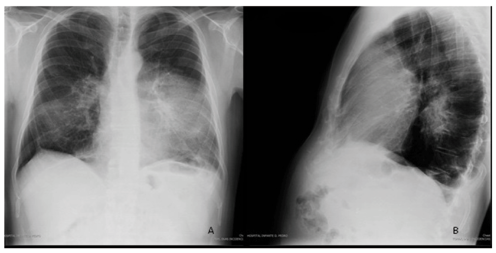
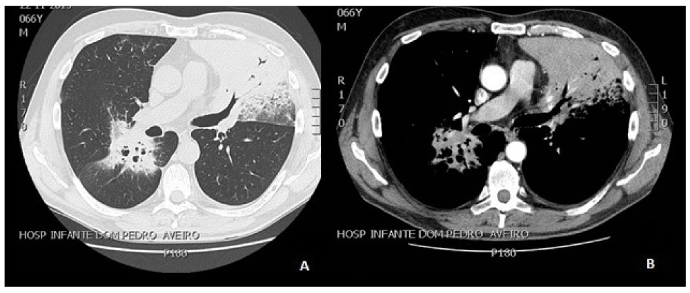
We present a 66-year-old man’s case, with subfebrile temperature, two months’ history of persistent cough, purulent sputum, occasional small haemoptysis, anorexia and non-quantified weight loss. He reported no clinical response to previous antibiotherapy with amoxicillin (500 mg orally 3 times a day for 7 days) and after moxifloxacin (400 mg orally once a day for 7 days). The patient reported a gastric lymphoma at the age of 40, treated with chemotherapy for which we have no detailed information. He had a 32-pack-year tobacco smoking history. He had no changes on physical examination. Initial chest radiography showed a bilateral heterogeneous opacity, covering the lower two thirds of the left hemithorax and the lower third of the right hemithorax (Figure 1). Inflammatory parameters, such as eritrocit sedimentation rate and C-reactive protein were normal as well as blood gas. Pneumonia was considered and antibiotherapy with amoxicillin/clavulanic acid (1000 mg/200 mg intravenously every 8 hours) and clarithromycin (500 mg orally twice a day) was initiated, which held during seven days. Later chest CT showed multiple foci of parenchymal consolidation with air bronchogram on the left upper lobe, lingula, apical segment of the left lower lobe, posterior segment of the right upper lobe and apical right lower lobe (Figure 2). Mantoux test was negative. He had elevated IgM (1451 mg/dl) as well as negative serology for HIV, B and C hepatitis. No endoscopic abnormalities were found on flexible bronchoscopy. Gastric endoscopy showed an antral gastritis with negative Helicobacter pylori biopsy test. Bacteriologic analysis of three sputum specimens were negative for either aerobic and anaerobic bacteria or acid fast bacilli, but positive for Mycobacterium tuberculosis complex (MTC) using polymerase chain reaction (PCR) technology, in two out of three samples, bronchial aspirate was bacteriologically negative. The patient was initiated on antituberculous treatment (ATT) with isoniazid (300 mg/day), rifampicin (600 mg/day), pyrazinamide (1500 mg/day) and ethambutol (1200 mg/day), orally, and was forwarded to an outpatient centre for respiratory diseases. Six weeks later, Lowenstein-Jensen cultures showed the presence of NTM. ATT was maintained due to clinical improvement while waiting for NTM identification. The lack of imagiologic improvement in the meantime, led to transthoracic lung biopsy realization. Immunohistochemistry showed positive lymphocyte identification to CD20 and BCL2, and CD23, CD10, CD5 and a negative CD3, consistent with the diagnostic of BALT lymphoma. There was no evidence of other extranodal or nodal involvement in complementary imagiological study. Haematologists decided to revaluate the patient’s condition after the end of ATT, taking into account his clinical stability. In the meantime, NTM was identified as M. chelonae and the laboratory informed us that the initial positive PCR for MTC was a laboratorial contamination. The drug resistance patterns were not performed. At this time new sputum samples were bacteriologically negative and ATT was stopped, with six months of treatment.

Chest radiography: bilateral heterogeneous opacity, covering lower two thirds of the left hemithorax and the lower third of the right hemithorax (A: posteroanterior view; B: lateral right view).

Thoracic computed tomography, axial view: pulmonary consolidation with air brochogram, in the left upper lob, lingual, apical segment of the left lower lobe, posterior segment of the right upper lobe and apical right lower lobe (A: pulmonary window; B: with contrast).
One month later the patient resumed his previous respiratory symptoms. Specific therapy for M. Chelonae with tobramycin (150 mg intramuscularly once a day) and clarithromycin (500 mg orally twice a day) was initiated, but discontinued due to nephro and hepatic toxicity, after seven weeks of treatment. Haematologists at this time decided to begin treatment with cyclophosphamide (50 mg orally once a day). After 6 months of cyclophosphamide treatment the patient showed significant clinical improvement, without any signs of pulmonary infection and partial imagiologic resolution. For this reason the patient remains in treatment, waiting for a revaluation with new clinical and imagiologic data.
The diagnosis of BALT lymphoma is challenging and frequently misdiagnosed as pneumonia, pulmonary tuberculosis or interstitial lung disease, because clinical and radiologic findings are nonspecific. According to retrospective analysis, average time to achieve diagnosis is around 20 months4. Chronic cough, sputum, progressive dyspnoea, fatigability, fever, night sweats and weight loss are the most common manifestations. Imagiologic findings are generally nonspecific, such as single or multiple nodules, consolidation areas, bronchiectasis, bronchiolitis phenomena or diffuse interstitial lung disease2,4,12.
Pulmonary tuberculosis was assumed initially with the identification of Mycobacterium tuberculosis complex by PCR, leading us to treat the patient with ATT. When the presence of an NTM was identified in two out of three sputum specimens together with clinical and imagiologic changes, an infection was assumed according to the criteria of American Thoracic Society (ATS) for NTM lung disease12. Nevertheless the doubt about M. chelonae infection or colonization emerged, when BALT lymphoma was diagnosed, because imagiologic changes could either be attributed to BALT or M. chelonae, which compromises ATS criteria for infection. Also the absence of clinical signs of pulmonary infection after stopping a short period of specific therapy, but with the beginning of chemotherapy, led us to consider that M. chelonae was possibly just a colonizer.
We were unable to establish if M. chelonae had a role as a chronic antigenic stimulus to BALT lymphoma or if it was BALT lymphoma that led to secondary infection or colonization by M. chelonae.
Optimal therapy is unknown. Chemotherapy with CHOP or R-CHOP (cyclophosphamide, doxorubicin, vincristine, and prednisone/prednisolone, with or without rituximab) is the most common treatment described by authors; however monotherapy with cyclophosphamide also presents a high rate of disease control and can be used as a single agent13,14. Some experts favour a “watchful waiting” option in early stages and on asymptomatic patients due to the indolent evolution and the good prognosis of BALT, generally expected to have more than 80–90% of a five year survival rate14,15.
In our case a less aggressive approach was decided due to the indolent evolution of BALT, the clinical stability of the patient, and to avoid a severe immunocompromised state, which could lead to a mycobacterial infection.
Written informed consent for publication of their clinical details and clinical images was obtained from the patient.
JN and LA conceived the study and carried out the research. All authors contribute in the diagnosis and follow up of the patient. JN prepared the first draft of the manuscript. All authors were involved in the revision of the draft manuscript and have agreed to the final content.
| Views | Downloads | |
|---|---|---|
| F1000Research | - | - |
|
PubMed Central
Data from PMC are received and updated monthly.
|
- | - |
References
1. Ye H, Liu H, Attygalle A, Wotherspoon AC, et al.: Variable frequencies of t(11;18)(q21;q21) in MALT lymphomas of different sites: significant association with CagA strains of H pylori in gastric MALT lymphoma.Blood. 2003; 102 (3): 1012-8 PubMed Abstract | Publisher Full TextCompeting Interests: No competing interests were disclosed.
Competing Interests: No competing interests were disclosed.
Alongside their report, reviewers assign a status to the article:
| Invited Reviewers | ||
|---|---|---|
| 1 | 2 | |
|
Version 1 21 Jan 16 |
read | read |
Provide sufficient details of any financial or non-financial competing interests to enable users to assess whether your comments might lead a reasonable person to question your impartiality. Consider the following examples, but note that this is not an exhaustive list:
Sign up for content alerts and receive a weekly or monthly email with all newly published articles
Already registered? Sign in
The email address should be the one you originally registered with F1000.
You registered with F1000 via Google, so we cannot reset your password.
To sign in, please click here.
If you still need help with your Google account password, please click here.
You registered with F1000 via Facebook, so we cannot reset your password.
To sign in, please click here.
If you still need help with your Facebook account password, please click here.
If your email address is registered with us, we will email you instructions to reset your password.
If you think you should have received this email but it has not arrived, please check your spam filters and/or contact for further assistance.
Comments on this article Comments (0)