Keywords
Myelolipoma adrenal, adrenal incidentaloma, benign adrenal tumor
Myelolipoma adrenal, adrenal incidentaloma, benign adrenal tumor
The term incidentaloma is derived from “incidental tumor,” describing a mass discovered on imaging by pure chance1. When discussing adrenal incidentalomas (AIs), this refers to a finding of a visible adrenal mass greater than 1cm in diameter found on imaging performed for other medical causes2. In general, adrenal tumors are detected in 0.4% of abdominal ultrasounds and occur with ten times greater frequency in those with a positive cancer history3. Exclusion criteria for AIs include patients who present with manifestations of adrenal dysfunction2 and those with extra-adrenal cancers in the process of stratification4.
Advances in modern diagnostic methods have produced a greater prevalence of AIs, especially due to advances in CT and MRI technology3. Incidental adrenal masses are found in 2–4% of abdominal CT scans and the frequency increases in correlation with the patient’s age - adding 0.2% in the third decade of life, up to 7% in those greater than 70 years old4. Among these, non-functional adenoma remains the most frequent (60–85%), while a minority present as functional adenomas (5–16%)5. Of functional masses, 6% consist of pheochromocytomas, 5% are subclinical Cushing Syndrome, 5% are adrenal carcinoma, 2% prove to be a metastasis, and the rest belong to other etiologies, such as myelolipomas, hematomas, cysts, or lymphomas6,7.
Nevertheless, the retroperitoneal location increases the difficulty of detection during a standard physical exam. This often leads to the late diagnosis of such tumors only when clinical systemic manifestation is present - in the case of functional incidentalomas - or the compromise of the adjacent tissues secondary to abnormal gland growth8. According to endocrinology guidelines, both hormonal and radiographic evaluation must be performed in order to rule out subclinical states9,10. In general, masses ≥4cm are removed surgically, independent of functionality. Furthermore, all functional tumors and those with malignant characteristics undergo an adrenalectomy under endocrinologic supervision. Non-functional adenomas, small myelolipomas, and benign asymptomatic cysts do not require surgical intervention10.
With this in mind, providers must remember two primary questions, first asking “Is the mass hormonally active?” as this differentiates between functional and nonfunctional masses5,6. Additionally, asking “Are there malignant characteristics?” proves equally important. This is determined by the radiologic imaging that look for heterogeneity, poorly delimited borders, the presence of necrosis, hemorrhage, calcification, or an attenuation coefficient greater than 20 Hounsfield Units7.
This case report describes a giant right upper quadrant incidentaloma in an asymptomatic patient that was initially thought to be a hepatic hemangioma, due to its size and location, which was later confirmed to be an adrenal tumor.
A 54 year old asymptomatic female patient was seen by her family physician in Marcaibo, Venezuela, for her annual health exam in January 2014 in a primary care center. She had no complaints, except for recent unintended weight gain. Her past medical and surgical history are notable for a left breast lumpectomy (1973), a salpingectomy (1994), a hysterectomy without oophorectomy for NIC III (2005), and a left unilateral oophorectomy for ovarian torsion (2007). The patient used no medications and has no known allergies, and denied tobacco, alcohol, or drug use. The patient is monogamous and happily married. Her family history is notable for a sister who died of Hodgkin Lymphoma.
On physical exam, the patient was afebrile with normal vital signs. Her weight was 92.5 kg, 1.74 meters tall, with a BMI of 30.6. She appeared well hydrated with moist mucous membranes. She had an unremarkable exam - no findings of violaceous striae, acanthosis, acrochordons, or signs of virilization.
Laboratory results showed a normal complete blood count, mixed dyslipidemia, fasting blood glucose levels >125 mg/dl (normal range, 70–100 mg/dl) on more than two occasions, and HOMA1-IR index >2.5 (normal index, ≤ 2.5) (Table 1); meeting the diagnostic criteria for type 2 diabetes mellitus (DM2) and metabolic syndrome. Initial recommendations were lifestyle changes, including 30 minutes walks five days a week, and a nutritionist consult. Additionally, pharmacotherapy, sitagliptin/metformin (Janumet®, 50/1000mg) 1 tab daily, ezetimibe/simvastatin (Vytorin®,10/40 mg) 1 tab daily, gemfibrozil (Lipontal®, 900 mg) 1 tab daily, and orlistat (Xerogras®, 120 mg) 1 cap daily, was initiated.
| Laboratory | October 2013 | November 2013 - January 2014 (initial treatment) | April 2014 (treatment control - pre-operative) |
|---|---|---|---|
| Cholesterol - Total | 264,30 mg/dl | 247 mg/dl | 121.30 mg/dl |
| HDL - C | 53 mg/dl | 41 mg/dl | 36 mg/dl |
| LDL - C | 164,64 mg/dl | 165 mg/dl | 67.64 mg/dl |
| Triglycerides | 236 mg/dl | 204 mg/dl | 86 mg/dl |
| Creatinine | 0.6 mg/dl | 0.7 mg/dl | 0.8 mg/dl |
| Uric Acid | 6.9 mg/dl | 5.0 mg/dl | 4.2 mg/dl |
| Urea | 23 mg/dl | 21 mg/dl | 29 mg/dl |
| AST | 19.28 UI/L | 9 UI/L | 21.92 UI/L |
| ALT | 17.00 UI/L | 10 UI/L | 21.11 UI/L |
| Blood Glucose | 148,80 mg/dl | 129 mg/dl | 94.35 mg/dl |
| Fasting Insulin | 11.50 uIU/ml | ||
| Postprandial Insulin | 103.30 uIU/ml | ||
| *HOMA | 3,5 |
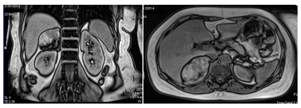
Simultaneously, a right upper quadrant ultrasound was ordered showing slight hepatic steatosis, as well as a round space occupying lesion with well-defined hyperechoic borders measuring 5.6×7.3cm in segment V of the right lobe suggestive of a hemangioma. Of note, a bilateral non-obstructive nephrolithiasis was observed (Figure 1). Due to these findings, the patient was referred to a local hospital diagnostic center for imaging studies, a triphasic hepatic MRI was performed as part of an additional workup. This identified a 7.0×6.0cm right adrenal space occupying lesion suggestive of a large adrenal adenoma (Figure 2). A hormone profile was performed with normal results - classifying this mass as a non-functional adenoma. Lack of reagents in local laboratories caused that the patients moved to Avila Clinic in Caracas (Capital of Venezuela) (Table 2). The work up was completed with a serologic evaluation to rule out fungal infection with negative results for mycoplasma IgM (0.15; normal range: 0.00 – 0.90).
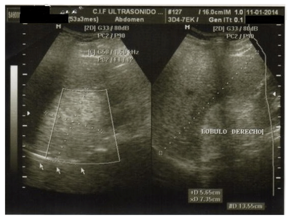
A hyperechogenic 5.6 × 7.3 cm image is observed in segment V of the right hepatic lobe suggestive of an incidental hemangioma.
Left panel, longitudinal cut; right panel, transverse cut. Performed using SIEMENS Magneton Essenza 1.5 TESLA.
| Adrenal cortex | |||
|---|---|---|---|
| Zona reticularis | Zona fasciculata | ||
| Hormone | Result | Hormone | Result |
| Testosterone - Total | 0.09 ng/ml (VN: 0,06-0,82) | Urine cortisol occasional | 12.30 ug/dl (VN: 0,20-50,00) |
| Free Testosterone | 0.79 pg/ml (VN: 1,20-6,60) | Cortisol (am) | 5.50 ug/dl (VN:5-25) |
| DHEA-S | 76.60 ug/dl (VN: 35,40-256,30) | Cortisol (pm) | 4.21 ug/dl |
| Androstenedione | 1,10 ng/nl (VN: 0,85-10,00) | ||
| Adrenal medulla (in urine) | |||
| Catecholamines* | Metanephrines** | ||
| Adrenaline | 13 mcg/24 hrs (VN: < 20) | Metanephrine/Urine | 43.0 mcg/L |
| Dopamine | 706 mcg/24 hrs (VN:<600) | Metanephrine/24 hrs | 166.0 mcg/24 hrs (25,0-312,o) |
| Noradrenaline | 10 mcg/24 hrs (VN: < 90) | ||
In April 2014, a right subcostal adrenalectomy was performed in at a level three hospital so as to ensure the presence of an intensive care unit due to the potential bleeding risk. The pathology report described a 4×7×6cm adrenal mass with a grey-yellow surface covered partially with a thick grey capsule with brown areas with a hemorrhagic and yellow adipose center. The microscopic evaluation showed an external layer of clear cortical cells of the adrenal granulosa; a center made of mature adipocytes and all three hematopoietic cell lines without calcifications or fibrosis. The final diagnosis was determined to be an adrenal myelolipoma (Figure 3).
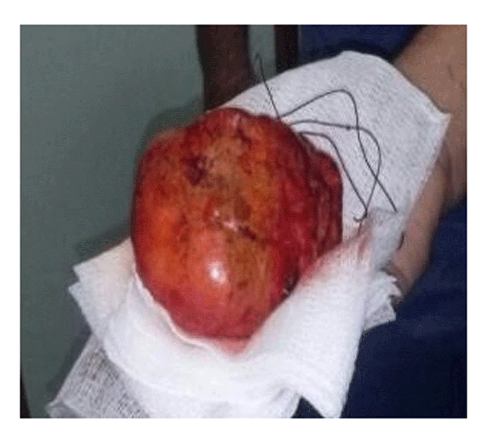
Surgical specimen, macroscopic. Amado Polyclinic, Maracaibo- Edo Zulia (10/04/2013).
The patient experienced no post-surgical complications. She has subsequently completed regular physical activity and continues with the same treatment at the same dosage. Standard laboratory checks at three months showed notable improvement in all parameters.
Adrenal myelolipoma is a rare encapsulated benign tumor described first in 1905 by Gierke11 and later named by the French pathologist Charles Oberling in 192912,13. These tumors are metabolically inactive - or nonfunctional - and composed of adipose and hematopoietic cells originating from the adrenal stroma. They are predominantly asymptomatic and tend to be discovered incidentally13–15.
The incidence of these tumors is between 0.08–0.4%12, although they comprise 15% of the AIs discovered due to advances in radiographic imagery13. They frequently present between the fifth and seventh decades of life without a predominance in either sex - though there is a greater incidence in the right adrenal gland15. Though the adrenal location predominates, there have been discoveries in other locations with a preference for the presacral region, and less frequently in gastric, hepatic, ganglionic lymphatics, cranium, and spleen locations16. These statistics are in accordance with this case report.
The etiology for adrenal myelolipoma is not clear with numerous theories being proposed. Some suggest a metaplasia of the adrenal and myeloid cells that migrated during embryogenesis, extramedullary hematopoiesis, and embolization of osseous medulla elements17. This metaplasia may occur as a response to necrosis, stress, infections, or prolonged adrenocorticotropic hormone (ACTH) stimulation11,18. For example, Al-Bahri et al.19 reported a case of a large bilateral myelolipoma in a 39 year old male with a history of congenital adrenal hyperplasia secondary to a 21-α hydroxylase deficiency treated with steroids starting in childhood. This was later stopped during adolescence with a subsequent myelolipoma development - supporting the theory that ACTH stimulation causes adrenocortical metaplasia. Finally, giant myelolipomas usually are associated with hematologic disorders, like hereditary spherocytosis, thalassemia, and falciform anemia, as a response to adrenal stimulation from erythropoietin20.
Recent cytogenetic analyses propose that myelolipomas are out-of-place masses of myeloid cells. Mitsui et al. described an extremely rare case with the presence of osseous tissue with cells similar to osteoblasts21. Upon immunohistochemical analysis, there were positive results for bone morphogenetic protein 2 (BMP2), which acts as an inductor for osseous formation and the β-catenin that intervenes in the signal pathway. This finding can help give insight into myelolipoma tumorigenesis.
Researchers have also identified (3,21)(q25;p11) chromosomal translocations in patients with myelolipomas and hematological neoplasias18. Because of this, some consider myelolipomas as variants of multiple endocrine neoplasias22, while others recommend that they be grouped with other tumors, such as lipomas, teratomas, liposarcomas, or angiomyolipomas23,24. Despite its benign characteristics, the pathological studies and immunohistochemical evaluation (not performed due to lack of reagents) was recommended, because of the patient's personal and family history that increased risk for malignant results.
Though these tumors are nonfunctional13–15,25, there may be the coexistence of myelolipoma with hyperplasia in any of the three adrenal cortical zones26,27. For these cases, treatment is adrenalectomy (just as in any case of myelolipomas >6cm) independent of its functionality, due to the risk of intratumoral necrosis, hemorrhage from rupture or compression of adjacent structures due to mass effect28. Alternatively, nonfunctional tumors ≤4cm with benign characteristics are recommended to be periodically monitored with radiological and biochemical evaluations. For masses between 4 and 6cm, the surgical intervention should be based on presenting characteristics, growth rate, and the patient’s preference7,29.
It is estimated that 20% of AIs will have subclinical hormone production and these patients represent an at-risk population with greater risk of metabolic disorders and cardiovascular disease7,19. In the present case, the patient’s hormone values were within normal parameters - ruling out subclinical states, including Conn's Syndrome (hyperaldosteronism), Cushing Syndrome or pheochromocytoma. Nevertheless, the presence of myelolipoma is associated with obesity, DM2 and dyslipidemia warranting pharmacological intervention30. This was further emphasized through a retrospective review of 34 AIs in patients of both sexes over the age of 50, where over half suffered from hypertension, 20.6% had DM2, and 37% had obesity. Of these, 80% were histopathologically confirmed to be adenomas with one being a myelolipoma25,30.
As strengths, we can point out the collaboration between different levels of medical attention and the shared effort of the family and the patient to travel to another state to complete this medical Care. Despite the Venezuelan medical assistance crisis, a relatively quick resolution of the case was achieved. Lastly, we emphasize the compliance with the protocol for proper management of adrenal tumors.
The limitations include the inability to perform the hormonal profile and determine whether the tumor was functional or not. Additionally, the choice of imaging could have been better. Specifically, the use of MRI instead of CT is not the first choice for the diagnosis of the myelolipoma; however, this occurred because the initial diagnosis was directed towards a hepatic hemangioma.
Adrenal myelolipomas are rare benign tumors that are generally asymptomatic, whose size ranges from a few millimeters to over a dozen centimetres. Much uncertainty exists surrounding the etiology of these masses with continued debate in the current literature on whether or not they are true neoplasms or manifestations secondary to a reactive process26. In general, surgical management depends on hormone production, tumor size, high risk features on imaging and patient consent. Yet additional studies and information are needed to better understand myelolipomas, their etiology, and clinical management.
Lastly, this case demonstrates how family physicians can manage various aspects of patient care through the facilitation of medical treatments, surgical interventions, and ensuring a proper multidisciplinary approach based on the endocrinology clinical guidelines.
Written informed consent was obtained from the patient for the publication of the patient’s details and accompanying images.
| Views | Downloads | |
|---|---|---|
| F1000Research | - | - |
|
PubMed Central
Data from PMC are received and updated monthly.
|
- | - |
Provide sufficient details of any financial or non-financial competing interests to enable users to assess whether your comments might lead a reasonable person to question your impartiality. Consider the following examples, but note that this is not an exhaustive list:
Sign up for content alerts and receive a weekly or monthly email with all newly published articles
Already registered? Sign in
The email address should be the one you originally registered with F1000.
You registered with F1000 via Google, so we cannot reset your password.
To sign in, please click here.
If you still need help with your Google account password, please click here.
You registered with F1000 via Facebook, so we cannot reset your password.
To sign in, please click here.
If you still need help with your Facebook account password, please click here.
If your email address is registered with us, we will email you instructions to reset your password.
If you think you should have received this email but it has not arrived, please check your spam filters and/or contact for further assistance.
Comments on this article Comments (1)