Keywords
Cholecystocutaneous, Fistula, cholethiasis, abdominal fistula, gallbladder stones, gallbladder cancer, Bile Ducts, Laparotomy
Cholecystocutaneous, Fistula, cholethiasis, abdominal fistula, gallbladder stones, gallbladder cancer, Bile Ducts, Laparotomy
Spontaneous cholecystocutaneous abscess or fistula is an extremely uncommon complication of gallbladder disease. Less than 100 cases have been described in the literature. The first descriptions of cholecystocutaneous fistula was made by Thelisus in 1670. Later Courvoisier reported 169 cases of biliary fistula in the 19th century1. The natural history of the disease has changed from suppurative cholecystitis with spontaneous rupture to operative external drainage of an abscess2. Early and effective medical and surgical management of biliary tract disease can prevent this rare condition. Stone obstruction of the biliary tree plays a crucial role in the pathophysiology of the development of this condition; intra-gallbladder pressure can increase dramatically due to the obstruction of the cystic duct. The unresolved obstruction of the bile outflow compromise gallbladder wall blood circulation, as well as lymphatic drainage, resulting in necrosis of the gallbladder wall with the fistula formation. Once pierced, the gallbladder may drain into the peritoneal cavity, causing peritoneal localized abscess or the abscess can lead into an external fistula due to its adherence to the abdominal wall1–3.
A 76 year old man was admitted to our University Hospital “Ospedale Vittorio Emenuele” and seen in our surgical department. He presented with a 3 cm tumefaction of the right hypochondrium, surrounded by an erythematous skin area, with small secretion of a yellowing-green material, attributable to a bile leaking (Figure 1). The patient’s medical history was clear from previous medical disease and surgery; he only referred to previous upper right quadrant pain and nonspecific dyspeptic disorder.
An abdominal ultrasound examination revealed the presence of a lesion in the aponeurotic muscle wall, but the possible underlying pathology was unknown. No other signs of pathology were observed. Routine blood work was normal.
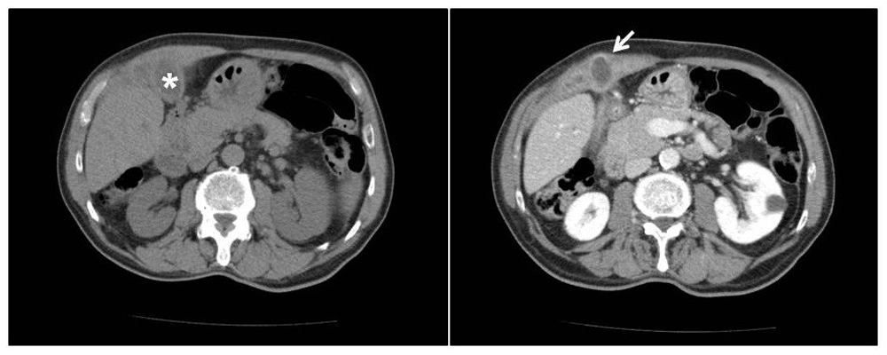
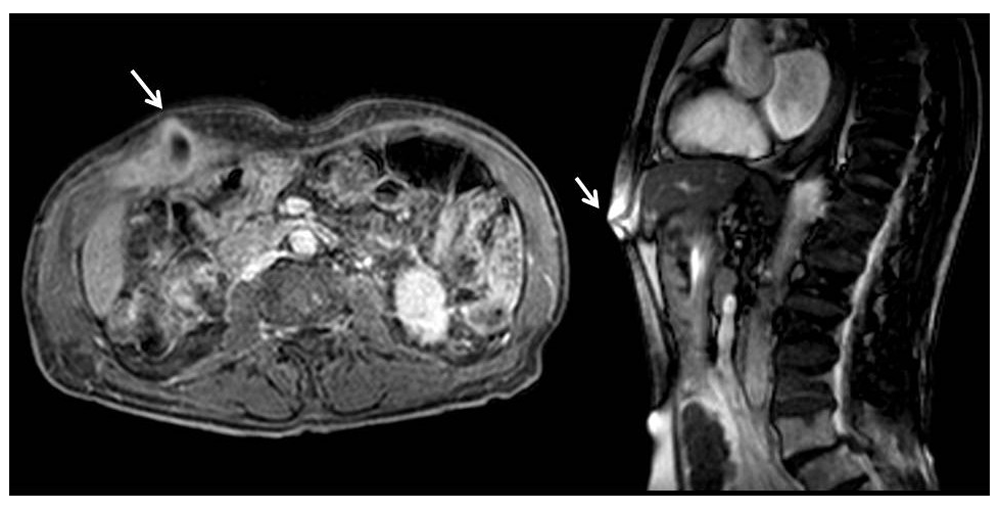
Abdominal computed tomography scan with contrast media showed the gallbladder walls had diffuse thickened and blurred edges, and the right and transverse abdominal muscles were almost covered and embedded with minute hypo-dense ailments compatible with relapsing phlogistic processes (Figure 2). Hepatobiliary MRI detected that the gallbladder had anteriorly shifted and adhered to the right abdominal muscles. The side wall showed a break through with consensual purulent collection, which extruded through the thick abdominal wall (Figure 3). Eventually, several different-sized stones were revealed inside the cholecyst. Consequently, a diagnosis of spontaneous cholecystocutaneous fistula was made.

Left image, * indicates the gallbladder; right image, the arrows indicates the fistula.

The white arrows show the fistula. From left to right: coronal and sagittal fistula view.
The patient underwent cholecystectomy surgery 10 days after diagnosis, with an open laparotomy with en block aponeurotic muscle, skin and fistula orifice excision. In order to have a good abdominal wall reconstruction, a properly shaped Prolene Prosthesis was placed using fibrin glue4–7. The patient received broad-spectrum antibiotics after surgery (Piperacillin/Tazobactam 4.5 g, 3 times a day for 5 days via IV).
Considering the patient’s good general condition and good post-operative course, discharge was on the seventh day post-surgery. Surgical wound re-dressing was made one week after discharge in our facility, where the surgical stitches were removed. Scar appearance was good and without concern. No other dressing was needed and the scar was covered with a bandage. The first follow-up was scheduled 15 days after discharge, second one 60 days from discharge. For both follow-ups, routine blood work and surgical scar checking were performed, the results of which were normal and the scar was healing normally.
A histological examination confirmed the diagnosis of chronic cholecystitis with gallstones and cholecystocutaneous fistula.
Thanks to the progress made with medical imaging and surgical techniques, biliary fistula is today a very rare pathology8–11. Fistulas often represent the result of post-surgical12 or post-traumatic15 complications that generally involve the duodenum (77%) and colon (15%)16.
Spontaneous cholecystocutaneous fistula represents a truly exceptional event, as confirmed by the analysis of the literature, which revealed only 28 cases published over the last 10 years (Table 1). This disease mainly affects female subjects over the age of 60. Etiology is generally due to an acute inflammatory process as a consequence of a cholecystitis or chronic gallstones disease17–20, although there are described cases of spontaneous cholecystocutaneous fistula in the absence of gallstones21. Rarely does cholecystocutaneous fistula evolve into a neoplastic process. Instead, fistula can be a sign of gallbladder cancer19,20. According to Sibakoti, polyarteritis nodosa with gallbladder vasculitis and prolonged use of high dose steroids can be considered predisposing factors21. Fistula primum movens is by cystic duct obstruction, which increases the pressure within the gallbladder, with wall distension and impaired vascularization, resulting in the formation of focal necrosis of the wall with perforation evolution and abscess formation to the surrounding area that will rupture in to the continuous structures. In the present case, the abscess drained through the abdominal wall and the fistulose pathway originated from the bottom of the gallbladder. This area is the most distant from the cystic artery and physiologically the least vascularized and therefore more susceptible to ischemia17.
| Author(s) | Year published | Number of cases | Country | Age | Gender | Treatment technique |
|---|---|---|---|---|---|---|
| Maynard et al.35 | 2016 | 1 | United Kingdom | 68 | F | Open |
| Jayasinghe et al.36 | 2016 | 1 | United Kingdom | >70 | F | Open |
| Guardado-B et al.37 | 2015 | 1 | Mexico | 30 | F | Open |
| Álvarez et al.38 | 2014 | 1 | Argentina | 79 | F | Open |
| Dixon et al.39 | 2014 | 1 | United Kingdom | 94 | F | Open |
| Pripotnev and Petrakos40 | 2014 | 1 | Canada | 85 | F | Open |
| Kim et al.41 | 2013 | 1 | Australia | Open | ||
| Jayant et al.42 | 2013 | 1 | India | 42 | F | Open |
| Sodhi et al.43 | 2012 | 1 | India | 66 | F | Open |
| Kapoor et al.44 | 2013 | 1 | India | 45 | M | Open |
| Ozdemir et al.45 | 2012 | 2 | India | 45 & 65 | M | Open |
| Ugalde Serrano et al.46 | 2012 | 1 | Spain | 83 | M | Open |
| Andersen and Friis-Andersen24,47 | 2012 | 1 | Denmark | 89 | F | Open |
| Ioamidis et al.48 | 2012 | 1 | Greece | 71 | M | Open |
| Cheng et al.49 | 2011 | 1 | China | Open | ||
| Gordon50 | 2011 | 1 | U.S.A. | 83 | F | Open |
| Sayed et al.51 | 2010 | 1 | United Kingdom | 85 | F | Open |
| Pezzilli et al.52 | 2010 | 1 | Italy | 90 | F | Open |
| Metsemakers et al.53 | 2010 | 1 | Belgium | 69 | M | Open |
| Tallón Aguilar et al.54 | 2010 | 1 | Spain | 83 | F | Open |
| Kahn et al.55 | 2010 | 1 | Ireland | 76 | M | Open |
| Hawari et al.56 | 2010 | 1 | United Kingdom | 84 | M | Open |
| Murphy et al.57 | 2008 | 1 | United Kingdom | 80 | M | Open |
| Ijaz et al.58 | 2008 | 1 | United Kingdom | 80 | F | Open |
| Chatterjee et al.59 | 2007 | 1 | India | 45 | F | Open |
| Malik et al.60 | 2007 | 1 | United Kingdom | 76 | F | Laparoscopic |
The external fistular orifice is usually on the right upper quadrant, but other locations have been described, including the left hypocondrium, umbilical scar, right lumbar, and right iliac fossa, and rarely the right gluteus and breast region19,24,47.
The diagnostic process always begins with upper abdomen ultrasound and ends with hepatobiliary MRI to visualize the biliary tree. Considering that 11% of cholecystitis have concomitant presence of gallstones in the main bile duct, it is advisable to perform endoscopic retrograde cholangiopancreatography (ERCP)2–29. In our case CT with CM and hepatobiliary MRI confirmed the fistula presence and it was not necessary to execute the ERCP before surgery. Although an intraoperative cholangiogram was performed to check that the bile ducts were clear from gallstones. Cholecystocutaneous fistula has always been treated by two different strategies. The first includes a two-step approach: percutaneous drainage and antibiotic therapy, and subsequently cholecystectomy. The second directly involves laparotomy cholecystectomy execution with en block aponeurotic muscles, as well as skin and fistula orifice excision.
The second strategy is the most commonly used since the two-step approach treatment is reserved for patients with sepsis and poor general condition12,15,29.
In 1998, Kumar described the first case of gynecological fistula treated with laparoscopic technique, proposing to the scientific community the feasibility of this innovative approach27,30.
Rarity of this pathology confirms the great quality of progress made by early diagnostic techniques and medical treatment to prevent complication of cholethiasis. Although cholecystocutaneous spontaneous fistula is not common, it can lead to a serious condition. If not quickly treated, it can rapidly evolve into a generalized septic state with severe impairment prognosis. In our case, the patient was in good health arguably because the fistula was draining the most of the abscess outside the body and not in the peritoneum space. Surgical treatment was, however, essential to restore the physiologic bile flow and adequate broad-spectrum antibiotic prophylaxis lowed the risk of post-operative infections. Although laparoscopic approaches have been described since 1998, this pathology is, in most cases, continuing to be treated with open technique, most likely because it is easier and with fewer risks of post-surgical complications31–34.
Written informed consent was obtained from the patient for the publication of the patient’s clinical details and related images.
| Views | Downloads | |
|---|---|---|
| F1000Research | - | - |
|
PubMed Central
Data from PMC are received and updated monthly.
|
- | - |
Is the background of the case’s history and progression described in sufficient detail?
Yes
Are enough details provided of any physical examination and diagnostic tests, treatment given and outcomes?
Yes
Is sufficient discussion included of the importance of the findings and their relevance to future understanding of disease processes, diagnosis or treatment?
Yes
Is the case presented with sufficient detail to be useful for other practitioners?
Yes
Competing Interests: No competing interests were disclosed.
Is the background of the case’s history and progression described in sufficient detail?
Yes
Are enough details provided of any physical examination and diagnostic tests, treatment given and outcomes?
Yes
Is sufficient discussion included of the importance of the findings and their relevance to future understanding of disease processes, diagnosis or treatment?
Yes
Is the case presented with sufficient detail to be useful for other practitioners?
Partly
Competing Interests: No competing interests were disclosed.
Reviewer Expertise: General and emergency surgery
Is the background of the case’s history and progression described in sufficient detail?
Yes
Are enough details provided of any physical examination and diagnostic tests, treatment given and outcomes?
Yes
Is sufficient discussion included of the importance of the findings and their relevance to future understanding of disease processes, diagnosis or treatment?
Yes
Is the case presented with sufficient detail to be useful for other practitioners?
Yes
Competing Interests: No competing interests were disclosed.
Alongside their report, reviewers assign a status to the article:
| Invited Reviewers | |||
|---|---|---|---|
| 1 | 2 | 3 | |
|
Version 1 27 Sep 17 |
read | read | read |
Provide sufficient details of any financial or non-financial competing interests to enable users to assess whether your comments might lead a reasonable person to question your impartiality. Consider the following examples, but note that this is not an exhaustive list:
Sign up for content alerts and receive a weekly or monthly email with all newly published articles
Already registered? Sign in
The email address should be the one you originally registered with F1000.
You registered with F1000 via Google, so we cannot reset your password.
To sign in, please click here.
If you still need help with your Google account password, please click here.
You registered with F1000 via Facebook, so we cannot reset your password.
To sign in, please click here.
If you still need help with your Facebook account password, please click here.
If your email address is registered with us, we will email you instructions to reset your password.
If you think you should have received this email but it has not arrived, please check your spam filters and/or contact for further assistance.
Comments on this article Comments (0)