Keywords
trans-illumination, translucent zone, frontal teeth
trans-illumination, translucent zone, frontal teeth
The term trans-illumination depicts tissue diagnostics with a high intensity light beam projected through an object. The object that is studied is positioned between the light source and the observer. The method is quick, accurate, precise, and can determine the current state of the tissues, which makes it a valuable tool1–3.
Trans-illumination is a well-known diagnostic concept used in dentistry. The index of light transmission of healthy tooth tissues is higher than those with caries, and differs from dental calculus. If a tooth is illuminated with sufficient intensity, those clinical findings would appear as dark spots surrounded by light, healthy tooth substance3,4. Using this method fractures lines and orifices of molars, and premolars can be visualized5,6. These are among the reasons why trans-illumination is most frequently applied in operative dentistry. The two devices commonly used are FOTI (fiber-optical trans-illumination) and DIFOTI (digital fiber-optic trans-illumination)7,8.
Currently for trans-illumination, white LED light that is transmitted to the surveyed field through a fiber-optical handpiece is used. This ensures direct contact, without the risk of overheating the structures or causing discomfort to the patient8,9. The small dimensions of the hand-piece allows direct access to all surfaces of the teeth – lingual, vestibular, occlusal - and the ability to assess hard to reach areas, for example the distal parts of the dentition3.
In comparison to X-ray imaging, the teeth can be lit from different angles, which enables the operator to assess more viewpoints. Another advantage of the method is the absence of distortion, which occurs in X-ray images because of the translation of three dimensional objects to two dimensions10. In addition, trans-illumination has better acceptance among patients because of its noninvasive nature11.
In prosthetic dentistry, trans-illumination is used as a proof tool for diagnosis of cracks in ceramic masses after sintering. In cases of full ceramic crowns, the light beam is projected from the intaglio surface of the construction, whereas in cases of metal-ceramic restorations, a tangential direction is used3,6.
In dentistry, translucency is defined as the gradient between transparent and opaque12. Furthermore, the translucent zones, especially in the upper frontal teeth, play a major role in the aesthetic perception and have an impact on the vital appearance of artificial prosthetic constructions. Therefore, their correct in-office registration and recreation by the dental technician are important prerequisites for successful treatment outcome. In standard clinical conditions these areas cannot be assessed. The only commercially available device for individual translucent zone registration currently is the MHT SpectroShade (Verona, Italy), which is based on spectrophotometry. The device presents the individual translucent zone as a scale, which was developed by the manufacturers13.
Therefore, the objective of this article is to propose a clinical protocol for obtaining the individual localization of the translucent zone of frontal teeth with the use of trans-illumination.
All experimental work associated with the current project was approved by the Ethical Committee of Plovdiv Medical University (protocol number P-1546; approved on 13th March 2014). Written informed consent was obtained from all individuals participating in this method, which has been used in clinical practice as part of routine care.
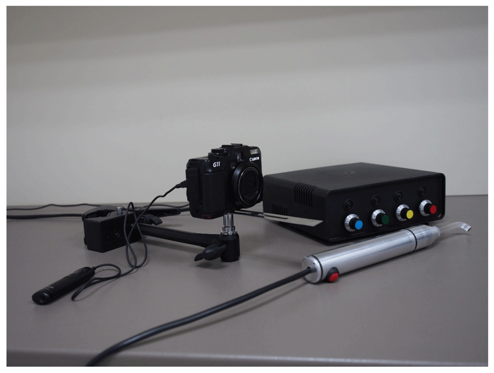
In order to utilize trans-illumination for individual transparent zone registration, a device called ‘Apparatus for registration of translucent individual zone’ (ARTIZ; shown in Figure 1) was developed by the authors. The main components of the apparatus are a power module and emitting module. The main component of the emitting module is a 10W, LZ4-00MA10 emitter (type RGBA LED, Led Engine, USA). The power module is constructed with separate drivers for each LED, based on ZXLD1360 (ZETEX) controller.
The proposed protocol for clinical registration of the individual transparent zone is separated into five steps:
1. Ensuring access to the structures undergoing translucent zone diagnosis
2. Powering and tuning of the apparatus
3. Positioning of the hand-piece
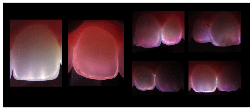
4. Registration of the observed translucent zone with a digital photograph
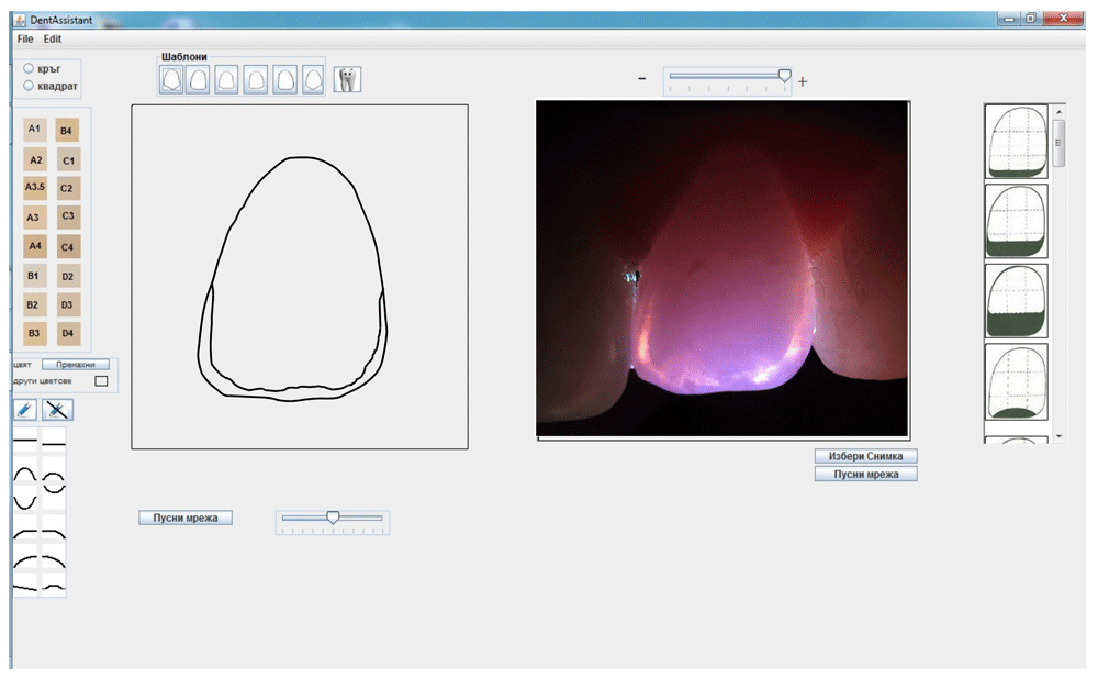
5. Creation of a ‘translucent map’ with the use of custom-made software developed by the authors – ARTIZ-soft.
The steps of the clinical protocol are visualized in Figure 1–Figure 5. Step 2, as well as different technical peculiarities are further mentioned in the next sections.
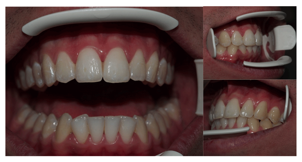
Step 1 (Figure 1)
The teeth undergoing translucent zone registration should be polished with a soft silicone brush and pumice prior to the procedure. If calculus, food leftovers or debris are present, a cleaning procedure is advised. The lip and cheek retractor should enable the operator to position the hand piece in the desired orientation and ensure direct visibility of the surveyed teeth.
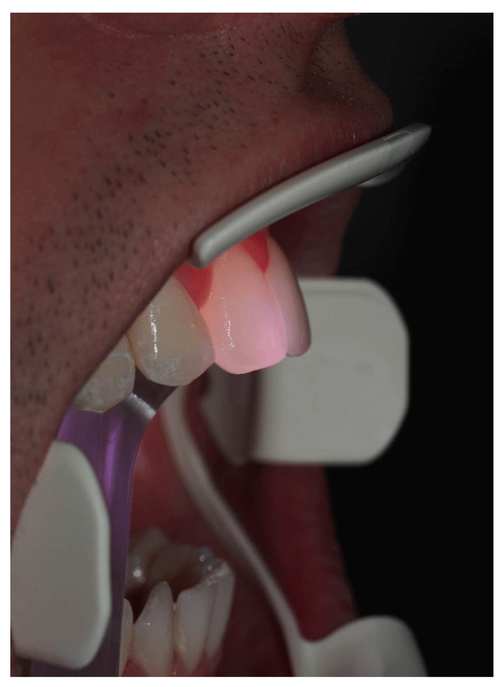
Steps 2 and 3 (Figure 2)
The optimal settings of the ARTIZ apparatus are white light with intensity 1 for all potentiometers. The orientation of the hand-piece depends on the position of the tooth undergoing translucent zone registration, and is up to 50° tangential to the long axis. The optimal angle is between 30–45° tangential to the lingual surface of the tooth, pointed apically.
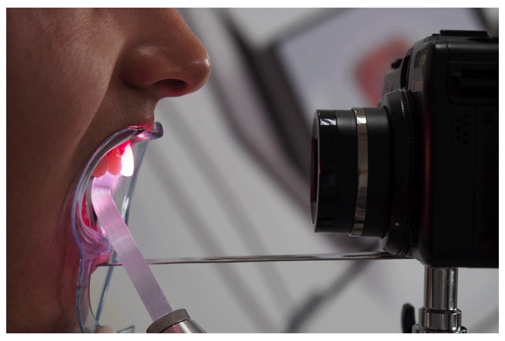
The camera is fixed to the reflector of the dental unit with a standard super-clamp at an angle approximately perpendicular to the vestibular surface. This will minimize the dimensional distortion caused by the transition of a 3D object to 2D. A remote shooter is used to negate vibration and movement during the image acquisition phase. We found out that the optimal camera settings for the Canon camera are as follows:
Exposition time: 1/260 to 1/500 s.;
ISO: 200;
Focal length: 6–7 cm from the vestibular surface of the diagnosed tooth.
Step 5 (Figure 6)
The image is then transported into ARTIZ-soft. After some in-software image manipulation, e.g. cropping, a template representing the tooth and the shape of the translucent zone is chosen (ARTIZ-soft includes digitized templates of the different types of translucent zones according to the classification of Ralin Ralev14) in order to create a ‘translucent map’ of the tooth.
The use of ARTIZ-soft is optional, since there are image manipulation tools freely available in common open-source graphical software, e.g. Inkscape, Gimp, ImageMagick, and using a free-draw tool the translucent zone can be recreated from Ralev’s classification, as depicted in Figure 5.
The translucent zone visualized with the help of the proposed method is clearly defined and easily visible. The registration itself is fast, straightforward and takes 5 to 10 minutes. As a result, a digital image of the individual translucent zone of the tooth is created. With the help of the custom developed software ARTIZ-soft, it is possible to transform the photograph into a ‘translucent map’, which can be used as a guideline for different restorative procedures.
In addition to the established concept of horizontal distribution of the translucent zone, parallel to the incisal edge, with the help of trans-illumination a vertical translucent zone is visualized. This finding complements the overall color registration procedure and gives the practitioner a more detailed information, making restorative procedures predictable.
ARTIZ-soft is currently still in development, with a release date of early 2018. However, the method can be performed with other freely available manipulation tools, as detailed above.
| Views | Downloads | |
|---|---|---|
| F1000Research | - | - |
|
PubMed Central
Data from PMC are received and updated monthly.
|
- | - |
Provide sufficient details of any financial or non-financial competing interests to enable users to assess whether your comments might lead a reasonable person to question your impartiality. Consider the following examples, but note that this is not an exhaustive list:
Sign up for content alerts and receive a weekly or monthly email with all newly published articles
Already registered? Sign in
The email address should be the one you originally registered with F1000.
You registered with F1000 via Google, so we cannot reset your password.
To sign in, please click here.
If you still need help with your Google account password, please click here.
You registered with F1000 via Facebook, so we cannot reset your password.
To sign in, please click here.
If you still need help with your Facebook account password, please click here.
If your email address is registered with us, we will email you instructions to reset your password.
If you think you should have received this email but it has not arrived, please check your spam filters and/or contact for further assistance.
Comments on this article Comments (0)