Keywords
Lumboperitoneal shunt, LP shunt, fluoroscopy
Lumboperitoneal shunt, LP shunt, fluoroscopy
Following feedback from our referees, changes to the article have been made to clarify the term misplacement with regards to position of the LP shunts and to the outcomes of patients with misplaced shunts. We have also further clarified the indication for LP shunt within our patient population.
To read any peer review reports and author responses for this article, follow the "read" links in the Open Peer Review table.
Historically, lumboperitoneal (LP) shunts were the mainstay of cerebrospinal fluid (CSF) diversion therapy for idiopathic intracranial hypertension (IIH). The traditionally cited advantage of LP shunts over ventriculoperitoneal (VP) shunts is the ease of insertion in IIH patients who usually have small and sometimes difficult to catheterise ventricles1–4.
Multiple studies have shown that when functional, LP shunts are effective in alleviating headaches and improving or stabilising visual symptoms in patients with IIH5–8. Studies have shown that IIH patients who underwent LP shunting had improvement in both visual acuity and visual fields with patients also reporting an improvement in headache symptoms post LP shunting6,9. In these previous studies, and in many others, the most common complication was shunt obstruction, with up to 65% of cases requiring revision in one study9. Other less frequent yet significant complications of LP shunts include infection, radiculopathy, shunt migration, syrinx, low pressure headaches, tonsillar herniation, subdural haematomas and potential damage of the distal end of the spinal cord.
The conus medullaris is the tapered, lower end of the spinal cord. Multiple cadaveric studies have demonstrated the level of the conus medullaris to be between T12 and L310. Other studies report that the conus reaches the adult level by two years of age and lies at an average position of L1 to L211. This position was also confirmed by a large radiological study performed in 199810. Due to the proximity of the distal end of the spinal cord, it is best practice to avoid the insertion of LP shunts higher than L2/3 level. The ideal position for this procedure is considered to be at L3/L4 level or below.
The primary advantage of a LP over a VP shunt is the ability to cannulate the CSF space, in this case the thecal sac, as opposed to having to cannulate the very commonly found slit ventricles associated with IIH when considering a VP shunt1–4. However, there are also a series of challenges associated with this procedure.
Generally LP shunt patients are positioned in the lateral position to provide simultaneous access to the lumbar spine and flank. Percutaneous cannulation of the thecal sac can be very challenging, often requiring specific long needles. Additionally, it is often difficult to get the flexion (“foetal position”) required in these patients to open the interlaminar space and allow for the needle to access the thecal sac. Once the proximal catheter enters the thecal sac it needs to be threaded cranially into position, which is at times challenging as the catheter often kinks within the significant tissue volume. Following placement of the proximal catheter, the remainder then needs to be tunneled through the subcutaneous tissue into the flank region. At this point, while in the lateral position and with the significant amount of adipose tissue, the surgeon then needs to identify the peritoneum. This can prove to be quite challenging given the non-anatomic patient’s position as well as the fact that gravity is working against the surgeon. At this point the catheter is then passed into the flank within the peritoneum.
The insertion of the catheter into the lumbar CSF space determines the success or failure of the LP shunt. Since this involves a manual manoeuvre with a “blind” tap, the catheter may be inadvertently placed incorrectly and migration of the shunt catheters is a common experience. The lumbar catheter can migrate relative to the thecal sac (usually into the subcutaneous space), and the peritoneal catheter can likewise come out of the peritoneum. The incidence ranges from 3–20%. Migration complications have been noted to be more common in the paediatric population4,12,13. When a catheter migrates out of the thecal sac, a subcutaneous collection of spinal fluid can be observed.
Newly onset radicular pain has been noted to occur with LP shunts. This may result from catheter migration or localised inflammation leading to arachnoiditis. The onset of symptoms may necessitate shunt revision. The incidence of developing newly onset radicular pain ranges from 5–6%13,14.
The efficacy of fluoroscopic guidance in the placement of a lumbar catheter in patients treated with an LP shunt has been reported9. The method includes using intraoperative portable fluoroscopy with contrast medium. The direction of the inserted catheter can be confirmed, and loop formation or absence thereof can be detected intraoperatively. It is possible to confirm that the catheter has not migrated into the extra-CSF space or the intervertebral foramen containing the spinal nerve roots. Improved visibility of the catheter in the spine, by filling it with contrast medium, is the key to the success of this procedure15. Intraoperative fluoroscopic guidance has become widely available in last two decades. Despite its efficacy, it exposes the patients and the staff to radiation. Moreover, there are possible side effects and restrictions related to the use of contrast medium, such as allergy, anaphylactic shock and acute renal failure. For these and other reasons, there is still a significant number of LP shunts operations that are performed without fluoroscopic guidance.
The National Hospital for Neurology and Neurosurgery offers dedicated hydrocephalus services and receives quaternary referrals from centres situated in the UK and abroad. The present study describes the accuracy of LP shunt placement without intraoperative fluoroscopic guidance over a 10-year period.
An analysis of the hospital electronic records identified 163 LP shunt procedures performed without fluoroscopic guidance on patients with a diagnosis of idiopathic intracranial hypertension (IIH). They were performed between 2006 and 2016. Cases with no post-procedural imaging were excluded (56), due to the impossibility to identify the level of the proximal catheter.
Post-procedural imaging was reviewed and reported by two independent operators who were blinded to each other’s results and verified using visible anatomical landmarks on the x-ray, the location of the proximal end of the catheter was recorded. In all cases, the imaging used was lumbar x-ray. Clinical notes were also reviewed for those patients who had incorrect positioning of the proximal catheter to identify potentially related signs and symptoms, such as lumbar radiculopathy.
The data collection and analysis was carried out using Microsoft Excel 2010.
Between 2006 and 2016, 163 LP shunt procedures were performed on patients with IIH without intraoperative fluoroscopic guidance. After exclusion of the cases without post-procedural imaging (56), a total of 107 procedures performed on 73 patients were selected. A total of 57 patients were female and 16 were male (M:F, 1:3.5), the mean age was 41 years (± 13 SD) ranging from 16–69 years.
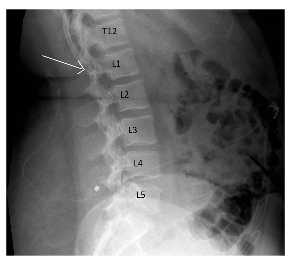
The review of all post-procedural imaging showed that in 17 cases (15.8%) patients had the proximal catheter placed at the level of L1/L2 or above (T12/L1, 1.8%; L1/2, 14.0%) (Figure 1). On the other hand, in 94 cases (84.2%), patients had the proximal tip of the catheter placed at the level of L2/L3 or below (L2/3, 33.0%; L3/4, 37.4%; L4/5, 12.0%; L5/S1, 1.8%) (Table 1).
| Level of insertion | Number of cases (%) |
|---|---|
| T12/L1 | 2 (1.8) |
| L1/L2 | 15 (14.0) |
| L2/L3 | 35 (32.7) |
| L3/L4 | 40 (37.4) |
| L4/L5 | 13 (12.0) |
| L5/S1 | 2 (1.8) |
An analysis of the clinical notes of the patients who had mispositioned LP shunts was carried out for a minimal post-operative period of one year. None of the patients complained of signs or symptoms related to possible distal spinal cord damage.
This study demonstrates that, without intraoperative fluoroscopic guidance, an LP shunt insertion procedure can lead to a mispositioned proximal catheter in 15.8% of cases. Despite none of our patients presenting with any signs or symptoms of spinal cord damage, this risk needs to be considered when performing this procedure “blindly”.
One of the biggest challenges in performing LP shunts in IIH patients is often related to their habitus. It is in fact recognised that a strong association between IIH and obesity exists16. Approximately 70–80% of IIH patients are obese and over 90% are overweight16. In this group of patients finding the anatomical landmarks, maintaining them and inserting the lumbar catheter at the correct level, can represent a technical challenge; this is especially true when the procedure is performed without fluoroscopy guidance.
We suggest that the use of intra-operative imaging guidance should be adopted: this practice could reduce the incidence of mispositioned LP shunts and therefore decrease the risk of significant spinal cord damage, which may have serious, irreversible consequences.
The results of this series must be interpreted considering the limitations of the nature of any retrospective study. It could be argued that results achieved by our unit could vary markedly from those achieved at other units. We also do not take into account for operator experience, which may be partially responsible for the differences in success rate, and again may vary from individual to individual. Ultimately, to prove the efficacy and benefits of intraoperative imaging for LP shunt insertion, large, prospective, randomised controlled studies should be performed.
While this series is too small to conclude whether intraoperative imaging should be used to minimize the risk of misplaced proximal LP shunt catheters, it prepares the basis for further prospective studies. Our results suggest that LP shunt insertion without fluoroscopic guidance has a 15.8% risk of misplacement of the end position of the proximal catheter, and for this reason the use of intraoperative image guidance is suggested to reduce the risk of spinal cord damage and its potentially catastrophic consequences, although no spinal cord injury was noted as a result of misplacement in our patient population.
Ethical approval and registration was obtained from the National Hospital for Neurology and Neurosurgery.
This study was performed as part of an audit to analyse the current practice against the department policy standards.
Dataset 1: X-rays showing the final position of the lumboperitoneal (LP) shunt in patients that underwent LP shunt insertion without fluoroscopic guidance. Each page of the dataset indicates a different procedure. doi, 10.5256/f1000research.11089.d15468617
| Views | Downloads | |
|---|---|---|
| F1000Research | - | - |
|
PubMed Central
Data from PMC are received and updated monthly.
|
- | - |
Click here to access the data.
Spreadsheet data files may not format correctly if your computer is using different default delimiters (symbols used to separate values into separate cells) - a spreadsheet created in one region is sometimes misinterpreted by computers in other regions. You can change the regional settings on your computer so that the spreadsheet can be interpreted correctly.
Provide sufficient details of any financial or non-financial competing interests to enable users to assess whether your comments might lead a reasonable person to question your impartiality. Consider the following examples, but note that this is not an exhaustive list:
Sign up for content alerts and receive a weekly or monthly email with all newly published articles
Already registered? Sign in
The email address should be the one you originally registered with F1000.
You registered with F1000 via Google, so we cannot reset your password.
To sign in, please click here.
If you still need help with your Google account password, please click here.
You registered with F1000 via Facebook, so we cannot reset your password.
To sign in, please click here.
If you still need help with your Facebook account password, please click here.
If your email address is registered with us, we will email you instructions to reset your password.
If you think you should have received this email but it has not arrived, please check your spam filters and/or contact for further assistance.
Comments on this article Comments (0)