Keywords
Hepatopulmonary Syndrome, COPD, MAI Infection, Bronchiectasis
Hepatopulmonary Syndrome, COPD, MAI Infection, Bronchiectasis
In this revised version I have attempted to provide further clinical information about the state and nature of the patient's other pulmonary diseases so as to give further evidence as to what role HPS played in her clinical presentation. I have also tried to better characterize the gas-exchange abnormalities caused by HPS which I describe in the discussion.
To read any peer review reports and author responses for this article, follow the "read" links in the Open Peer Review table.
The triad of chronic liver disease, hypoxemia, and microvascular dilatations/malformations in the lungs make up hepatopulmonary syndrome (HPS), a well-known sequela of hepatic cirrhosis1. These aforementioned vascular abnormalities result in ventilation-perfusion mismatch, which yields the clinical findings of dyspnea (universally) as well as digital clubbing and/or cyanosis (both very common)2. We report a case of a patient presenting with HPS as their first clinical manifestation of suspected cirrhosis, and describe how it was distinguishable from other potential causes of her symptoms such as her comorbid chronic obstructive pulmonary disease (COPD), bronchiectasis, and mycobacterium avium-intracellulare (MAI) infection.
An 86 year old retired latina woman with a past medical history of COPD, bronchiectasis, MAI infection (not previously treated), tobacco dependence (40 pack-years, quit 25 years prior to presentation), diabetes mellitus, hyperlipidemia, and hypertension presented with two weeks of worsened dyspnea and non-productive cough. She reported a baseline of daily shortness of breath with an exercise tolerance of 3 blocks, but over the two weeks prior to her presentation it decreased to the point where she would feel dyspneic when walking around her apartment. Interestingly she stated that she also generally felt more short of breath while seated than when lying down, and also cited a worsening cough over this time course productive of green sputum. Her exam on presentation was significant for an oral temperature of 101.4 degrees Fahrenheit, oxygen saturation of 84% on room air, tachypnea and coarse crackles appreciated diffusely on lung examination. Her blood-work was notable for a white blood cell count of 19.8 k/µL, with multiple diffuse small nodular opacities seen on chest x-ray consistent with her known MAI infection. She was started on levofloxacin for treatment of a presumed bronchiectasis flare along with oxygen therapy via nasal cannula in addition to other supportive treatments. Although her fever, leukocytosis, and cough improved with antibiotics (further supporting a diagnosis of bronchiectasis flare), her dyspnea and hypoxemia persisted. Consequently, a chest computerized tomography (CT) scan was ordered which showed the same nodularities seen on chest x-ray, but also elucidated a nodular liver consistent with cirrhosis. While her platelet count, transaminases, bilirubin, and prothrombin time were all normal and she had no ascites or other edema on exam, she did however have spider angiomas. Further chart review done at that time revealed that she had known cirrhotic characteristics on liver imaging as they were incidentally seen almost five years prior, although she had never had any decompensations or serologic evidence of liver dysfunction since. Work-up back then elucidated no potential cause except for non-alcoholic fatty liver disease, given her histories of hyperlipidemia and diabetes. In light of this knowledge gained from deep chart review, the specter of hepatopulmonary syndrome was raised as a possible explanation for her persistent hypoxemia and dyspnea. In order to investigate this possibility, both seated and supine arterial blood gases were obtained which elucidated orthodeoxia (see Table 1). A transthoracic echocardiogram with bubble study was then performed which suggested an intrapulmonary shunt (see Figure 1), thereby confirming the diagnosis of HPS. While oxygen supplementation caused her dyspnea to improve and oxygen saturation to rise to a safe level, she interestingly was never able to reach a saturation of 100%. However given this improvement in her dyspnea and oxygenation, as well as the resolution of all signs and manifestations of the bronchiectasis flare that she initially presented with, the patient was discharged home with oxygen. Soon after discharge, she was seen in a pulmonology clinic where she was found to be in stable condition.
| Parameter | Supine | Seated |
|---|---|---|
| FiO2 | 21% (Room Air) | 21% (Room Air) |
| PaO2, Arterial | 65.5 mmHg | 53.1 mmHg |
| Direct O2 Saturation | 93.3% | 88.6% |
| PaCO2, Arterial | 45.0 mmHg | 43.1 mmHg |
| A-a gradient | 28.0 mmHg | 42.8 mmgHg |
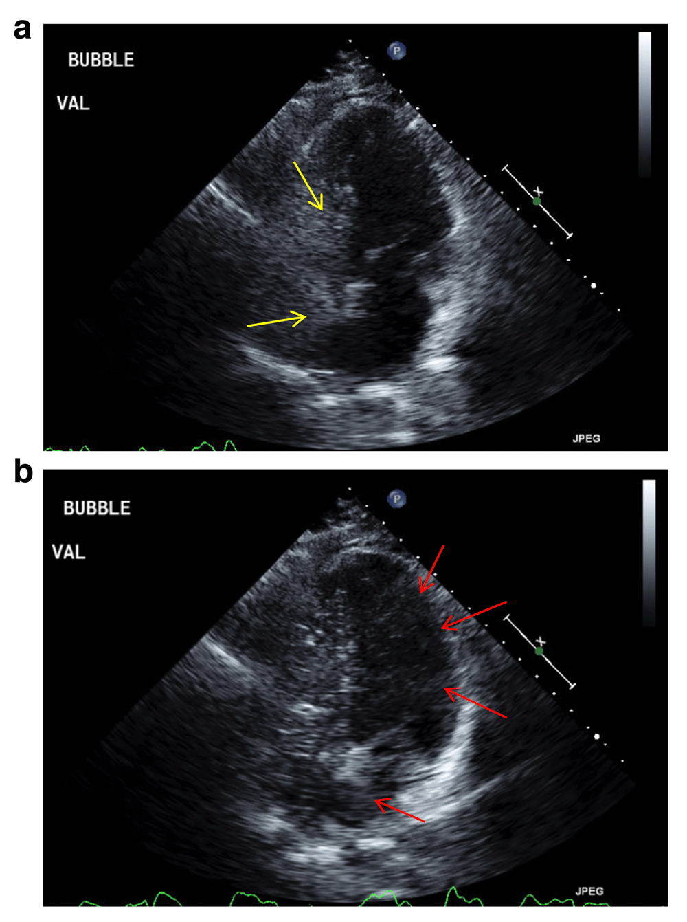
Still images from patient’s transthoracic echocardiogram showing (a) no early shunting with saline bubble (identified by yellow arrows) injection, followed by (b) late passage of bubbles (identified by red arrows) into the Left Atrium and Ventricle representing Intrapulmonary Shunting.
Our patient possessed all three of the cardinal findings of hepatopulmonary syndrome: chronic liver disease, hypoxemia, and evidence of pulmonary microvascular abnormalities. Her symptom of platypnea (shortness of breath worsened by going from a supine to seated position) and finding of orthodeoxia on arterial blood gas analysis also clearly pointed to HPS. But while most cirrhotic patients with hepatopulonary syndrome have only mild disease and its severity is often proportional to that of their cirrhosis, she presented with severe disease despite seemingly having compensated cirrhosis3. Moreover, it is very unusual for HPS to be the first symptomatically manifesting sequela of cirrhosis as it was in this patient; there are few other examples of this happening in the literature4.
As it is classically described, our patient's hypoxemia likely resulted from intrapulmonary vascular abnormalities causing ventilation-perfusion mismatch (primarily) and direct arteriovenous communications (to a lesser degree), which yielded her dyspnea5,6. More specifically this shunting caused her to have platypnea, which is the symptomatic manifestation of orthodeoxia, and has been shown to be very closely tied with HPS. In one prospective study comparing cirrhotics with HPS vs. those without the complication, platypnea was endorsed by 65.5% of the HPS group vs 6.2% of the non-HPS cirrhotic group7.
Given that they are also manifestations of cirrhosis-related vascular malformations, spider angiomas are also commonly seen in HPS as they were with our patient8. The intrapulmonary anomalies can be reliably detected via saline-enhanced transthoracic echocardiography, as well as with more advanced confirmatory tests such as technitium-99m macroaggregated albumin (MAA) nuclear scanning or pulmonary angiography3,5,9. Given the clear presence of lately-transmitted bubbles seen on her transthoracic echocardiogram though, these more advanced and expensive tests were not pursued in our patient's case.
Once the diagnosis is made with the aforementioned triad, disease severity is assessed via PaO2. Patients with mild disease have a PaO2 ≥ 80mmHg, those with moderate have PaO2 ≥ 60 < 80 mmHg, severe have PaO2 ≥ 50 < 60 mmHg, and those with very severe disease have a PaO2 of < 50 mmHg10. Our patient was found to have severe disease based upon her PaO2, although much of her arterial hypoxemia was likely related to her other pulmonary diseases. The pathogenesis of HPS is thought to involve increased serum levels of nitric oxide (although correlations with elevated carbon monoxide and tumor necrosis factor α have also been seen) resulting from cirrhosis, which is postulated to cause pulmonary vascular dilatation, and to a lesser degree arteriovenous malformations (AVMs)5,11. Autopsy studies have shown that the number of dilated precapillary and capillary vessels in the lungs far outnumbers the number of pulmonary AVMs in these patients, but the end result of each is the same: passage of mixed-venous blood into the pulmonary veins, resulting in V/Q mismatch and hypoxemia5. Where those vessels dilated as a result of HPS are concerned, the hypoxemia is a result of diffusion-limited gas exchange. Cirrhosis itself (independent of HPS) is associated with impaired autoregulation of pulmonary vascular tone in a heterogenous distribution throughout the lung, which in HPS results in orthodeoxia as alveoli in dependent areas see alterations in ventilation for which blood flow cannot be adequately accommodated for with respect to gravitational changes when patient position is adjusted5,6,12.
The only definitive treatment for HPS is liver transplantation13. While those with this condition are more prone to post-operative complications than other patients post-liver transplant, ultimately those who survive have resolution of the hypoxemia caused by their pre-transplant HPS14. Given her age, comorbidities, and otherwise well-compensated liver disease, liver transplant was not considered in our patient.
In addition to HPS, there are other similar clinical entities which are worth discussing as part of the differential diagnosis for patients who present like ours. Similar to HPS, portopulmonary hypertension (PPH) is also a sequela of cirrhosis which is characterized by pulmonary hypertension (as defined by elevated mean arterial pressure of >25mmHg at rest, and pulmonary vascular resistance greater than 240 dynes/sec/cm-5) in the setting of portal hypertension and/or cirrhosis with other causes of PH having been excluded10. Patients with PPH typically present with clinical findings consistent with other causes of pulmonary hypertension, namely external dyspnea, fatigue, chest pain, and/or syncope, with progression to cor pulmonale in severe cases15. PPH was excluded in our patient by the fact that her transthoracic echocardiogram showed no evidence of pulmonary hypertension.
Hereditary hemorrhagic telangiectasia (HHT) is a genetic (autosomal dominant) disease in which arteriovenous malformations (AVMs) and mucocutaneous telangiectasis form throughout the body16. The predominant symptom with which people present is paroxysmal epistaxis related to AVMs in the nasal mucosa, although 15–35% of patients with HHT have pulmonary AVMs which cause dyspnea in a manner analogous to those with HPS17.
While it was tempting at first to presume that our patient's untreated MAI (and/or her other lung diseases, see Table 2 below) could be causing her symptoms, the clinical presentations which result from this infection are not typically mimickers of HPS. Those with symptomatic pulmonary MAI infection typically present in one of two ways, depending on whether they have underlying lung disease or not. Those with underlying lung disease (most commonly COPD or bronchiectasis) typically have a tuberculosis-like (albeit milder) presentation: chronic cough, weakness, malaise, weight loss, dyspnea, and upper-lobe predominant infiltrates and/or cavitation on chest imaging18. Patients with underlying bronchiectasis will usually have their MAI infection develop in bronchiectatic areas, not necessarily in the upper lobes19. Those without underlying lung disease tend to present with months of productive cough, without the other “tuberculosis-like” constitutional symptoms20. A subset of these patients classically present with “Lady Windermere syndrome:” lingular/right-middle lobe infiltrates in elderly women without predisposing lung disease who suppress their cough21. Regardless of which way a patient presents, diagnosis is made via the combination of radiographic findings indicative of pulmonary disease along with either MAI-positive sputum or an MAI-positive bronchial wash in a patient with respiratory symptoms, according to the Infectious Disease Society of America and the American Thoracic Society22.
The patient’s lack of basilar-predominant nodularities on CT scan made MAI the unlikely cause of her platypnea and/or orthodeoxia. As Table 2 also illustrates though, many of her signs and symptoms could be ascribed to her COPD and/or bronchiectasis. Obstructive airway diseases such as these are a known cause of ventilation-perfusion defects similar to those seen in HPS, as the distribution of alveolar ventilation is increased whereas that of pulmonary blood flow is not, with this effect positively correlated with disease severity but significantly hastened during acute exacerbations23. However neither of these conditions are known to cause orthodeoxia, and her COPD was stable (FEV1 was 0.61 liters on recent spirometry) at the time of her hospitalization. In the end, the clinical and diagnostic characteristics of HPS are so specific as a result of its entirely unique gas-exchange pattern that it can be reliably diagnosed even in the setting of other diseases that cause arterial hypoxemia, as it was in our patient24.
Hepatopulmonary syndrome belongs on the differential diagnosis for dyspnea in any patient with chronic liver disease, even those like our patient without any previous decompensations of cirrhosis. While it can present in a way which is symptomatically similar to that of other chronic lung diseases, it can be definitively diagnosed via a relatively simple work-up which should be performed in any dyspneic patient with evidence of liver disease. While the only definitive cure is liver transplant, it can be conservatively managed via oxygen therapy when transplant is contraindicated, as it was in our patient's interesting case.
Written informed consent for publication of their clinical details and/or clinical images was obtained from the patient.
All data underlying the results are available as part of the article and no additional source data are required.
| Views | Downloads | |
|---|---|---|
| F1000Research | - | - |
|
PubMed Central
Data from PMC are received and updated monthly.
|
- | - |
Provide sufficient details of any financial or non-financial competing interests to enable users to assess whether your comments might lead a reasonable person to question your impartiality. Consider the following examples, but note that this is not an exhaustive list:
Sign up for content alerts and receive a weekly or monthly email with all newly published articles
Already registered? Sign in
The email address should be the one you originally registered with F1000.
You registered with F1000 via Google, so we cannot reset your password.
To sign in, please click here.
If you still need help with your Google account password, please click here.
You registered with F1000 via Facebook, so we cannot reset your password.
To sign in, please click here.
If you still need help with your Facebook account password, please click here.
If your email address is registered with us, we will email you instructions to reset your password.
If you think you should have received this email but it has not arrived, please check your spam filters and/or contact for further assistance.
Comments on this article Comments (0)