Keywords
Atrial septal defect, pulmonary stenosis, echocardiography, angioplasty, cardiac catheterization.
Atrial septal defect, pulmonary stenosis, echocardiography, angioplasty, cardiac catheterization.
Atrial septal defect (ASD) is a very common congenital heart disease1,2. It can be associated with other cardiovascular abnormalities; the most common is pulmonary stenosis2. The latter usually concerns the valve or the right outflow tract but rarely pulmonary artery branches. This unusual association can be suspected by careful analysis of complementary exams. Currently, advances in interventional treatment make possible reliable and effective treatment of ASD even when associated with other lesions, in particular pulmonary stenosis3.
The aim of this article is to report, from a case report and literature review, diagnostic challenges and the contribution of simple complementary exams, such as chest X-ray, for the diagnostic orientation of an ASD associated with peripheral pulmonary artery stenosis, as well as therapeutic particularities.
We report the case of a girl born in 2007, with history of dyspnoea and recurrent bronchitis in whom a systolic murmur was detected in our outpatient office at the age of two years. The physical examination at this age noted a non-dysmorphic child with normal growth and psychomotor development. Auscultation of the pulmonary area noted loud 3/6 systolic ejection-type murmur, splited second heart sound with a marqued pulmonary component. An electrocardiogram recorded sinus rhythm, incomplete right bundle branch block and right ventricular hypertrophy.
Chest X-ray showed moderate cardiomegaly with cardio-thoracic ratio of 0.53, convex mid-left arch, and above all marked hypervascularity of the left lung contrasting with reduced blood flow to the right lung (Figure 1).
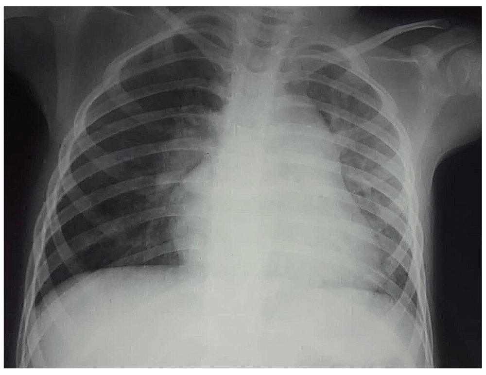
Cardiomegaly, convex middle arch and unilateral left lung hypervascularity contrasting with reduced blood flow to the right lung is shown.
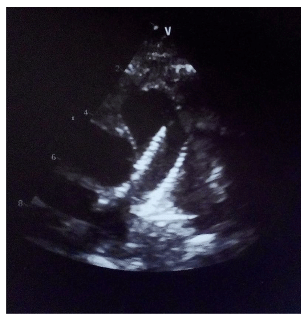
Doppler echocardiography noted a 20 mm diameter ostium secundum ASD with right chamber volume overload associated with right pulmonary artery (RPA) stenosis. Diagnostic confirmation of peripheral pulmonary branch stenosis was made by CT scan and right heart catheterization (Figure 2).

(A) Tight stenosis at the origin of the right pulmonary artery (red arrow); (B) small right pulmonary artery (15 mm) contrasting with (C) dilated left artery (27 mm).
A two-step percutaneous treatment for these lesions was decided. RPA stenosis was treated firstly with 8 mm × 2 cm balloon in 2012 with poor initial result. A novel attempt in the same year with a balloon and stent placement (Express™ Vascular LD 10*37mm) was successful (Figure 3).
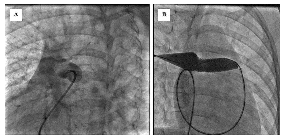
(A) Selective angiography of the right pulmonary artery and (B) stent deployment.
This result was optimized 6 months later through a 18 mm × 20 mm balloon leaving mild residual gradient of 10 mm Hg between pulmonary trunk and RPA. The ASD was closed successfully 1 year later in July 2013 with a 24 mm Figulla Flex II prosthesis. The procedure was uneventful and fluoroscopic control at the end noted ASD prosthesis in place and stent at RPA level (Figure 4). Control chest X-ray showed symmetrical bilateral vascularisation of the two lungs (Figure 5). Outcome was favourable. Control echocardiography performed at 4 years of regular follow-up noted mild residual pulmonary stenosis (maximal residual gradient of 15 mmHg), no stent restenosis and a well-sealed ASD prosthesis (Figure 6). Systolic right ventricle function indices were normal.
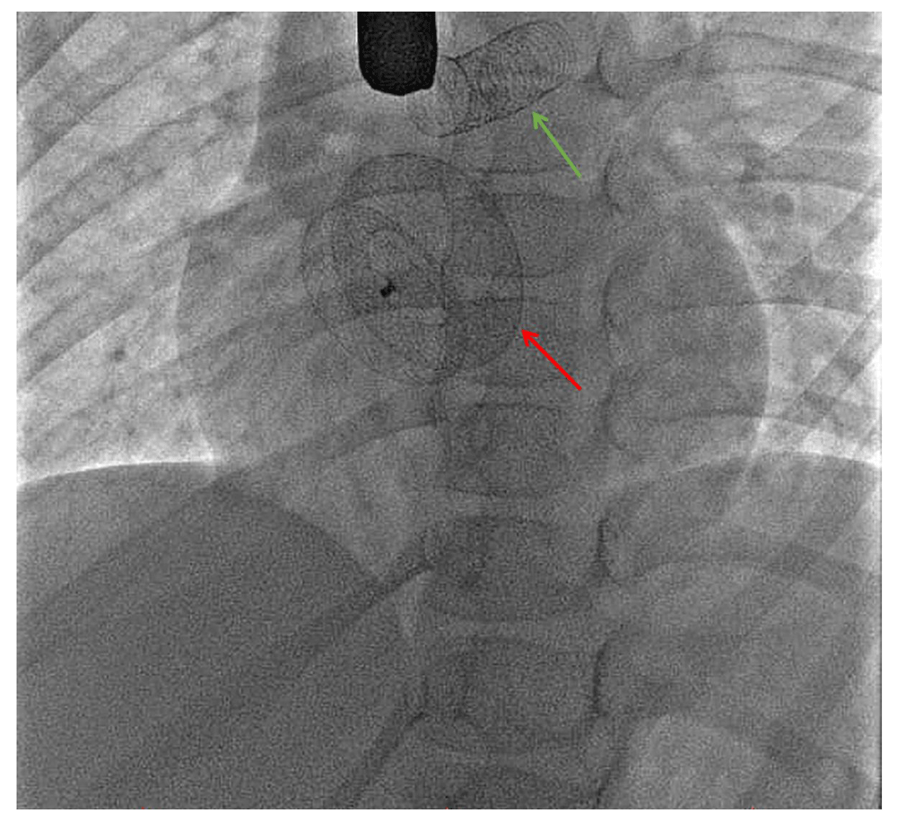
Atrial septal defect prosthesis (red arrow) and the right pulmonary artery stent (green arrow).
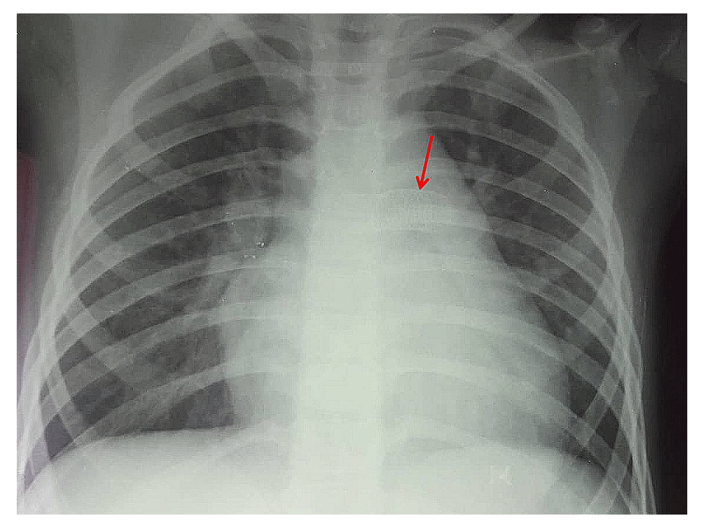
Symmetrical pulmonary vasculature and right pulmonary artery stent shown by red arrow.
Our case emphasizes perfectly the importance of careful basic semiology analysis in the diagnosis process of congenital heart disease. In fact, heart murmur characteristics and asymmetric pulmonary vasculature in chest X-ray oriented the diagnosis of ASD associated with pulmonary branch stenosis4. Confirmation was made by appropriate investigations, particularly Doppler echocardiography, thoracic CT angiography and finally cardiac catheterization with selective angiograms. Chest roentgenogram still retains great value for the diagnostic process in cardiology. Thus in our patient, the finding of an unbalanced pulmonary vasculature especially when associated with an intense pulmonary murmur oriented the diagnosis of pulmonary artery branch stenosis. RPA stenosis caused pulmonary flow deviation mainly to the healthy side resulting in increased vascularisation of the left lung contrasting with a hypovascularity of the right one. This unilateral hyper-flow was quite marked because of its association to relevant left to right shunt related to wide-associated ASD. This suspicion of RPA stenosis was easily confirmed by echocardiography, CT scan, and right-heart catheterization with measure of pressure in right heart chambers, pulmonary branches and finally selective pulmonary artery branches angiographies.
Conventionally, treatment of this condition was surgical with ASD closure and pulmonary artery branch plasty. Currently, balloon dilatation with stent placement has revolutionized management of pulmonary stenosis especially those involving branches5,6. Pulmonary artery stenosis can complicate the course of many congenital heart diseases. Percutaneous treatment can be performed as a surrogate or adjunct to surgery and it is considered as standard of care for proximal stenosis. For distal stenosis, it allows treatment of lesions inaccessible to the surgeon, often in addition to repair surgery of right ventricular outflow tract3. Angioplasty of the pulmonary arteries has evolved considerably since its introduction in the early 1980s. High pressure balloons usually are 2 to 4 times larger than the diameter of the stenosis are used. Stents used are still currently most often not premounted and have the advantage of being expandable to a diameter sufficiently close to vessel size in adulthood7. This type of stent was successfully used in our patient and allowed restoration of pulmonary vasculature by removing the peripheral pulmonary stenosis. The success of pulmonary dilatation authorized percutaneous closure of the ASD, which was performed successfully one year later by prosthesis. Percutaneous closure is currently the standard treatment for ostium secundum ASD with adequate rims and diameter less than 38 mm with a success rate close to 100% and lower morbidity compared to surgery.
Our case is very rare and to our knowledge, no similar cases have been reported. It proves feasibility and reliability of percutaneous treatment for such a case. The sequence of the lesion treatment is dictated by lesions complexity whose failure can shift the case to surgery. This is the reason why we waited obtaining a satisfactory and stable result on pulmonary artery stenosis before treating ASD.
Pulmonary stenosis can be associated with ASD limiting pulmonary hyper-flow. In our case, this stenosis was tight and sat on the origin of the right branch, which resulted in reducing significantly blood flow to the ipsilateral lung. Therefore, ASD-related pulmonary hyperflow was directed to the left lung field, explaining the radiological aspect particularly unilateral hypervascularity. Careful chest X-ray analysis can allow suspicion of pulmonary artery branch stenosis. Confirmation can be made by Doppler echocardiography and, if necessary, by further examination allowing treatment adaptation. Management of this association benefited from interventional techniques progress allowing successfully treatment with stable long-term outcome. Indeed, with a follow-up of four years, the atrial septum was tight and there was no residual pulmonary stenosis with a normalized RV function.
Written informed consent for publication of the clinical details and images was obtained from the patient's father.
All data underlying the results are available as part of the article and no additional source data are required.
| Views | Downloads | |
|---|---|---|
| F1000Research | - | - |
|
PubMed Central
Data from PMC are received and updated monthly.
|
- | - |
Provide sufficient details of any financial or non-financial competing interests to enable users to assess whether your comments might lead a reasonable person to question your impartiality. Consider the following examples, but note that this is not an exhaustive list:
Sign up for content alerts and receive a weekly or monthly email with all newly published articles
Already registered? Sign in
The email address should be the one you originally registered with F1000.
You registered with F1000 via Google, so we cannot reset your password.
To sign in, please click here.
If you still need help with your Google account password, please click here.
You registered with F1000 via Facebook, so we cannot reset your password.
To sign in, please click here.
If you still need help with your Facebook account password, please click here.
If your email address is registered with us, we will email you instructions to reset your password.
If you think you should have received this email but it has not arrived, please check your spam filters and/or contact for further assistance.
Comments on this article Comments (0)