Keywords
Papillon – Lefèvre syndrome, Periodontitis, Premature tooth loss, Palmoplantar keratosis.
Papillon – Lefèvre syndrome, Periodontitis, Premature tooth loss, Palmoplantar keratosis.
Papillon-Lefèvre syndrome (PLS) is an autosomal recessive disorder that typically becomes apparent from one to five years of age, which coincides with the timing of eruption of primary dentition. The estimated prevalence of the syndrome is 1–4 cases per million individuals1.
The exact etiopathogenesis of the syndrome is relatively unclear and different etiological factors have been suggested, such as immunologic, genetic or bacterial, but recently it was suggested that mutations of cathepsin C gene, which results in deficiency of cathepsin C enzymatic activity, to be the possible etiological factor. This was supported by the fact that expression of the cathepsin C gene occurs mainly in epithelial regions, such as the soles, palms and keratinized oral gingiva, which are the most affected areas in patients with PLS2.
An important feature of the syndrome is the presence of palmoplantar hyperkeratosis; its onset usually occurs between the ages of one to four years and usually involves the palms of the hands and soles of the feet3. Another major feature is severe gingivostomatitis and periodontitis. Deciduous teeth usually erupt in normal sequence, timing and with normal structure and form, although it was reported that some cases may have microdontia and incomplete root formation4.
First, the gingiva becomes inflamed and then rapid destruction of periodontium occurs. This is manifested in the form of redness and swelling in the gingiva with severe bone resorption and periodontal pockets. Patients usually suffer from looseness, drifting, migration, and exfoliation of teeth so that by the age of 4–5 years all primary teeth are prematurely exfoliated and the same cycle is repeated with permanent teeth5.
A multidisciplinary approach for the management of cases with PLS is usually required and periodontal treatment, if started early, will decrease the rate of periodontal destruction6.
We hereby report a rare case that, to the best of our knowledge, may be the first for a child with PLS together with peripheral ossifying fibroma lesion that is not a characteristic feature for the syndrome.
A four-year-old Egyptian boy presented to the Pediatric Dental Clinic, Faculty of Dentistry, Cairo University, suffering from premature loss of anterior teeth, friable and bleeding gums and swelling related to the upper anterior region. Medical history revealed absence of any medical problems; family history revealed that neither parents nor siblings had the same problem and the parents were not of consanguineous marriage.
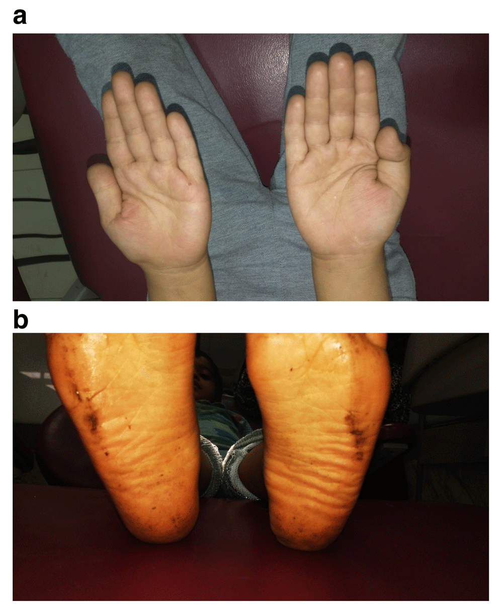
Examination of the palms of the hand revealed normal skin, while the soles of the feet revealed very slight hyperkeratosis (Figure 1a,b). Intraoral examination revealed severe gingival recession; inflammation especially in anterior region; aggressive periodontitis; mobility of maxillary left central incisor and canine, with swelling related to the maxillary right missed canine region extending toward occlusal surface. The swelling appeared as a solitary rounded lesion, with onset gradual for 2 months. The size of the swelling was 4×4 mm, and upon palpation it was not tender but slightly hemorrhagic (Figure 2a,b).
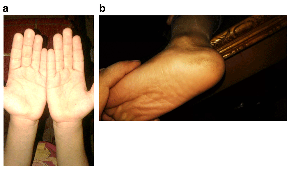
Photographs of (a) the palms of the hands showing normal skin and (b) the soles of the feet showing very slight hyperkeratosis.

Intraoral photographs showing (a) severe gingival recession and inflammation, especially in anterior region, and aggressive periodontitis; (b) swelling related to the maxillary right missed canine region extending toward occlusal surface.
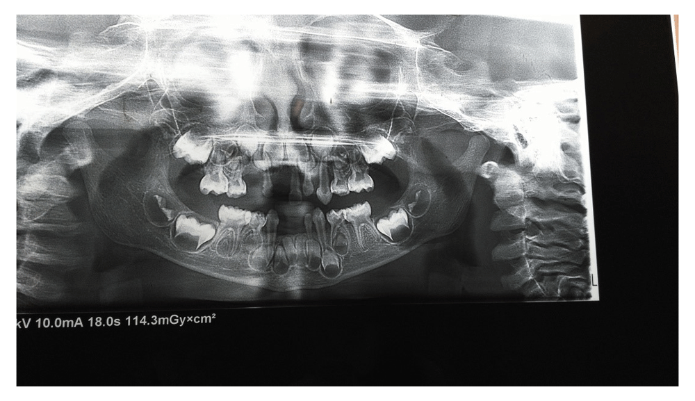
Radiographic examination showed severe destruction and loss of alveolar bone (Figure 3). Lab investigations were normal (Table 1).
Taking into consideration the clinical features and investigations, a diagnosis of PLS was confirmed.
Conventional periodontal treatment in the form of scaling and root planning was performed. Antibiotic amoxicillin and metronidazole (250 mg, 3 times daily) for one week along with a mouth rinse (0.2% chlorhexidine gluconate, 10 mL twice daily) was prescribed to the patient7.
Extraction of the maxillary left central and canine teeth was advised, but the parent refused even after the risk was explained of not extracting these loose teeth.
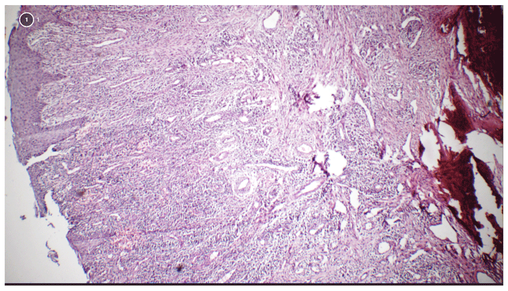
After laboratory investigations, excisional biopsy of the swelling was done under antibiotic coverage and local anesthesia. Thorough curettage of the adjacent periodontal ligament and periosteum was carried out to prevent recurrence (Figure 4 a,b). Histopathological examination revealed the lesion as peripheral ossifying fibroma (Figure 5).
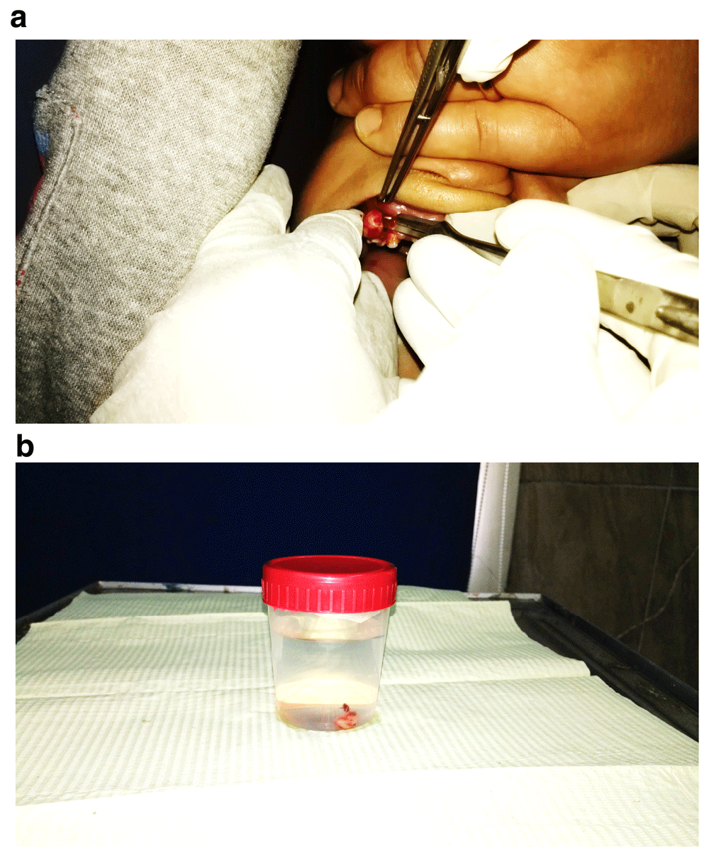
Photograph showing (a) removal of the swelling and (b) excisional biopsy of the swelling.
The patient was educated for oral hygiene and scheduled for a follow-up visit every month for scaling and checking the condition of the patient.
The patient was followed up for 2 years during which loss of maxillary left central incisor occurred and extraction of loose upper left canine was done with no recurrence of the lesion (Figure 6). The palms of the hands revealed no change, while examination of the soles of the feet showed slight increase in keratosis (Figure 7 a,b).

Follow-up photographs after 2 years showing (a) absence of change in the palms of the feet and (b) slight increase in keratosis in the soles of the feet.
Papillon-Lefèvre syndrome (PLS) is inherited as an autosomal recessive disorder where the parents of the patient with PLS should have the autosomal gene for the syndrome in order to manifest in their offspring. However, in the present case the parents are clinically healthy with no family history of the disorder. Studies have shown that when carrier parents for the affected gene mate, there is a 25% chance that they have an affected offspring8. This could explain the reason that the child had the syndrome although his parents were clinically healthy.
The intraoral appearance of severe aggressive periodontitis, which appears at the age of 3–4 years following complete eruption of primary teeth as seen in this case, concurred with observations in similar reported cases in the literature where primary teeth develop normally but eruption is accompanied with severe gingivitis followed by periodontal destruction, resulting in early loss of primary teeth9.
Ullbro et al.10 suggested that the two major components of PLS (palmar-plantar hyperkeratosis and aggressively progressing periodontitis) are not related to each other, as these authors found absence of association between the degree of hyperkeratosis and severity of periodontitis. This is in accordance with our case as the degree of hyperkeratosis is slight although periodontitis is severe.
Acrodynia, hypophosphatasia and cyclic neutropenia are differential diagnoses of PLS. This case is not acrodynia due to absence of erythrocyanosis, insomnia, and teeth erupting prematurely with dystrophic enamel. It is not hypophosphatasia due to normal level of alkaline phosphatase and it is not cyclic neutropenia, as in cyclic neutropenia the palmoplantar hyperkeratosis is absent11.
Management of cases with PLS should be multidisciplinary with dentists, dermatologists and pediatricians. Early diagnosis and management of oral problems help in reducing the undesirable sequelae of the syndrome. Following the treatment protocol for periodontal therapy proposed by Ullbro et al.10 periodontal deterioration can be minimized. This includes: scaling and polishing; giving systemic antibiotics aimed at eliminating the reservoir of causative organisms; extraction of teeth having poor prognosis; giving instructions for maintenance of oral hygiene; and continuous monitoring and frequent recall appointments.
In the present case an early diagnosis of PLS and a treatment protocol minimized the periodontal deterioration and prevented further loss of other teeth. The parents were satisfied by these results.
Written informed consent for publication of the clinical details and images was obtained from the patient's mother.
All data underlying the results are available as part of the article and no additional source data are required.
| Views | Downloads | |
|---|---|---|
| F1000Research | - | - |
|
PubMed Central
Data from PMC are received and updated monthly.
|
- | - |
Provide sufficient details of any financial or non-financial competing interests to enable users to assess whether your comments might lead a reasonable person to question your impartiality. Consider the following examples, but note that this is not an exhaustive list:
Sign up for content alerts and receive a weekly or monthly email with all newly published articles
Already registered? Sign in
The email address should be the one you originally registered with F1000.
You registered with F1000 via Google, so we cannot reset your password.
To sign in, please click here.
If you still need help with your Google account password, please click here.
You registered with F1000 via Facebook, so we cannot reset your password.
To sign in, please click here.
If you still need help with your Facebook account password, please click here.
If your email address is registered with us, we will email you instructions to reset your password.
If you think you should have received this email but it has not arrived, please check your spam filters and/or contact for further assistance.
Comments on this article Comments (0)