Keywords
Mucormycosis, Reverse Halo sign, PCR, Isavuconazole, Liposomal amphotericin B
Mucormycosis, Reverse Halo sign, PCR, Isavuconazole, Liposomal amphotericin B
Mucormycoses are life-threatening fungal infections mostly occurring in hematology, solid organ transplant, or diabetic patients, it may also affect immunocompetent patients following a trauma or burn1. Nosocomial or community outbreaks have been described2. Mucormycosis is characterized by host tissue infarction and necrosis resulting from vasculature invasion by hyphae starting with a specific interaction with endothelial cells. Most common clinical presentations are rhino-orbito-cerebral and pulmonary. Multicenter and single-center studies have reported an increasing incidence probably due to an increase in the at-risk population and improved diagnostic tools3,4. In a French study, mucormycosis incidence increased by 7.3% per year, especially in patients with neutropenia5.
These infections are difficult to manage for several reasons6. Firstly, diagnosis is difficult because of clinico-radiological similarities with invasive aspergillosis and historical lack of diagnostic tools. However, new tools in serum and tissue as well as the recognition of highly suggestive radiological signs recently modified diagnostic possibilities. Secondly, treatment is an emergency and combines surgery, which is frequently required owing to the angioinvasive and necrotic character of infection7, and antifungal treatment. Primary in vitro resistance to several antifungal drugs limits therapeutic options8. However, recent data enlarge the antifungal armamentarium with the US Food and Drug Administration’s and European Medicines Agency’s approval of the new triazole isavuconazole. However, comparative clinical data are lacking, and the respective places of polyenes and different azoles need to be discussed.
Human mucormycoses are caused by a wide range of pathogenic species. Mucormycosis location is linked to the mucorales species; Rhizopus arrhizus (Figure 1) is present in 85% of rhino-cerebral forms, compared with only 17% of non-rhino-cerebral forms in the French RetroZygo study9. This finding could be explained by virulence differences between Mucorales species. In experimental studies, ketoacidosis has been found to predispose mice to Rhizopus spp. but not Lichtheimia spp. infection10,11. In parallel, corticosteroid treatment enhanced the susceptibility of mice to lung infections caused by Lichtheimia corymbifera or Lichtheimia ramosa10,11.
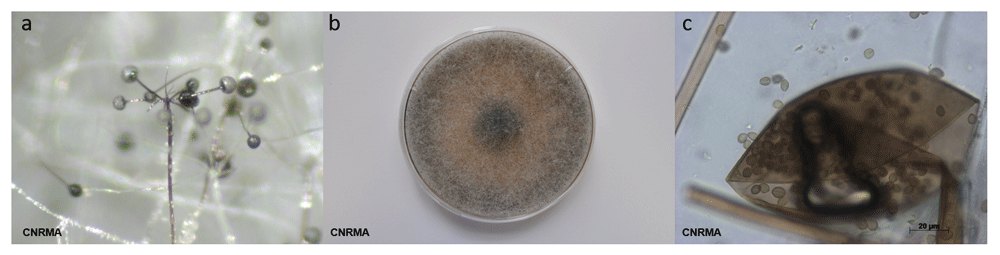
(a) Sporangiophore branching and rhizoids (stereomicroscope); (b) grey-brownish colony on malt 2% medium; (c) melanized sporangium and sporangiospores.
Mucormycosis’ clinical presentation is also related to underlying conditions. Rhino-cerebral mucormycosis is the most common form in patients with diabetes mellitus1, while pulmonary mucormycosis occurs most often in patients with hematological malignancies12. Radiological findings in patients with pulmonary mucormycosis are also related to immunological status13. Although unusual, lately there have been diagnoses made in the gastrointestinal system. The stomach is more frequently involved, and then the colon. Symptoms are abdominal pain and gastro-intestinal bleeding14. Diagnosis is suspected on endoscopic findings with necrotic lesions that can lead to perforation and peritonitis15.
Mucorales can gain entry to a susceptible host through inhalation, ingestion of contaminated food, or abraded skin. These routes result in rhino-orbito-cerebral, pulmonary, gastrointestinal, or cutaneous/wound infections. One of the characteristic features of mucormycosis is its angioinvasive property, resulting in vascular thromboses and ultimately tissue necrosis. Ketoacidosis and deferoxamine are known to predispose to mucormycosis, revealing the importance of hyperglycemia, iron, and acidifying ketone bodies in mucorales virulence. Angioinvasion was reported to be related to the interaction between a spore-coating protein family (CotH) on Rhizopus spp. surface and endothelium glucose regulator protein 78 (GRP78) expressed at the surface of endothelial cells. This interaction triggers host cell injury and subsequent fungus hematogenous dissemination16. Elevated levels of serum glucose, iron, and ketone bodies increase fungal growth and induce the expression of GRP78 and CotH, resulting in increased ability of Rhizopus to invade host tissues and explaining the susceptibility of diabetic and deferoxamine-treated patients to mucormycosis. However, it should be noted that the majority of studies on virulence and the association between ketoacidosis and the occurrence of mucormycosis have been conducted with Rhizopus species17. Decreased numbers and impaired function of monocytes and neutrophils are important mucormycosis risk factors, since they are known to inhibit Mucorales spore germination. This includes patients with hematological disorders, AIDS, or liver cirrhosis, those who have undergone solid organ transplant, and those being treated with high-dose steroids18,19. Finally, victims of natural disaster are also at risk20 owing to wounds contaminated with water, soil, or debris21, such as after the 2004 Indian Ocean tsunami22 or after the 2011 Missouri tornado23.
The most common radiological pattern of lung mucormycosis on initial computed tomography (CT) scan is a halo sign and then nodule or mass13,24. However, when studied very early and on serial follow-up, sequential morphologic changes could be observed as (i) reversed halo sign (Figure 2) followed by (ii) consolidation or nodule or mass with halo sign and, finally, (iii) central necrosis and air-crescent sign. For pulmonary mucormycosis, a recent study showed that there was a significant increase in the prevalence of reversed halo sign in neutropenic (79%) and non-neutropenic (31%) patients (P <0.05)25.
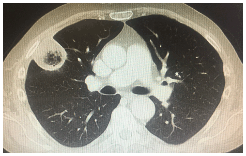
Inversed halo sign.
Mucormycosis diagnosis is challenging, as it is associated with high mortality, especially in hematological patients. Early distinction from invasive aspergillosis is of utmost importance, as antifungal treatment may differ, whereas underlying conditions and clinical presentation are often similar. Until recently, mucormycosis diagnostic tools were based on limited basic microbiology and frequently led to diagnosis delay.
PCR, polymerase chain reaction.
Unlike invasive aspergillosis, the detection of circulating antigen such as galactomannan and β-D-1,3-glucan provides no help for mucormycosis diagnosis. Therefore, samples from the infection site are highly required to diagnose mucormycosis based on the microscopic detection of typical hyphae or on a positive culture26. Recently, the development of molecular biology tools has allowed the non-invasive diagnosis of mucormycosis. Million et al. designed a quantitative multiplex polymerase chain reaction (qPCR)-based 18S rRNA targeting Mucor/Rhizopus, Lichtheimia, and Rhizomucor. This PCR assay was evaluated with the aim to detect Mucorales DNA early in the course of the infection in the blood (serum)27. The authors were able to detect Mucorales DNA in serum samples from 90% of patients up to three days before mucormycosis diagnosis27. Negative serum PCR was also associated with better outcome as compared to patients with persistently positive PCR. Furthermore, a study among severely ill burn patients found that circulating Mucorales DNA was detected 11 (4.5–15) days before standard diagnosis for invasive wound mucormycosis28. Other studies have evaluated the use of real-time PCR targeting Mucorales on tissue or respiratory samples in patients with hematological malignancy suffering from proven and probable mucormycosis29–32.
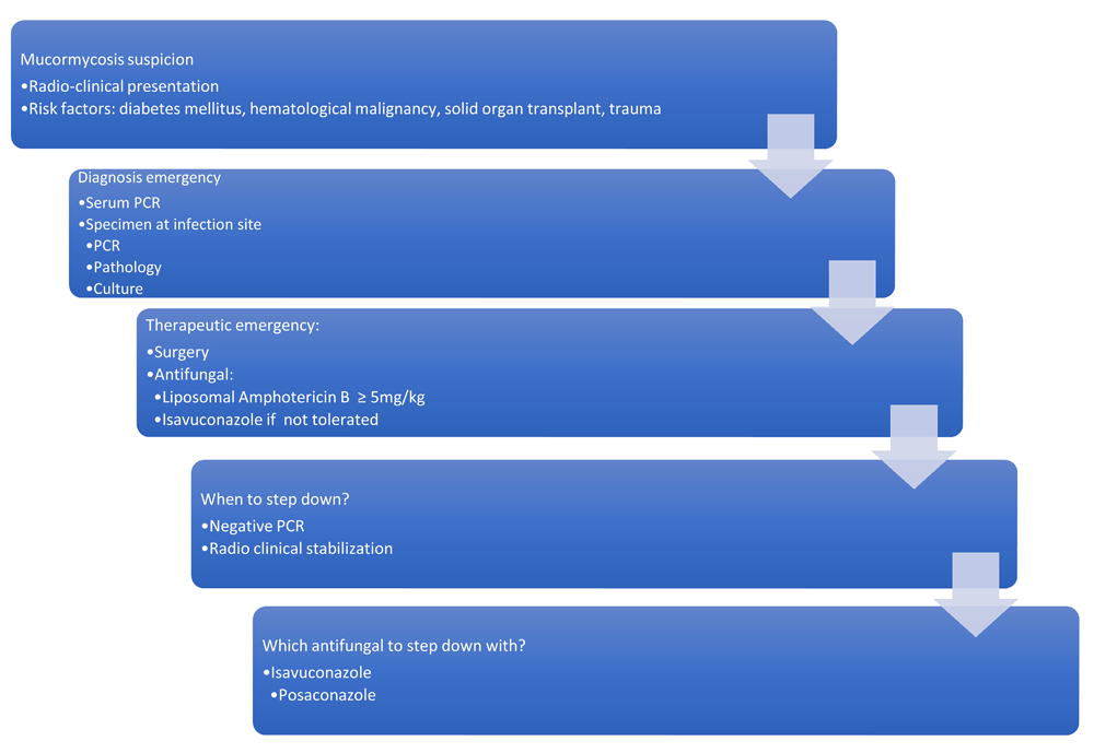
Consequently, it is currently necessary in patients with hematological malignancies to include the value of reverse halo sign on CT combined with serum qPCR targeting Mucorales in the early diagnosis of pulmonary mucormycosis33.
Current guidelines recommend antifungal treatment, surgical debridement, and correction of risk factors34,35. Surgical debridement has to be extensive, involving all necrotic areas for rhino-oculo-cerebral infection, and repeated surgical procedures are recommended to achieve local control and improve outcome36. For pulmonary mucormycosis, the indication and timing of surgical management outside emergency care (hemoptysis) is still unclear37. In a European series of 230 patients, surgical treatment reduced mortality by 79%38, leading to discuss surgery when feasible for any localization, however mandatory for rhino-cerebro-oculo-cerebral and post-traumatic mucormycosis36,39.
Amphotericin B (Amb) and its lipid formulations and posaconazole were the only antifungal drugs available with in vitro activity against mucorales40,41. The antifungal armamentarium recently enlarged with the development of isavuconazole.
The first-line recommended antifungal agent is liposomal Amb (L-Amb) or Amb lipid complex (ABLC)35. Studies in mice proposed that the efficacy of L-Amb and ABLC was dependent on the dose given and that 10 mg/kg yielded the best outcomes. A prospective French phase II multicenter study (AmBizygo trial) evaluated the efficacy and tolerance of high-dose (10 mg/kg/day) L-Amb in association with surgery when recommended for the treatment of 34 mucormycosis cases. A favorable response was seen in 45% of patients at week 12. However, serum creatinine doubled in 40% of patients, but in 63% of cases, once treatment had ended, creatinine levels normalized after three months42. According to this study, ECMM/ESCMID and ECIL-6 guidelines recommend the use of L-Amb with a daily dosage of at least 5 mg/kg/day for mucormycosis34,35, and dosages at 10 mg/kg/day are strongly supported by ECMM/ESCMID for cerebral infections35. Moreover, because of better diffusion, L-Amb should be favored in central nervous system infections.
The duration of the first-line antifungal treatment is still a matter of debate and should be determined on an individual basis and adjusted based on the underlying condition. Some authors proposed a lipid Amb treatment for at least three weeks, and, when there is clinical and radiological improvement, a consolidation by posaconazole can be started43. However, it could possibly be guided by negative PCR and therefore shortened for some patients.
Isavuconazonium sulfate is a water-soluble pro-drug, which is quickly hydrolyzed to the triazole isavuconazole after oral or intravenous administration. Isavuconazole has high oral bioavailability, linear pharmacokinetics, and a broad antifungal spectrum. The in vitro activity of isavuconazole minimum inhibitory concentration (MIC) ranges were 0.125 to 4 mg/L across L. corymbifera, L. ramosa, Rhizomucor pusillus, Rhizomucor microspores, and R. arrhizus but somewhat higher against Mucor circinelloides (1 to 16 mg/L). The MICs were in general one- to three-fold higher than those for posaconazole44. In the recently published Vital study, 21 patients were treated with isavuconazole as first-line treatment; 42-day response rate was only 14% and week 12 response was 10% (compared to 45% in the AmBizygo study) with 43% deaths45. The results of this study found a mortality rate at day 42 comparable to that observed in the AmBizygo study42. In the VITAL study, isavuconazole was well tolerated and toxic effects were an uncommon cause of discontinuation. The place of isavuconazole has not yet been specified in the most recent guidelines34. Finally, a cost-effectiveness study demonstrated the positive economic impact of the use of isavuconazole compared to Amb in the treatment of mucormycosis46.
Posaconazole has been shown to have in vitro and in vivo activity against mucorales, but there are no data for the use of first-line posaconazole therapy. Posaconazole, therefore, finds its place in the therapeutic armamentarium for prophylaxis or consolidation after induction treatment with L-Amb. No study on the efficacy of posaconazole intravenous or tablet formulations in mucormycosis treatment were conducted. Finally, mucormycosis cases have been reported in patients undergoing posaconazole prophylaxis despite satisfactory serum concentrations47.
However, it seems important to note that there are no current validated MIC breakpoints for any of the available antifungals and thus the determination of susceptibility categories is not possible for the agents of mucormycosis.
A case report has recently reported the benefit of a treatment with the checkpoint inhibitor nivolumab and interferon-Υ for an immunocompetent patient with extensive abdominal mucormycosis unresponsive to conventional therapy48.
Mucormycosis is a life-threatening fungal infection characterized by host tissue infarction and necrosis that occurs mostly in immunocompromised patients and is associated with an increasing incidence and mortality despite the availability of therapeutic tools. Determining whether the patient has invasive aspergillosis or mucormycosis could be challenging at the bedside. In this context, new tools of molecular biology have been developed to obtain earlier diagnosis and start optimal medico-surgical treatment. Comparative studies are needed to better optimize induction and consolidation treatment.
| Views | Downloads | |
|---|---|---|
| F1000Research | - | - |
|
PubMed Central
Data from PMC are received and updated monthly.
|
- | - |
Provide sufficient details of any financial or non-financial competing interests to enable users to assess whether your comments might lead a reasonable person to question your impartiality. Consider the following examples, but note that this is not an exhaustive list:
Sign up for content alerts and receive a weekly or monthly email with all newly published articles
Already registered? Sign in
The email address should be the one you originally registered with F1000.
You registered with F1000 via Google, so we cannot reset your password.
To sign in, please click here.
If you still need help with your Google account password, please click here.
You registered with F1000 via Facebook, so we cannot reset your password.
To sign in, please click here.
If you still need help with your Facebook account password, please click here.
If your email address is registered with us, we will email you instructions to reset your password.
If you think you should have received this email but it has not arrived, please check your spam filters and/or contact for further assistance.
Comments on this article Comments (0)