Keywords
ectopic calcification, pulp stone, denticles, recombinant parathyroid hormone 1-34.
ectopic calcification, pulp stone, denticles, recombinant parathyroid hormone 1-34.
Ectopic calcification is pathologic deposition of minerals within soft tissues as dental pulp or periodontal ligaments (PDL)1,2. Pulpal ectopic calcification may manifest as generalized, linear calcification, or as circumscribed calcification (also known as pulp stones or denticles). Pulp stones can be seen free within the pulp tissues, partially associated with dentin wall or completely embedded in dentin. They may manifest as false concentric calcification or true pulp stones3. The etiology of pulp calcification may be idiopathic4, although it may also be associated with pulp injury or degeneration5, orthodontic or physical forces6–8 or chemical stimuli9,10. Its incidence tends to increase with age3,11.
Parathyroid hormone (PTH) is a naturally occurring hormone, important for calcium homeostasis12,13. Its level in the blood dictates its effect on the skeletal system, with bone catabolic effect upon chronic increase in PTH level associated with hyperparathyroidism13,14 and bone anabolic effect upon administration of small intermittent dosage15,16. PTH secreted by the parathyroid gland (native PTH) is a polypeptide chain composed of 84 amino acids (PTH 1-84). While PTH 1–34, is a fragment of PTH molecule “synthetised through recombinant DNA technology using a strain of Escherichia coli bacteria”17,18. Intermittent PTH 1–34 administration, owing to its bone anabolic effect, is successfully used for the management of osteoporosis19–22. Its bone anabolic effect has been linked to elevated osteoblast differentiation, number and activity23–29.
This research was conducted as a part of a study examining the effect of PTH 1–34 on microarchitecture of alveolar process of osteoporotic rats.
In the current research, 18 male Wistar rats of the species Rattus norvegicus weighing 175–200 gm, aged between 3 to 4 months were used. The animals were acquired from and maintained in the Animal House, Faculty of Medicine, Cairo University under the care of a specialized veterinarian. Each animal was kept in a separate cage. They were maintained under controlled temperature at 25±2°C with 12 h light/dark cycle and had ad libitum access to standard rats’ chow and water. This study was approved by the Research Ethics committee faculty of Dentistry, Cairo University (approval number 151028).
Osteoporosis was induced in all experimental animals (n=18) by five weekly doses, of 7 mg/kg body weight dexamethasone sodium phosphate (Decadron® 4 mg/ml, Eipico Egypt), administered intramuscularly30,31. The animals were then randomly distributed by random sequence generator program (randomizer.org) into two groups each including 9 animals, matching of the animals with the numbers was done blindly through the primary investigator. Animals received either a daily subcutaneous injection of 60 μg/kg body weight PTH 1–34 (Forteo®; Eli lilly Pharmaceuticals) (n=9)32 or an equal volume of saline (control group) (n=9). Drugs were administered in the early morning hours (8–9 am). The body weight of the animals was measured weekly, and drug dosages were adjusted accordingly. Animals were euthanized with an intra-cardiac overdose of sodium thiopental (80 mg/kg) 4 weeks after initiation of Forteo administration. Mandibles were dissected and separated into two halves, only one hemi-mandible from each rat was utilized for histological examination. The experimental unit was the hemimandible of rats. The primary investigator was blinded.
Hemi-mandibles (n=18) were fixed in 10% calcium formol solution for 48 hours. The specimens were then washed and soaked in 10% EDTA for 4–5 weeks for decalcification. After decalcification was completed, the specimens were dehydrated in ascending grades of alcohol, cleaned in xylol, and then embedded in paraffin blocks. Next, 6-µm-thick paraffin sections were cut and mounted on a clean glass slide, then stained with haematoxylin and eosin stain31. The specimens were examined using Leica DM300 light microscopic (Leica Microsystems, Inc., Switzerland). Histological examination was done through blinded primary investigator. Dental pulp and surrounding periodontal ligaments of all teeth within hemimandible of both experimental groups (n=18) were examined for the presence of ectopic calcification.
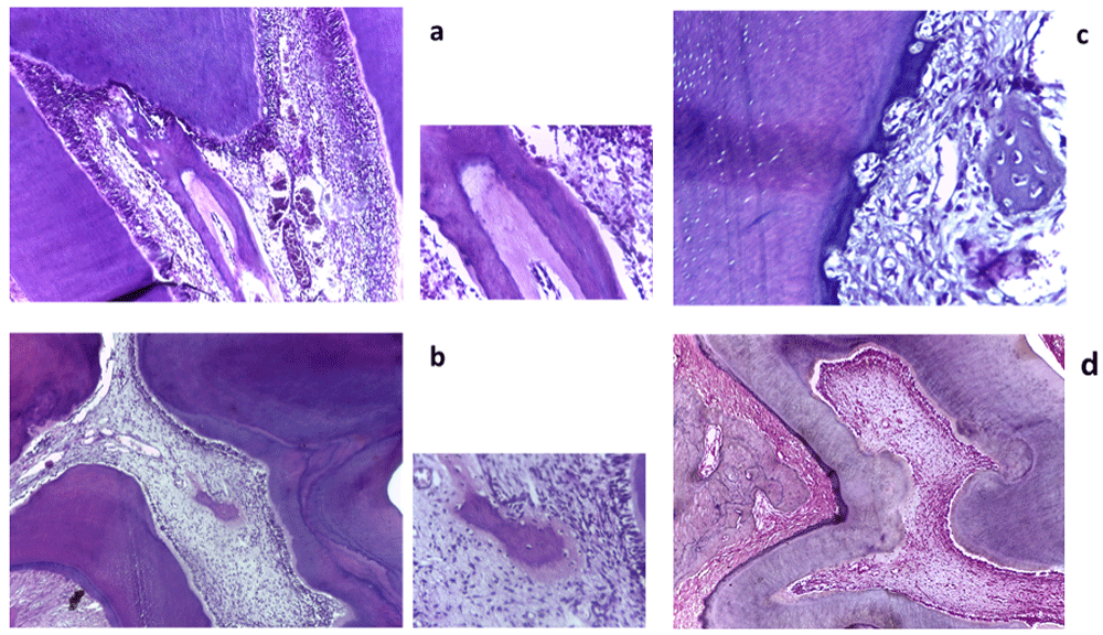
Upon histological examination of the Forteo group specimens, six rats showed normal pulp and periodontal ligaments with no ectopic calcifications, while ectopic calcifications were detected in three specimens (Dataset 1)33. Where true pulp stone with pre-dentin and dentin surrounding a central cavity lined by cells was detected in one specimen (Figure 1a). Another specimen showed the presence of intra-pulpal calcified structure with entrapped cells (Figure 1b). Meanwhile, one specimen displayed the presence of intra-periodontal bone-like calcified structure with entrapped cells (Figure 1c). On the other hand, no ectopic calcification was perceived in the control group specimens (Dataset 1)33, which showed normal pulp and periodontal ligaments (n=9) (Figure 1d).

(a) A true pulp stone with dentin, pre dentin and central cavity lined by cells (original magnification, x100 (left) and x400 (right)). (b) Intrapulpal calcification with entrapped cells (original magnification, x100 (left) and x400 (right)). (c) Intra-periodontal ectopic calcification with entrapped cells surrounded with disorganized periodontal ligaments (original magnification, x400). (d) Light microscope image of the control group showing normal pulp and periodontal ligaments with no ectopic calcification (original magnification x 100).
Despite the fact that PTH 1–34 can successfully lower blood calcium level, and prevent vascular calcification34, in the current work, PTH 1–34 was associated with ectopic calcifications within the pulp and PDL, while none was observed in the control group specimens.
Guimaraes et al. observed increased dentin deposition rate and elevated level of serum alkaline phosphatase in PTH 1–34 treated rats35. In a subsequent research, Guimaraes et al. elucidated that PTH 1–34 can regulate odontoblast like cells via protein kinase A- and protein kinase C-dependent pathways, with increases in odontoblast-like cells proliferation upon short PTH exposure and increases in their apoptosis upon longer exposure36.
Wang et al. demonstrated the ability of PTH to induce human PDL stem cells to differentiate into osteoblasts, which was associated with increased alkaline phosphatase activity and increased mineralization capacity37. Moreover, Li et al. described the ability of PTH 1–34 to induce the formation of calcified nodule in cementoblast cell line, which was attributed to the ability of the drug to increase cementoblast activity, alkaline phosphatase level and subsequently calcification38.
The stimulatory effect of PTH 1–34 on odontoblast, cementoblast and osteoblasts function can help explain the findings of the current research.
Dataset 1. Images captured from each mouse in each group not shown in Figure 1.DOI: https://doi.org/10.5256/f1000research.16298.d21852333.
| Views | Downloads | |
|---|---|---|
| F1000Research | - | - |
|
PubMed Central
Data from PMC are received and updated monthly.
|
- | - |
Is the work clearly and accurately presented and does it cite the current literature?
No
Is the study design appropriate and is the work technically sound?
Partly
Are sufficient details of methods and analysis provided to allow replication by others?
Yes
If applicable, is the statistical analysis and its interpretation appropriate?
Not applicable
Are all the source data underlying the results available to ensure full reproducibility?
Partly
Are the conclusions drawn adequately supported by the results?
No
References
1. Chen Y, Wang S, Cheng F, Hsu P, et al.: Intermittent parathyroid hormone improve bone microarchitecture of the mandible and femoral head in ovariectomized rats. BMC Musculoskeletal Disorders. 2017; 18 (1). Publisher Full TextCompeting Interests: No competing interests were disclosed.
Reviewer Expertise: Bone Biology
Is the work clearly and accurately presented and does it cite the current literature?
Yes
Is the study design appropriate and is the work technically sound?
Yes
Are sufficient details of methods and analysis provided to allow replication by others?
Yes
If applicable, is the statistical analysis and its interpretation appropriate?
Not applicable
Are all the source data underlying the results available to ensure full reproducibility?
Yes
Are the conclusions drawn adequately supported by the results?
Yes
Competing Interests: No competing interests were disclosed.
Alongside their report, reviewers assign a status to the article:
| Invited Reviewers | ||
|---|---|---|
| 1 | 2 | |
|
Version 1 26 Sep 18 |
read | read |
Click here to access the data.
Spreadsheet data files may not format correctly if your computer is using different default delimiters (symbols used to separate values into separate cells) - a spreadsheet created in one region is sometimes misinterpreted by computers in other regions. You can change the regional settings on your computer so that the spreadsheet can be interpreted correctly.
Provide sufficient details of any financial or non-financial competing interests to enable users to assess whether your comments might lead a reasonable person to question your impartiality. Consider the following examples, but note that this is not an exhaustive list:
Sign up for content alerts and receive a weekly or monthly email with all newly published articles
Already registered? Sign in
The email address should be the one you originally registered with F1000.
You registered with F1000 via Google, so we cannot reset your password.
To sign in, please click here.
If you still need help with your Google account password, please click here.
You registered with F1000 via Facebook, so we cannot reset your password.
To sign in, please click here.
If you still need help with your Facebook account password, please click here.
If your email address is registered with us, we will email you instructions to reset your password.
If you think you should have received this email but it has not arrived, please check your spam filters and/or contact for further assistance.
Comments on this article Comments (0)