Keywords
Pseudoangiomatous stromal hyperplasia, pash, lactational adenoma, breast
Pseudoangiomatous stromal hyperplasia, pash, lactational adenoma, breast
Pseudoangiomatous stromal hyperplasia (PASH) is a rare, benign breast lesion that is usually discovered as an incidental finding in breast biopsies1. Despite usually being an incidental finding, there have been many reports in the literature of PASH presenting clinically as a discrete mass, and several reports of it presenting as bilateral diffuse breast enlargement2. We present a case report of PASH presenting as a large clinically-evident breast mass in a pregnant woman which was initially diagnosed as a lactational adenoma on the core biopsy. The subsequent excision specimen demonstrated a large, discrete tumour composed of PASH with a central galactocele, as well as prominent but only patchy lactational change. Following this, PASH was retrospectively identified as being present in the initial core biopsy.
Clinically, PASH has a wide range of signs and symptoms, ranging from presenting as a large breast mass, to being an incidental finding. Indeed, non-tumour-forming PASH has been reported to be an incidental microscopic finding in 23% of breast biopsies3. Tumour-forming PASH occurs predominantly in premenopausal women and usually presents clinically as a palpable, mobile, firm, painless, intra-mammary mass4. Cases of tumour-forming PASH have also been described in post-menopausal women, men, adolescents and even in the paediatric age group5. In our case, the patient was a twenty three-year-old female who presented with a large central mass in the left breast 24 weeks into her pregnancy (Figure 1). She first noticed the mass at around week 10 of her pregnancy but it then massively increased in size around week 24. She had a history of a biopsy-proven lactational adenoma six years previously during her first pregnancy. An ultrasound-guided core biopsy was performed which was reported as a lactational adenoma, B2, and due to the significant size of the mass it was excised in week 25 of her pregnancy as a suspected giant lactational adenoma.
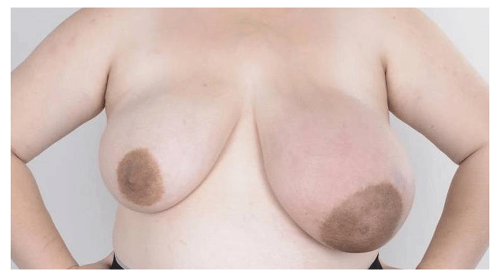
The patient first noticed a left breast mass in week 10 of her pregnancy but it then massively increased in size around week 24.
Several publications of case series6–9 show the typical ultrasound appearance of PASH to be a well-circumscribed oval or round mass encompassed by a thin echogenic capsule and usually indistinguishable from a fibroadenoma, with size ranging from 3 to 70 mm. Internal echotexture is most commonly hypoechoic, sometimes isoechoic, and occasionally complex containing cysts or vascular channels. In this case, the 140 mm mass within a breast hypertrophied through pregnancy changes presented a challenge for imaging with conventional ultrasound due to the extremely large size. The subcutaneous breast tissue was oedematous and there was skin thickening present up to 5.5 mm in more dependant areas of the breast. The mass had well-defined superficial and radial margins with multiple large gentle lobulations, and a thin echogenic pseudocapsule pointing towards a benign diagnosis (Figure 2). The internal echotexture was predominantly bland, homogenous and hypoechoic. Multiple prominent internal vessels were visualised on doppler imaging; PASH lesions do not commonly have internal blood flow however, which therefore pointed away from the diagnosis in this case. It is likely the imaging features were confounded by the pregnant state. The differential diagnosis based on the ultrasound appearances included a fibroadenoma, a lactational adenoma and a phyllodes tumour.

The mass had a thin echogenic pseudocapsule pointing towards a benign diagnosis and the internal echotexture was predominantly bland, homogenous and hypoechoic.
The classical histological appearance of PASH is inter-anastomosing channels lined by slender spindle cells in the interlobular breast stroma10. As the name suggests, ultrastructural observations have determined that these channels are not true vascular spaces and the distinction from an angiosarcoma is obviously important11. Immunohistochemistry shows the cells are positive for vimentin and CD3412 but are negative for CD31. Macroscopically, the lesion in this case consisted of a large lobulated red mass measuring 170 x 170 x 75 mm and weighing 838 g with a central area containing yellow cream-like material measuring 25 x 20 mm (Figure 3). Histologically the breast tissue showed prominent gynaecomastoid-like lobules with intervening oedematous stroma showing florid pseudoangiomatous hyperplasia (Figure 4). There was prominent but only patchy lactational change. The central cyst was lined by a single layer of apocrine-like cells in keeping with a galactocoele. There was no evidence of atypical hyperplasia, in-situ or invasive carcinoma. The diagnosis was given as pseudoangiomatous stromal hyperplasia with prominent but only patchy lactational change and a galactocoele. Similar changes were seen in the initial core biopsy of this lesion and on review of the lesion cored during the first pregnancy.

The lesion consisted of a large lobulated red mass measuring 170 x 170 x 75 mm and weighing 838 g with a central area containing yellow cream-like material.

The breast tissue showed prominent gynaecomastoid-like lobules, with intervening oedematous stroma showing florid pseudoangiomatous hyperplasia and prominent but only patchy lactational change.
PASH can often be an incidental finding and is commonly found in combination with other diagnoses. It is therefore possible for PASH to be overlooked in biopsy specimens and it is important to analyse the breast stroma carefully for evidence of PASH, even if the biopsy contains an alternative lesion that could account for the mass seen clinically. As highlighted well by this case, the presence of lactational change on its own in the core biopsy could have accounted for the mass lesion identified clinically and radiologically. This would have been further reinforced by the history of a previous biopsy-proven lactational adenoma during her last pregnancy. This highlights the potential for PASH to be overlooked in core biopsy specimens and therefore not appropriately treated. Fortunately, in this case the mass was so large and impacting upon the patient that it was excised anyway. PASH discovered incidentally does not require any specific additional treatment; however, tumour-forming PASH should be treated with local surgical excision and has an excellent prognosis with minimal risk of recurrence if adequately surgically excised according to some reports in the literature13. Other papers have reported recurrence rates between 15% and 22% after surgical excision14. The long-term prognosis for this patient would be expected to be excellent but we do not yet have long-term follow up data available. At her initial follow-up clinic appointment no problems were reported and her pregnancy was progressing as planned.
In summary we present a case report of tumour-forming PASH mimicking a giant lactational adenoma in a young pregnant patient. We feel it highlights the importance of looking carefully for PASH in core biopsy specimens, even if a concurrent lesion is also present which would account for the mass identified radiologically.
Written informed consent for publication of their clinical details and/or clinical images was obtained from the patient according to the Declaration of Helsinki.
| Views | Downloads | |
|---|---|---|
| F1000Research | - | - |
|
PubMed Central
Data from PMC are received and updated monthly.
|
- | - |
Provide sufficient details of any financial or non-financial competing interests to enable users to assess whether your comments might lead a reasonable person to question your impartiality. Consider the following examples, but note that this is not an exhaustive list:
Sign up for content alerts and receive a weekly or monthly email with all newly published articles
Already registered? Sign in
The email address should be the one you originally registered with F1000.
You registered with F1000 via Google, so we cannot reset your password.
To sign in, please click here.
If you still need help with your Google account password, please click here.
You registered with F1000 via Facebook, so we cannot reset your password.
To sign in, please click here.
If you still need help with your Facebook account password, please click here.
If your email address is registered with us, we will email you instructions to reset your password.
If you think you should have received this email but it has not arrived, please check your spam filters and/or contact for further assistance.
Comments on this article Comments (0)