Keywords
Penile Diseases, Urologic Surgical Procedures, Injuries, Penile induration
Penile Diseases, Urologic Surgical Procedures, Injuries, Penile induration
“Universidad Continental, Lima, Peru” has been added as an affiliation for J. Antonio Grandez-Urbina in this version.
We realize and mention that there are other etiologies related to penile fracture such as, “Taqaandan”, which is practiced in Middle Eastern countries. We have edited the discussion adding the percentage and absolute number of patients that present double penile fracture in a case series. Also, we mentioned the role of imaging in the study of penile fracture and added techniques used to detect urethral injuries.
We recognized that more research is needed on this topic to make firm recommendations.
To read any peer review reports and author responses for this article, follow the "read" links in the Open Peer Review table.
Fracture of the penis, or faux pas du coit, is an uncommon surgical emergency that occurs as a result of trauma to the erect penis1,2. The incidence of penile injuries is underreported because many patients do not seek medical attention3. Penile fracture is defined as the rupture of the tunica albuginea and the corpora cavernosa4. Most cases occur during sexual intercourse, usually due to hitting the symphysis pubis or the perineum after the penis slips out of the vagina; less commonly reported is during masturbation2,3.
It is manifested by a cracking or popping sound accompanied by immediate detumescence, followed by rapid swelling, widespread ecchymosis, sharp pain and deformity (away from the trauma site)2,5.
This condition can be quickly diagnosed after history taking, physical examination and imaging. Prompt diagnosis and early surgical repair are essential to ensure a successful outcome6,7.
It is uncommon for a penile fracture to involve both of the corpora cavernosa. We report a rare case of a double penile fracture and describe its presentation and management.
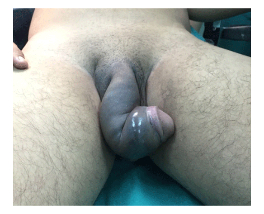
A 32-year-old patient presented to the emergency department two hours after having penile trauma during vigorous sexual intercourse. He was having sexual intercourse in a 'woman-on-top' position when he heard a ‘snap’ sound followed by severe pain and immediate loss of tumescence. On physical examination, his penis was swollen, deformed, and with signs of ecchymosis. On admission, two ventral irregularities were found during penile palpation (Figure 1). There was no urethral bleeding, nor voiding difficulties suggesting an uncompromised urethra. During the patient's consultation, no previous sexual disorders were found and the patient denied the usage of PDE5 inhibitors or intra-carvernosum injections. Labs results were within normal limits and radiological images were not obtained prior to surgical intervention due to lack of resources.
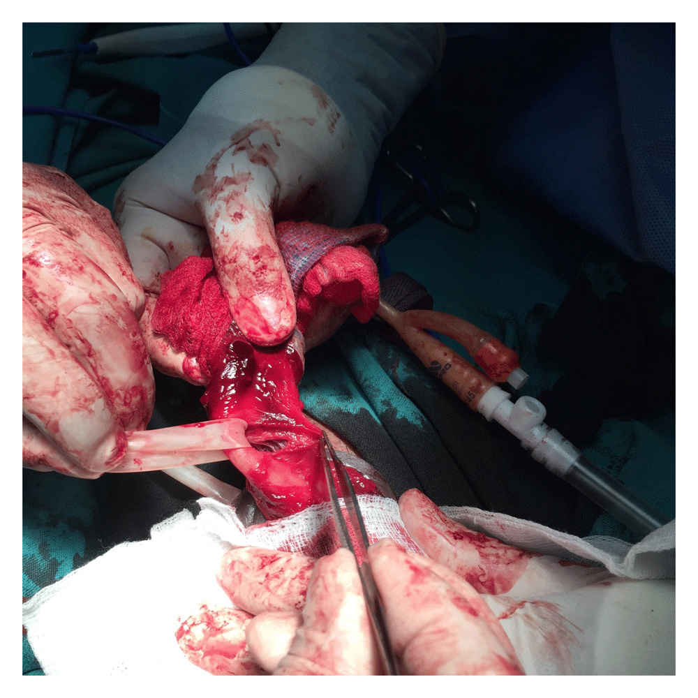
Cefazoline 2gr IV was administered prophylactically before surgery. Emergency exploration was performed 4 hours post-penile injury. A 16 Fr. Foley catheter was placed without difficulties. Then a circumcision was made, and surgical exploration was performed. On degloving the penile skin, a fracture on each corpora cavernosa was found. The length of the right and left defects were 25mm and 35mm, respectively (Figure 2). In order to protect the urethra, a Penrose drain was used as a vessel loop through the double fracture separating it from the site of injury (Figure 3). Repair of the two lacerations was done using 3-0 Vicryl in a continuous suture pattern. The Foley catheter was removed the next day without adverse events. The patient had a favorable recovery and was discharged three days after the surgical procedure without any complication (Figure 4).

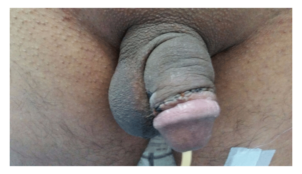
At a follow-up period of 90 days, the patient had a proper erectile function, no deformity, no pain during sexual intercourse, and no voiding difficulties.
The penis is composed of erectile tissue arranged in a columnar fashion. Two dorsolateral corpora cavernosa and one ventral corpus spongiosum, each enclosed by the tunica albuginea. The urethra traverse the corpus spongiosum throughout its length. The distal expansion of the corpus spongiosum forms the glans penis. Buck’s fascia encloses the corpus spongiosum ventrally, and splits dorsally to surround the two corpora cavernosa1,4,5.
Penile fracture, or faux pas du coit, is an underreported but emergent urological condition8. It’s underreported because many patients do not seek medical attention due to embarrassment, shame, or lack of guidance3. Activities that can result in penile fracture includes self-manipulation to achieve tumescence, sexual intercourse, turning over in bed, a direct blow to the erect penis and interrupting the erection due to a violent bending of their penis called "Taqaandan"2,4,9. The most common cause is sexual intercourse, with injuries often caused by different sexual positions. In an original article published in 2017, Barros et al. reported that ‘doggy style' was more commonly associated with double fractures out of 67 patients (10%) presented double corpus cavernosum lesión10. In fact, any activity associated with tumescence can increase the chance of penile fractures. In 2002, Blake et al. reported the first case of a fracture related to pharmacologically induced erections11. The increased risk of penile fractures during tumescence is related to the tunica albuginea stretching and thinning. The thickness of the tunica reduces from 2.4mm, in the flaccid state, to 0.25-0.5mm in the erect form 12.
The most extensive review of cases was made by Eke between 1935 and 2001. He analyzed 1331 cases from 183 publications. Most reports were from the Mediterranean region; the median age of patients were 35 years. Clinical features included sudden penile pain, detumescence, voiding difficulties, penile swelling and deviation. Associated injuries included urethral rupture. Complications of the rupture included coital difficulty, urethral fistula, penile plaque and erectile dysfunction1.
The diagnosis is mainly clinical, although in the absence of typical signs it can pose a real challenge. Therefore, an adequate history and physical examination are cornerstones of the diagnostic process. In our patient, radiologic investigations were not performed due to lack of resources and the diagnosis was based solely on physical examination findings. Most cases reported in literature describes imaging modalities being used to exclude the presence of a concomitant urethral injury and to delineate the exact location of the albuginea rupture13. Various imaging modalities have been used to aid in the diagnosis, such as cavernosography, retrograde urethrography, ultrasonography-colour Doppler, and magnetic resonance. Cavernosography is an invasive method that is rarely used. Retrograde urethrography is also an invasive method, but is required only if associated urethral injury is suspected7. Ultrasonography is the modality most frequently used because of its cost and availability. Magnetic resonance imagine is the most accurate method to localize the lesions, but its availability is limited due to its cost14. We would prefer to use MRI in this case in order to support the surgical decision, but the role of imaging is largely limited to unclear cases with equivocal findings or unreliable history15.
Most studies report a solitary fracture of the corpora cavernosa. In our patient, both corpus were found to have fractures. Although no imaging modalities were performed, the omission of careful surgical exploration for the second site of injury might have led to unsatisfactory outcomes. Therefore, it seems essential to make a complete surgical exploration, degloving the penile shaft, while operating upon such cases.
Urgent surgery is the recognized gold standard approach. However, recent studies have revealed that long-term outcomes of early versus delayed repair in patients without urethral involvement are similar16,17. In 2011, Kozacioglu et al. evaluated 56 patients who underwent early and delayed penile fracture repair. Their study noted no serious deformities nor erectile dysfunction in the long term as a result of a delay in surgery in patients without urethral involvement5. It would be useful to use intra-operative manoeuvres like urethral injection of methylene blue in order to detect occult urethral injuries. In our case, surgical repair was offered early in the course of the presentation. The outcome was favorable and no complications were noted on follow-up.
Double rupture of the tunic albuginea is a rare finding in penile fractures. The diagnosis is clinical; however imaging should be used to help delineate the exact location and extent of injury. Degloving of the entire penis is recommended to identify possible multiple fractures, urethral damage, and damage to nearby structures. In our case, surgical repair was performed early which led to a successful recovery without complications. This case report is intended to further increase the literature helping physicians in search of experiences managing this low incidence condition.
Written informed consent for publication of their clinical details and/or clinical images was obtained from the patient.
All data underlying the results are available as part of the article and no additional source data are required.
| Views | Downloads | |
|---|---|---|
| F1000Research | - | - |
|
PubMed Central
Data from PMC are received and updated monthly.
|
- | - |
Provide sufficient details of any financial or non-financial competing interests to enable users to assess whether your comments might lead a reasonable person to question your impartiality. Consider the following examples, but note that this is not an exhaustive list:
Sign up for content alerts and receive a weekly or monthly email with all newly published articles
Already registered? Sign in
The email address should be the one you originally registered with F1000.
You registered with F1000 via Google, so we cannot reset your password.
To sign in, please click here.
If you still need help with your Google account password, please click here.
You registered with F1000 via Facebook, so we cannot reset your password.
To sign in, please click here.
If you still need help with your Facebook account password, please click here.
If your email address is registered with us, we will email you instructions to reset your password.
If you think you should have received this email but it has not arrived, please check your spam filters and/or contact for further assistance.
Comments on this article Comments (0)