Keywords
Salivary gland tumors, frozen section, differential diagnosis, Warthin’s tumor, pleomorphic adenoma
Salivary gland tumors, frozen section, differential diagnosis, Warthin’s tumor, pleomorphic adenoma
Salivary gland tumors are rare lesions that occur mainly in the major salivary glands; 80% of tumors occur in the parotid gland, and these are prevalently benign.
The parotid gland histology is complex: there are i) abundant intralobular and extralobular adipose tissues, which increase in relative volume with age, ii) randomly distributed lymphoid aggregates, and iii) lymph nodes that occasionally contain ducts or salivary acini. Therefore, it is often difficult to distinguish a neoplastic lesion from a non-neoplastic lesion, as well as a benign lesion from a malignant lesion, especially in view of the morphological variability of salivary tumors. However, a correct differential diagnosis, safely and promptly executed, is crucial for the entire patient’s management, both clinical and surgical, including possible tissue regeneration1–5.
The clinical approach to salivary lesions is supported by imaging techniques, such as ultrasound, computed tomography or magnetic resonance imaging; these can provide greater definition of the lesion, but are not always sufficient to formulate a definitive diagnosis. Therefore, it is necessary to resort to pre-surgical techniques to better define a salivary lesion.
Fine needle aspiration (FNA), which can be performed at the time of the initial clinical consultation, can be used both as a diagnostic test and as a guideline in selecting the patient’s management: surgical vs follow-up without surgery. The FNA technique demonstrates high sensitivity and specificity (80% and 97%) for benign tumors, but is not very sensitive for malignant neoplasms (sensitivity ranges from 54% to 92%; specificity 87% to 98%). False-negative rates range from 2% to 31% and false positive rates from 0% to 7%6–11.
FNA is a simple, safe procedure that does not require the use of local anesthesia, and can be performed either blinded or under ultrasound guidance. It is important to remember that the methodological approach, clinical needle aspiration skills and experience of the pathologist are the elements that affect the definitive diagnosis. Frozen section (FS) is a less rapid, more invasive intraoperative diagnostic procedure, but precisely because of this, it is a guarantee of better histological results. It allows surgery to be performed in a targeted treatment continuum1,9–10.
Once again, it should be emphasized that salivary tumors are a heterogeneous group of lesions that necessitate the examination of many sections. Early diagnosis is essential, to establish the correct histological type of salivary glands lesion, in order to achieve proper planning of the surgical treatment, which may also involve the regional lymph nodes, and adjacent tissues11. Variable percentages of effectiveness of the FS method applied to the diagnosis of salivary glands lesions are reported in the literature. The reliability rate ranges from 40% to 100%; this variability is often attributed to the pathologist's experience or to the technician responsible for slide preparation12–16.
Between May 2005 to October 2014, 499 patients (275 males and 224 females, mean age 54±17.2 years) suffering from localized masses in the salivary glands were recruited at the Complex Unit of Otolaryngology at the University of Bari (Italy).
Inclusion criteria were related to the clinical aspects of the first access diagnosis: only patients with localized masses developed in the major salivary glands regions were included. Exclusion criteria were a previous history of cancer or any suspicion of infectious disease as main noxa of the mass. Also, patients reporting smoking habits were excluded as well (Figure 1).
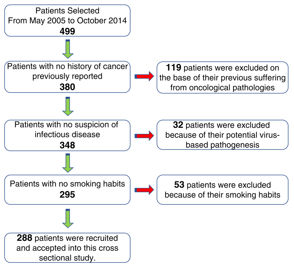
We selected 288 salivary lesions (out of the total 499) operated on by the same team of surgeons and pre-analyzed with FS.
To reduce bias, in 90% of cases, the intraoperative examination was performed by the same team of pathologists. In all cases, the radiological examination posed an indication for surgical treatment and suggested a provisional diagnosis (i.e. benign vs malignant). FNA was not considered because if done at all, it was at non-dedicated centers and there was a high number of "non-diagnostic" results.
Our study was aimed to assess if and how many were the neoplastic lesions, how many were benign and/or malignant, and if some non-neoplastic lesions were also diagnosed. Our attention was also directed towards the identification of some false positive/negative results. The accuracy of FS procedure was compared, in all the reported cases, with the traditional histological assay. Our null-hypothesis was aimed to assess that FS of the salivary glands may be proposed as a routine procedure and should be used in the decision-making process.
The FS procedure, also called cryosection, is a commonly used procedure to perform rapid microscopic analysis of a specimen: it is mostly used as a first-look diagnostic tool in intraoperative oncological surgery. The protocol adopted for performing intraoperative FS was a standardized method: i) the biopsy was fixed and cut in a cryostat at -25°-28°C, ii) the samples were cut into 5 micrometers thick slices, iii) the samples were subjected to haematoxylin-eosin staining. (Figure 2).

(A–C) Left parotidectomy surface that includes a well-demarcated nodule measuring 3cm in diameter and showing translucent appearance of the cut surface; (D) The intraoperative frozen section (FS) technique and (E) corresponding hematoxylin-eosin section; (F–G) Pathology preliminary FS report compatible with pleomorphic adenoma.
The medical report was obtained, in all patient cases, within 10–15 minutes. Each sample was stored with labels detailing the biodata and the macroscopic characteristics of the excised lesion.
We reported 288 patients with salivary lesions operated on by the same team of surgeons and pre-analyzed with FS, in order to make a comparative analysis between the two different techniques.
FS was useful to indicate the correct surgical treatment of 269 nodules (93.4%) of the parotid gland, 14 nodules (4.9%) of the submandibular gland and 5 lesions (1.7%) that involved the minor salivary glands located in the palatal mucosa. Using the FS method, a correct diagnosis was obtained in 280 cases (97.2%) (Table 1).
The highest concordance between the FS and the definitive diagnosis was for pleomorphic adenoma (98%), Warthin’s tumor (97%), inflammatory processes (99%) and malignant neoplasms (96%). In 8 cases (2.8%), the "provisional diagnosis" with intraoperative FS and the definitive diagnosis were discordant. The false negative results for FS consisted of the following: 1 inflammatory process, actually diagnosed as Warthin’s tumor; 1 normal tissue, actually diagnosed as an arteriovenous malformation; 1 reactive lymphoid tissue, actually diagnosed as a non-Hodgkin’s lymphoma; and 1 sialometaplasia, actually diagnosed as a squamous cell carcinoma. Two false positive results were obtained: in both these cases squamous metaplasia in pleomorphic adenoma was interpreted as malignant (Table 2). Thus, the sensitivity and specificity for malignancy were assessed to 97%. Patients with a positive FS diagnosis underwent lymphadenectomy. The FS diagnosis of malignancy was not confirmed at histology in two cases: 1 carcinoma, actually a NH-Lymphoma; 1 NH-Lymphoma, actually a small cell carcinoma. In these cases, only one patient received overtreatment.
Analyzing the definitive diagnosis vs FS, the false positive cases were found to have been diagnosed by a non-dedicated pathologist, as also one case of Warthin’s cancer, defined as an inflammatory process.
This study was conducted in order to assess the diagnostic accuracy, sensitivity and specificity of FS examination, used as a preoperative method to guide the surgeon in the choice of treatment of salivary lesions. A further aim of the study was to analyse the diagnostic accuracy of FS examination to pose the diagnosis of malignancy. Previous studies have compared the accuracy of FS examination with FNA cytology, observing a greater reliability of the FS technique13. In fact, FNA shows too high a number of false negatives due to factors related to: i) triage errors, ii) hypocellularity of the material, iii) interpretation errors10,11. Some studies have demonstrated an excellent effectiveness of a FS examination, obtaining a specificity of 99% and a sensitivity of 90%, other authors have even reported maximum specificity and sensitivity, equal to 100%16,17.
In our study, we excluded FNA as a perioperative examination because the number of "inadequate" tissues was too high. In our experience, FS could replace FNA, reducing the risk of inappropriate surgery, of unnecessary adjuvant radiotherapy, as well as reducing National Health System costs18.
Based on the results described in this study, the FS examination shows a high reliability in the diagnosis of salivary gland tumors. FS is particularly useful in cases of differential diagnosis between neoplastic and non-neoplastic lesions; it also shows a good reliability in the differential diagnosis between benign and malignant neoplasms.
Despite our study limitations, related to the small sample size and related to the single unique center participating to this study, our results are in agreement to those reported by various authors, and they also highlight the need to be able to rely on a dedicated team of clinicians and pathologists15–20.
“Misinterpretation” was observed in those cases diagnosed by pathologists with no specific experience of head and neck tumors. Our work group experience emphasizes the importance of intraoperative examination in order to define the histotype and the cancer margins, permitting the performance of effective, predictable surgery of the salivary glands.
In Italy, directors of clinical research units and those responsible for clinical services may access data records for research purposes if patients have previously signed a consent form that confirming that they allow this use of their data. Therefore, no specific ethical approval is required for this study, and all patients included in the study signed written informed consent allowing their data to be used in future research.
Dataset 1: Raw data underlying the study, including final diagnosis and FS diagnosis. DOI, 10.5256/f1000research.13043.d19293521
| Views | Downloads | |
|---|---|---|
| F1000Research | - | - |
|
PubMed Central
Data from PMC are received and updated monthly.
|
- | - |
Click here to access the data.
Spreadsheet data files may not format correctly if your computer is using different default delimiters (symbols used to separate values into separate cells) - a spreadsheet created in one region is sometimes misinterpreted by computers in other regions. You can change the regional settings on your computer so that the spreadsheet can be interpreted correctly.
Provide sufficient details of any financial or non-financial competing interests to enable users to assess whether your comments might lead a reasonable person to question your impartiality. Consider the following examples, but note that this is not an exhaustive list:
Sign up for content alerts and receive a weekly or monthly email with all newly published articles
Already registered? Sign in
The email address should be the one you originally registered with F1000.
You registered with F1000 via Google, so we cannot reset your password.
To sign in, please click here.
If you still need help with your Google account password, please click here.
You registered with F1000 via Facebook, so we cannot reset your password.
To sign in, please click here.
If you still need help with your Facebook account password, please click here.
If your email address is registered with us, we will email you instructions to reset your password.
If you think you should have received this email but it has not arrived, please check your spam filters and/or contact for further assistance.
Comments on this article Comments (0)