Keywords
Reliability, accuracy, three dimensional, 3D, scanner, anthropometry, torso, children, adolescents
Reliability, accuracy, three dimensional, 3D, scanner, anthropometry, torso, children, adolescents
Three-dimensional (3D) scanning is a time saving procedure and due to minimum discomfort it has become acceptable to use in children when body components and disease risk are being studied1,2. Moreover, body mass index or head circumference may also be evaluated accurately through 3D scanners that capture three-dimensional images3–5.
Scanning devices that scan the body surface generating 3D images originated in the garment industry6. When adapted for computers or personal devices, 3D scanners can measure and display with precision the size and shape of a person's body and the surface of the skin, and offer great potential for medical applications7,8.
Currently, there are several safe, accurate and reliable portable devices that perform their function in a few seconds4,9. However, despite technological advances, there are few studies using 3D images in children. Pfeiffer et al.10 reported in 2006 the first study of prevalence of flatfoot in children using 3D measurements. Prieto et al.11 reported a study measuring burnt skin area on different ages and Djordjevic et al.12 reported on facial symmetry in adolescents.
Torso 3D measurements are reported in relation to breast position assessment for plastic surgery13 and scoliosis follow-up without x-ray exposure14,15. Clinical or public health relevance for torso 3D measurements in relation to the obesity pandemic still requires additional research to define its usefulness.
This is a sub-sample of a larger project called South American Youth/Child cARdiovascular and Environmental Study (SAYCARE), an observational multicentre feasibility study based at public and private schools, aimed at developing methods for collecting reliable, comparable and validated data on cardiovascular health biomarkers, lifestyles, and environmental, social and family risk factors in children and adolescents. A detailed description of the SAYCARE sampling and recruitment methodology, data collection and quality control activities has been published elsewhere16.
Sample size calculation was performed considering a comparison between observed body surface area (BSA) mean obtained by a 3D scanner (2,139; SD=224) and a calculated BSA mean value using a mathematical formula (2,225), as reported by Schloesser et al.17. We included a type I error α of 0.05 and a type II error β of 0.95. The estimated sample size was 72, and was increased to 86 allowing an anticipated loss up to 20%. Thirty-six female and 46 male participants were recruited. There were 66 adolescents (29 girls and 37 boys) and 16 children under 10 years old (7 girls and 9 boys).
For data collection, the schools were initially contacted and received a formal invitation with detailed information about the study. The schools were selected for their proximity to the institute and researchers in charge of the study, for being public or private, and because they had students in the required age groups. For the schools that agreed to participate, an information letter and a verbal explanation were provided to the potential participants and their parents or legal guardians.
Images were captured using a portable scanner (iSense, Cubify, USA) attached to a Tablet 128Gb with OSX (Ipad-Air Apple, USA). Special training was not required to use these devices. The training in the use of the scanner was carried out with the support of the local dealer technician during one morning. The training included information on safety, assembly, calibration and how to scan and export images with the scanner attached to the tablet. The scanner was operated by two authors (CD and EA). It allows scanning objects from 30cm to 3 meters in size. Images were capturing by rotating around the subject with the device focused towards the centre. Some training practical sessions by scanning objects were conducted before actually scanning people. The acquired images were rebuilt as objects without texture and processed with software ad-hoc for analysis and processing of digital images. The images were manually reshaped using the 3D design software Rhinoceros for OSX, v5.3.2 (Robert McNeel & Associates, USA) in order to exclude hair in girls or arms in boys and girls, retaining only the torso (Figure 1). Area and volume were measured using 3D design software (Rhinoceros for OSX, v5.3.2).
Body surface scanning was performed in a room with daylight, and with doors and windows closed. Girls were evaluated in a standing position, with their arms over their heads, holding their hair. Boys were evaluated in a standing position with the arms at the sides and the palms forward.
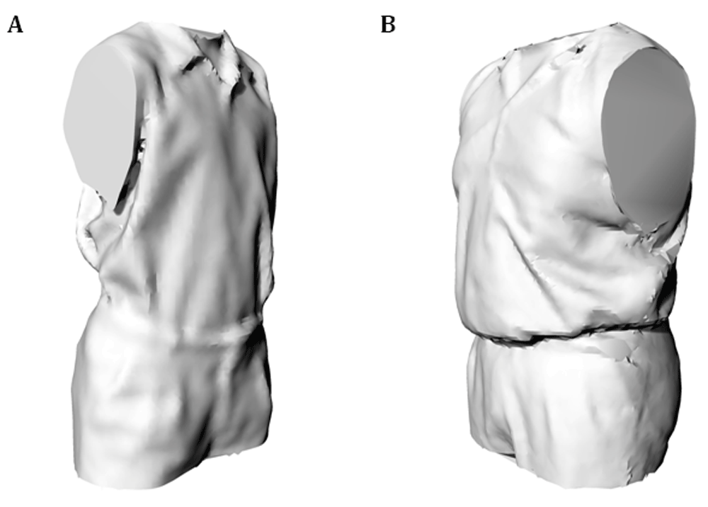
A: Waist to Height Ratio=0.4; B Waist to Height Ratio=0.6. In addition a 3D scan example is presented for Figure 1 B - Torso 3D scan: Model ID 3DPX-008635. Available from https://3dprint.nih.gov/discover/3dpx-008635.
Descriptive analysis included mean, standard deviation and coefficient of variation. Reliability for area in m2 and volume in litres1 was made comparing the first and the second measurement, through the concordance correlation coefficient (rho_c). A new variable was constructed by dividing volume over area in order to apply a curve ROC analysis and estimate the accuracy, sensitivity and specificity for certain values of this method. The Waist-to-Height Ratio (WHtR) was considered as a “gold-standard” to measure obesity, considering a 0.5 as the cut-off point for abdominal obesity. We used a WHtR > 0.5 as gold standard for obesity classification, because this ratio has been reported as accurate in cross-sectional studies for children and adults18. Waist and height measurements were obtained by conventional anthropometric measurements during fieldwork. Expert anthropometrist hired for fieldwork took the anthropometric measures. The size was measured with a stadiometer with the feet not raised from the ground and with the head in the Frankfort plane. The waist was measured with a non-elastic and flexible tape measure, at the midpoint between the last rib and the iliac crest.
The statistical analyses were performed using the Stata software version 12.1 (StataCorp, College Station, Texas) and were stratified by sex and age group.
Table 1 shows the descriptive values for first measurements of area, volume, waist, height and waist to height ratio (WHtR). The original sample comprised 54 girls and 46 boys. Images from 18 participants were excluded because they were incomplete or scanning could not be repeated twice. We obtained complete images for analysis from 36 girls and 46 boys. All 82 children studied were from two private schools in Lima.
Table 2 shows a descriptive analysis that includes the mean values for torso scanned area and volume measurements. It also shows the reliability coefficients for first and second area and volume measurement by sex and age group. In boys the reliability coefficients were strong (rho_c > 0.80) in every comparison between first and second area and volume measurements at any age group. In girls, this coefficient was moderate (rho_c > 0.70) only for area comparison in adolescents older than 10 years of age.
Table 3 shows mean values for torso scanned area and volume measurements classified by WHtR and age group. It also shows the reliability coefficients for first and second area and volume measurement. The reliability coefficients for obesity were strong (rho_c> 0.80) in every comparison between first and second area and volume measurements at any age group.
Figure 1 shows examples of 3D scanned images of the torso captured from two male teenagers with opposite values of WHtR. Both adolescents shown in Figure 1 have similar age (both are 16 years-old) and height (both had a height around 1.7 m), but dissimilar WHtR (Image A=0.4 and Image B=0.6).
Figure 2 shows that the first and second three-dimensional torso area and volume measurements have a monotonic correlation.
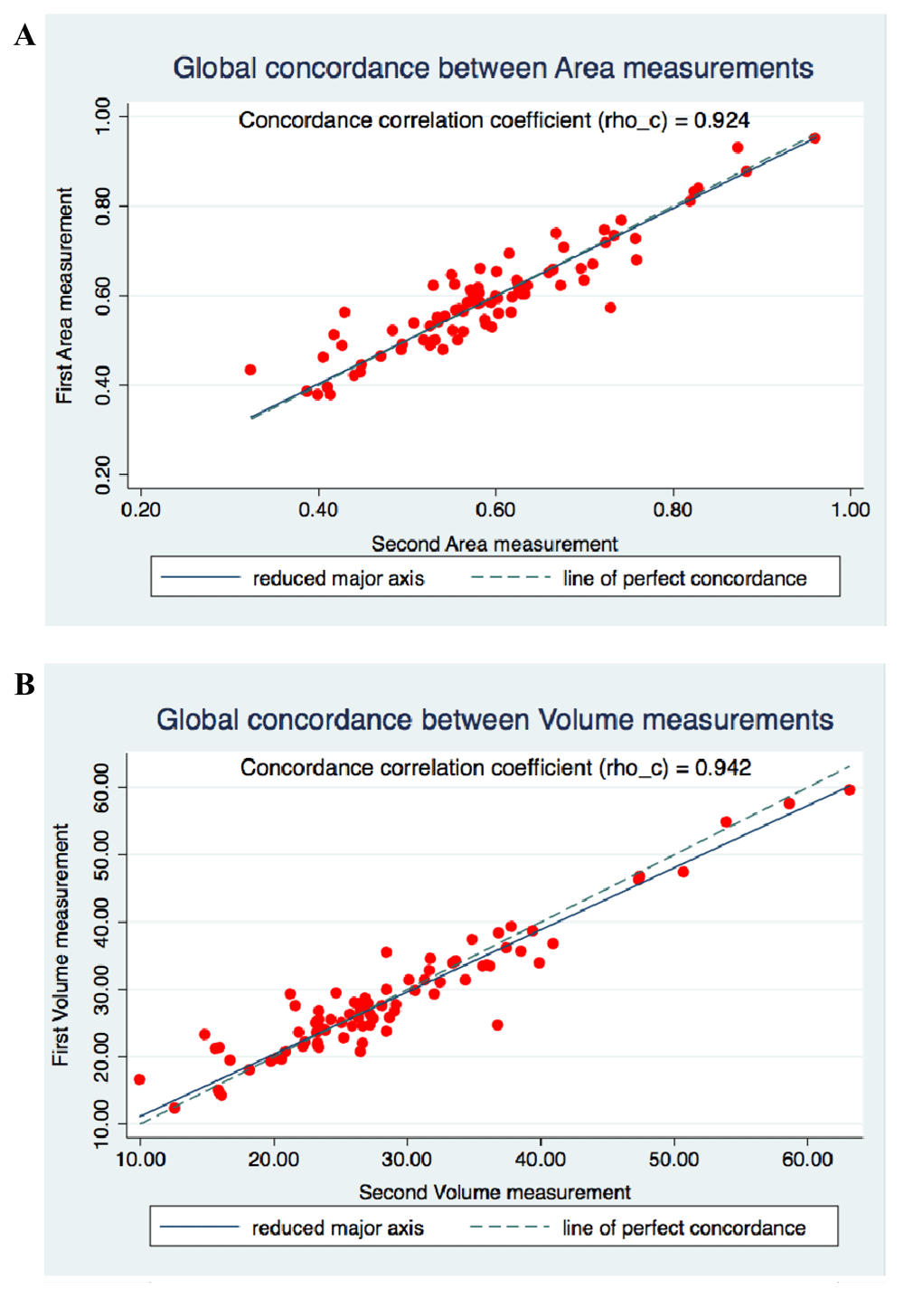
A: Area measurements; B: Volume measurements. Syntaxes used by STATA program included the command: concord to graph area and volume concordance.
Figure 3 shows the ROC curve for different cut-off points for volume/area ratios of 44 and 48. The area under the ROC curve ranged between 0.5707 and 0.6383 when volume/area ratio was compared to the cut-off point used as compared to the “gold standard” (WHtR > 0.5). In order to measure accuracy volume (in l)/area (in m2) ratio selected cut-off were 44 and 48. Sensitivity was higher (75%) than specificity (39%) when using the volume/area ratio = 44. Specificity was higher (80%) than sensitivity (47%) when using volume/area ratio = 48.

A: Volume to Area ratio = 44; Sensitivity 75%; Specificity 39%. B: Volume to Area ratio = 48; Sensitivity 47%; Specificity 80%. Syntaxes used by STATA program included the command: roctab to graph area and volume area under the curve.
Portable scanners are reliable, time-saving devices, and are applicable in childhood nutritional research. Torso 3D measurements obtained by using low cost, portable scanners may increase unconventional anthropometric assessment in children and adolescents.
In this study, we showed strong reliability of 3D scanning images for torso area and volume measurements, particularly in boys and obese children. Our results are in line with previous reports showing that 3D scanner devices are reliable for different anthropometric measurements2,17,19, and pave the way for further studies with larger numbers of participants.
To the best of our knowledge, we did not find published papers that used the iSense hand-held technology. However, Knoops et al.20 compared four 3D scanning systems for describing facial form, including the Structure Sensor (Occipital Inc., San Francisco, CA, USA) that is similar to the scanning device used in our study. Those authors found that Structure Sensor performance was within a clinically acceptable range of 2 mm, showing fair agreement with systems more than tenfold its cost, therefore being of great promise for clinical use. For our study, we invested less than $2,000 USD for each scanner attached to a tablet when purchased from local dealers. Portability and user-friendly performance are also important assets for fieldwork.
Of note, the reliability was higher in obese children and adolescents than in non-obese children and girls. As mentioned above in the Methods, images were manually reshaped to exclude head, hair, arms and legs, and then area and volume were measured using 3D software. It may be possible that the reshaping done after the capture of images introduced a bias, which can be more evident when dealing with smaller images.
Several authors assessed various 3D scanners in order to understand its usability for unconventional anthropometry.1,17. Santos et al.1 studied 3350 Brazilian children at 6 years old with the aim to describe variation in childhood body shape and size by using three-dimensional photonic scanner using TC2 Three-Dimensional Photonic Scanner (TC2, Cary, NC, USA; www.tc2.com), traditional anthropometry and dual X-ray absorptiometry. These authors found that the component termed corpulence showed strong correlations with traditional anthropometric and body composition measures. Schloesser et al.17 determined the body surface area (BSA) in healthy term and near-term neonates by 3D scanning and compared their results with those from five mathematical formulae for each subject. These authors found that scanned BSA for a full-term new-born was slightly lower than that calculated by mathematical formulae.
In our study, Area and Volume 3D measurements have strong reliability, but Area to Volume ratio, which was tested as an empirical approach to a 3D diagnostic tool for obesity shows low accuracy. A possible explanation is related to bias linked to manually reshaping of images. Area and volume are not directly measurable by conventional anthropometry but could be well-calculated using reconstruction algorithms from 3D surface imaging systems to assess obesity21. Obesity in children is a tractable condition, and if it is labelled as an epidemiologic pandemic22 or part of a bigger picture23, highly sensitive tools including automatic processing for early diagnosis are required.
The capture of three-dimensional images using a low-cost portable scanner can be done in approximately 100 seconds with high reliability between measurements. However the scanner is extremely sensitive to movements, and if this happens, it is necessary to repeat the whole procedure. In addition, when scanning people with comparison purposes, it was observed that some uniformity is required in the amount of clothes that can be used to perform the body surface scans.
A major strength in our study is that we were able to assess the performance of a portable, low cost device to evaluate unconventional torso anthropometry in youths in a middle-income country, so we are adding to scientific literature with results from people and places not well studied.
The use of portable and low cost 3D scanners provides a reliable but inaccurate alternative for area and volume as unconventional anthropometric torso measurements in children and adolescents.
Dataset 1: 3D-Scanner-Unconventional-Anthropometry_Database.
Available from: https://dataverse.harvard.edu Dataset Persistent ID doi:10.7910/DVN/BLH6BS
Data are available under the terms of the Creative Commons Zero "No rights reserved" data waiver (CC0 1.0 Public domain dedication).
Institutional Research Ethic Committees approved the study protocol in each center where the SAYCARE team collected data: University of Buenos Aires (Argentina), National Institute for Child’s Health at Lima (Peru), University of Antioquia (Colombia), Catholic University of Uruguay (Uruguay), University of Talca (Chile), University of Sao Paulo (Brazil), and Federal University of Piauí (Brazil).
For those who agreed to participate, informed written consent had to be signed by the parent. This had to be signed by a parent or legal guardian and by adolescent participants, before the enrolment. Adolescents, under 18 years-old are not legally able to consent alone. In addition, in Lima, children over 8 years of age were also asked to give their consent.
In Lima - Peru, the study was approved by the Review Board of the National Institute for Child’s Health (Document number 00222-CEI-INSN-2015, February 25th, 2015). The informed consent was obtained from all participants and guardians of the children, clarifying doubts when needed, through written communications, telephone or face-to-face conversations.
| Views | Downloads | |
|---|---|---|
| F1000Research | - | - |
|
PubMed Central
Data from PMC are received and updated monthly.
|
- | - |
Provide sufficient details of any financial or non-financial competing interests to enable users to assess whether your comments might lead a reasonable person to question your impartiality. Consider the following examples, but note that this is not an exhaustive list:
Sign up for content alerts and receive a weekly or monthly email with all newly published articles
Already registered? Sign in
The email address should be the one you originally registered with F1000.
You registered with F1000 via Google, so we cannot reset your password.
To sign in, please click here.
If you still need help with your Google account password, please click here.
You registered with F1000 via Facebook, so we cannot reset your password.
To sign in, please click here.
If you still need help with your Facebook account password, please click here.
If your email address is registered with us, we will email you instructions to reset your password.
If you think you should have received this email but it has not arrived, please check your spam filters and/or contact for further assistance.
Comments on this article Comments (0)