Keywords
Corpus callosum,apraxia,disconnection syndrome,stroke
Corpus callosum,apraxia,disconnection syndrome,stroke
Corpus callosum contains densely packed white matter tracts essential for coordination and integration of the two sides of brain. Pathology affecting the corpus callosum is very rare. Clinically it manifests with apraxia, cognitive dysfunction, disturbances of memory and may be mistaken for psychosis. Tumours (glioma, lymphoma, meningioma, metastasis), demyelinating diseases, trauma and congenital malformations can involve the corpus callosum. However, due to its rich collateral blood supply, isolated vascular lesions are extremely uncommon1. Here we report the case of a 50 year old woman who presented with features of corpus callosum apraxia, initially mistaken as a psychiatric symptom.
A 50 year old woman presented with a history of abrupt onset of numbness on the left side of her body. She had no history suggestive of raised intracranial tension, cranial nerve dysfunction or motor involvement. The patient was admitted with a provisional diagnosis of sensory stroke considering the rapidity of onset. On the second day of hospitalisation, she found it hard to transfer objects from her left hand to the right hand, and found it hard to execute finer activities with left hand. Her family mistook these manifestations as psychiatric symptoms. The patient had a past medical history of systemic hypertension for 12 years and was on tab. amlodipine 5 mg once daily.
On examination on the second day of admission, the patient was conscious, oriented to time, place and person. She had a body mass index of 22.1 kg/m2 and vital signs were stable. Tests for attention, registration, recall were normal. Her immediate and remote memory was intact. Cranial nerves were normal. She had grade 5 power in all limbs. Sensory system examination revealed apraxia of the left hand, which was diagnosed as ideomotor apraxia: the patient was unable to perform pantomime commands with her left hand and also had astereognosis of the left hand. The patient did not have any involuntary movements in the same limb and could perform all activities with her right hand, but was unable to do the same with the left. There were no cerebellar signs nor signs of meningeal irritation. These features were suggestive of callosal disconnection syndrome.
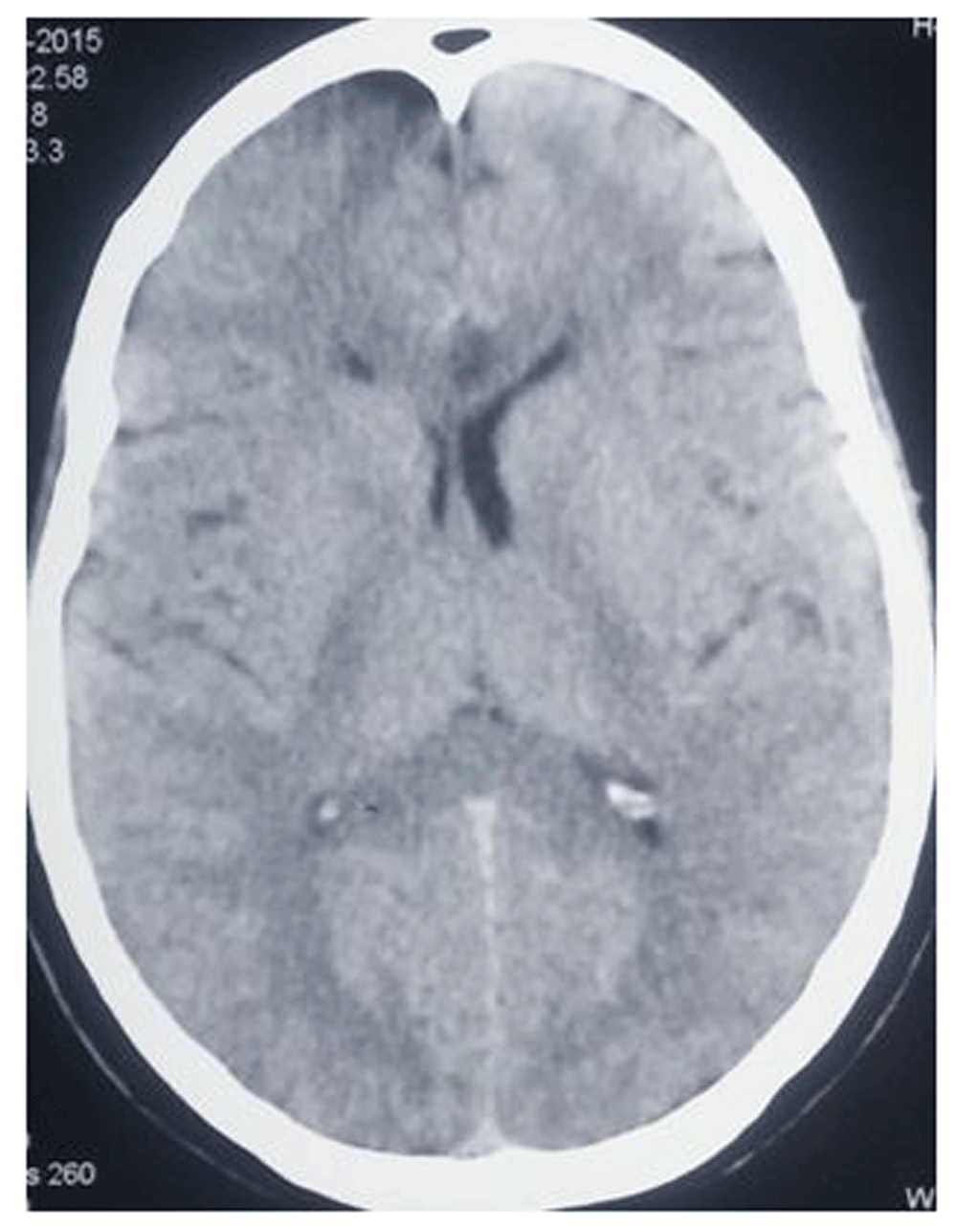
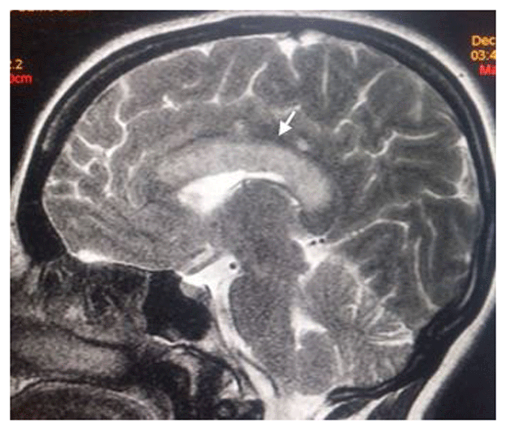
Computed tomography of brain revealed an acute infarct in the body of the corpus callosum (Figure 1). Magnetic resonance imaging (MRI) of the brain showed hyperintense signals in the body of corpus callosum in T2 weighted FLAIR images (Figure 2). Diffusion weighted images showed diffuse restriction confirming the presence of an acute infarct. Electroencephalogram was normal. Further workup was done to look for the cause of thrombosis. Echocardiography was normal. Arterial Doppler of the cerebral vessels did not reveal any abnormality. The patient’s complete blood count, erythrocyte sedimentation rate and peripheral smear were normal. Antinuclear antibody test and retroviral screening were negative.
The patient was treated with antiplatelets (tab. Ecosprin 150 mg once daily and tab. Clopidogrel 75 mg once daily), statins (tab. Atorvastatin 40 mg once daily), and antihypertensives (tab. Amlodipine 5mg once daily). She showed mild clinical improvement at 4 months of follow up (MRI showed resolution of edema and chronic infarct), although apraxia of the left hand persisted.
The corpus callosum contains the white matter bundle that connects the interhemispheric neurons and serves to connect the cortical and subcortical structures of the brain. The corpus callosum is present only in placental mammals and reflects the transition towards increased degree of myelination seen in higher animals. Anatomically it is divided into the rostrum, genu, body and splenium from anterior to posterior2. The corpus callosum receives a blood supply from three arterial networks. The anterior communicating artery (medical callosal and subcallosal branches), anterior cerebral artery (through the pericallosal artery), posterior cerebral artery (through the posterior pericallosal branch); each supplies different parts of the corpus callosum3.
Clinical manifestations of callosal infarcts include callosal disconnection syndrome (as in our case), neuropsychiatric symptoms, gait disorders and alien hand syndrome4. Callosal disconnection manifests in different ways based on the part of the callosum affected. Lesions in the anterior part of callosum presents as unilateral unresponsiveness to touch (tactile anomia), difficulty in calculation, difficulty in copying drawings, and unilateral agraphia (inability to write with one hand). Posterior lesions cause visual unresponsiveness on one side (visual anomia) and a phenomenon called “alexia without agraphia” (able to write, but unable to read). Lesions in the body of the callosum results in unilateral ideomotor apraxia, difficulty to use objects with one hand, and inability to transfer objects from one hand to the other. This occurs in the left hand in majority of the cases, as the left is the dominant hemisphere in most individuals5. MRI is the diagnostic test of choice for the localisation of a callosal infarct. Differential diagnoses for callosal infarcts include Marchiafava Bignami syndrome (callosal demyelination in chronic alcoholism) and multiple sclerosis6,7.
Written informed consent for publication of their clinical details and clinical images was obtained from the patient.
All data underlying the results are available as part of the article and no additional source data are required.
| Views | Downloads | |
|---|---|---|
| F1000Research | - | - |
|
PubMed Central
Data from PMC are received and updated monthly.
|
- | - |
Provide sufficient details of any financial or non-financial competing interests to enable users to assess whether your comments might lead a reasonable person to question your impartiality. Consider the following examples, but note that this is not an exhaustive list:
Sign up for content alerts and receive a weekly or monthly email with all newly published articles
Already registered? Sign in
The email address should be the one you originally registered with F1000.
You registered with F1000 via Google, so we cannot reset your password.
To sign in, please click here.
If you still need help with your Google account password, please click here.
You registered with F1000 via Facebook, so we cannot reset your password.
To sign in, please click here.
If you still need help with your Facebook account password, please click here.
If your email address is registered with us, we will email you instructions to reset your password.
If you think you should have received this email but it has not arrived, please check your spam filters and/or contact for further assistance.
Comments on this article Comments (0)