Keywords
bronchogenic, cyst, back, pain
bronchogenic, cyst, back, pain
Bronchogenic cysts are congenital malformations of the bronchial tree. They result from the anomalous development of ventral foregut and tracheobronchial tree1. They can present as a mediastinal mass that may enlarge and cause local compression. They are the most common primary cysts of the mediastinum with a prevalence of 6%2. Approximately 79% of cysts are located in the middle mediastinum, 17% in posterior mediastinum and 3% in the anterior mediastinum3.
Almost 75% of bronchogenic cysts are asymptomatic. Symptoms vary with age at presentation and with size and location of the cyst. The common symptoms include chest pain (22%), dyspnea (12%), cough (7%), stridor (7%) and respiratory compromise due to tracheal/bronchial compression (10%). Unusual manifestations are dysphagia (1%), pneumothorax (1%), and superior vena cava syndrome (1%)3.
Diagnosis is made by chest X-Ray and computed tomography (CT) chest although magnetic resonance imaging (MRI) chest and endobronchial ultrasound are highly sensitive and specific4. MRI chest can provide additional information about the consistency and nature of the cyst depending upon the presence of proteinous contents in the fluid. In general, bronchogenic cyst appears hypo-intense on T1-weighed images and hyper-intense on T2-weighed images5. Endoscopic ultrasound (EUS) is a relatively invasive procedure for the diagnosis of the bronchogenic cyst.
Treatment options depend on patient’s age and symptoms. Symptomatic bronchogenic cyst are managed surgically with resection, Endobronchial ultrasound (EBUS) guided aspiration, and video-assisted thoracoscopic surgery being a minimally invasive procedure6. Thoracotomy is performed for difficult cases. Asymptomatic cysts in younger patients should be removed due to low surgical risk and potential late complications such as infection, hemorrhage or neoplasia. Watchful waiting has been recommended for asymptomatic adults or high-risk patients. Percutaneous drainage or alcohol ablation has been performed in selected cases4. We present a case of a mediastinal bronchogenic cyst in a 44-year-old female presenting in the form of severe back pain, epigastric distress and nausea.
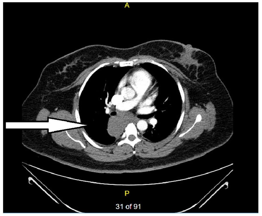
A 44-year-old Hispanic female presented with a three-week history of recurrent sharp interscapular pain radiating to the mid-sternal and epigastric region associated with refractory nausea and vomiting. She underwent cholecystectomy for intermittent epigastric pain two years ago. CT abdomen at that time showed a subcarinal mass measuring 5.4 X 5.0 cm (Figure 1). Subsequent EUS diagnosed it as a bronchogenic cyst. EBUS guided aspiration resulted in an incomplete drainage and she was discharged after partial improvement.
Current physical examination showed a heart rate of 126/min (normal range: 60–100/min) and respiratory rate of 20/min (normal range: 12–20/min). Initial labs showed white cell count of 10.58X10³/uL (normal range: 4000–11X103uL), elevated inflammatory markers [ESR of 63mm/hr (normal range: 0–20 mm/hr); CRP of 116 mg/L (normal range: <3.0 mg/L)], and hypokalemic metabolic alkalosis. Electrocardiogram showed non-specific T wave changes. Chest X-ray showed right posterior mediastinal mass (Figure 2).

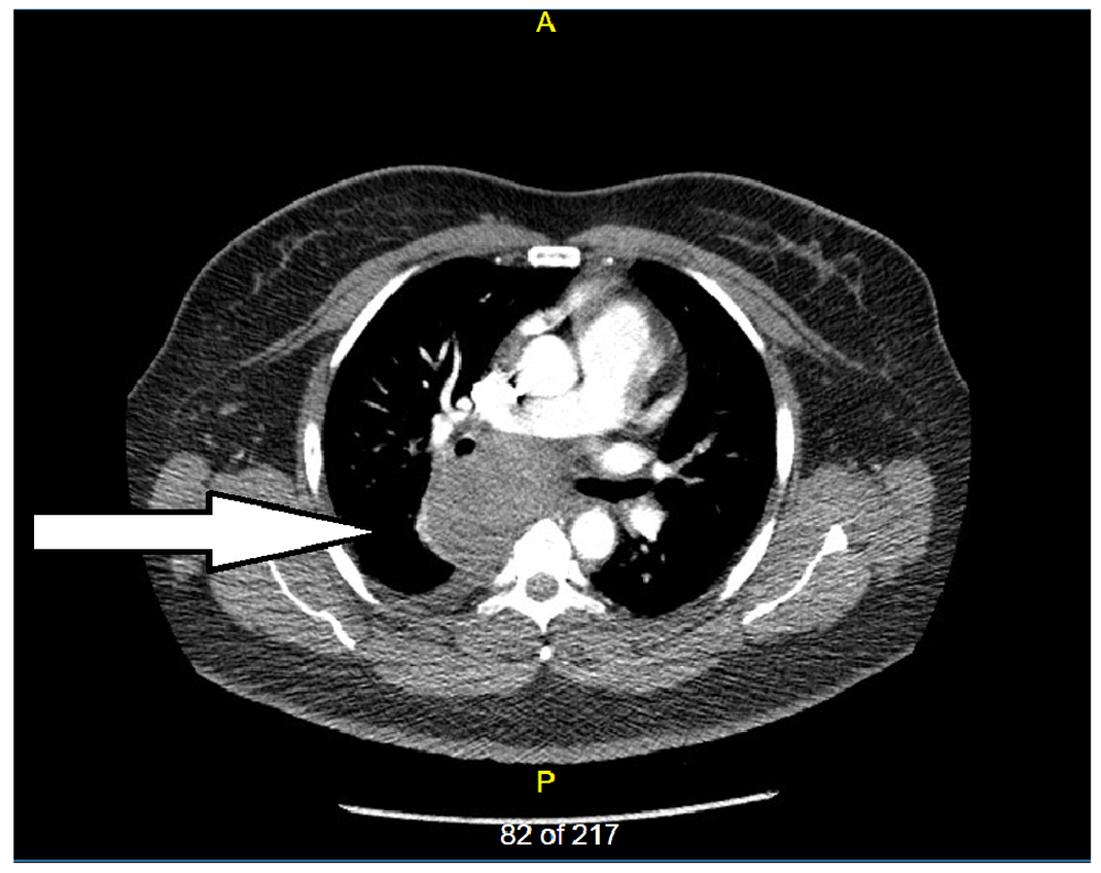
CT chest showed an increase in the size of the bronchogenic cyst (9.64 X 7.7 cm) with small right pleural effusion (Figure 3).
The X-ray and CT findings were consistent with partial cyst rupture or an infected cyst. X-ray esophagogram ruled out esophageal compression or contrast extravasation. The patient’s symptoms were refractory to conservative analgesic and antiemetic measure like Dilaudid (hydromorphone) 1 mg IV every 3 hourly and Zofran (Ondansetron) 4 mg IV every 4 hourly for pain and nausea/vomiting respectively. Cardiothoracic surgery was consulted and the patient underwent right thoracotomy and surgical cyst excision. Cyst pathology was consistent with severe inflammatory changes. Within 24–48 hours after the surgery, the resolution in the patient’s symptoms were noted in terms of decrease in need of pain and nausea medications. Repeated labs showed resolution of leukocytosis.
Bronchogenic cysts are the rare benign congenital malformation resulting from the anomalous budding of ventral foregut and tracheobronchial tree1. They are part of the bronchopulmonary foregut malformations. They are more commonly found in the mediastinum in the paratracheal and subcarinal regions. Less commonly they are found in the lung parenchyma.
Bronchogenic cyst may present with unusual symptoms posing a diagnostic challenge. Signs and symptoms of bronchogenic cyst mainly depend upon its location, size, and compression of surrounding structures like esophagus, trachea, and bronchus7. Most common presentation in adult patients includes chest pain, cough, dyspnea and dysphagia8.
Our patient presented with unusual symptoms of severe backache, epigastric discomfort, refractory nausea, and vomiting. It is believed that the back pain is caused by stretching of the nerves supplying the parietal pleura while the epigastric distress is caused by the stimulation of the vagal nerve4,9.
In our case, repeat CT chest confirmed an increase in the size of a bronchogenic cyst with small right pleural effusion. Considering that approximately 10% of the patients develop respiratory problems due to tracheal or bronchial compression, we performed X-ray esophagogram and ruled out esophageal compression or contrast extravasation.
At present management of symptomatic bronchogenic cyst is surgical as discussed. Management of asymptomatic cyst is controversial. It has been suggested that as most of the cysts eventually cause some symptoms or serious complications like respiratory distress from airway compression, infection and airway fistulae, surgical resection in asymptomatic patients is recommended. Also, postoperative surgical complications are more common in patients with symptomatic cysts as compared to asymptomatic cysts further implying the benefits of surgical resection of asymptomatic cysts10.
This case highlights the importance of recognizing bronchogenic cyst as a cause of severe back pain, refractory nausea, and vomiting. Back pain is caused by stretching of nerves supplying the parietal pleura; while nausea is caused by stimulation of vagus nerve. Prompt surgical excision can lead to complete symptom resolution and avoidance of future complications.
Written informed consent was obtained from the patient for the publication of this case report and any accompanying images.
All data underlying the results are available as part of the article and no additional source data are required.
| Views | Downloads | |
|---|---|---|
| F1000Research | - | - |
|
PubMed Central
Data from PMC are received and updated monthly.
|
- | - |
Provide sufficient details of any financial or non-financial competing interests to enable users to assess whether your comments might lead a reasonable person to question your impartiality. Consider the following examples, but note that this is not an exhaustive list:
Sign up for content alerts and receive a weekly or monthly email with all newly published articles
Already registered? Sign in
The email address should be the one you originally registered with F1000.
You registered with F1000 via Google, so we cannot reset your password.
To sign in, please click here.
If you still need help with your Google account password, please click here.
You registered with F1000 via Facebook, so we cannot reset your password.
To sign in, please click here.
If you still need help with your Facebook account password, please click here.
If your email address is registered with us, we will email you instructions to reset your password.
If you think you should have received this email but it has not arrived, please check your spam filters and/or contact for further assistance.
Comments on this article Comments (0)