Keywords
CNS lymphoma, Meningioma, Collision tumor
CNS lymphoma, Meningioma, Collision tumor
Additional details regarding the workup, intra- and post-operative management of the patient were included as well as further details on discussing the association between primary CNS lymphoma and meningioma.
To read any peer review reports and author responses for this article, follow the "read" links in the Open Peer Review table.
The incidence of two distinct primary intracranial pathologies is an exceedingly rare phenomenon. The reported incidence of such an occurrence is approximately 1 in a million annually (Lee et al., 2002). Although meningiomas, given their benign and slow-growing nature, are well known to coexist with other primary intracranial malignancies such as glioblastoma, metastases, adenomas, there are only nine reported cases of a meningioma occurring simultaneously with primary CNS lymphoma (PCNSL) in the literature (Gordon et al., 2011; Slowik & Jellinger, 1990). Here, we report a case of a woman who sustained multiple injuries due to two distinct intracranial pathologies, however, lateralizing signs were unrecognized for two weeks prior to her final diagnosis.
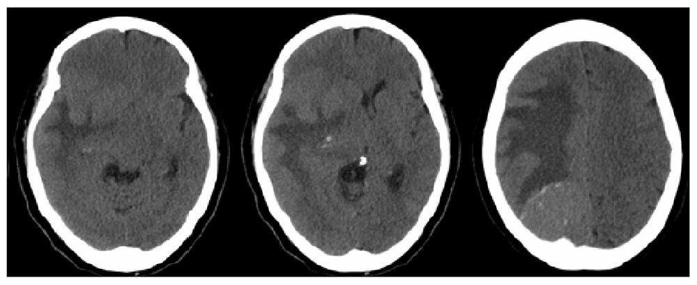
A 64-year-old Hispanic female with a past medical history of type 2 diabetes mellitus and hypertension presented with a chief complaint of left hemiparesis and paresthesias and was activated as a code stroke. The history appeared to be limited due to the patient being Spanish-speaking only. (Although the use of translators is routine, in the emergency department, they are not always readily available in time-sensitive situations.) She did not receive tPA because she stated her left-sided symptoms were not new and she had progressively worsening clumsiness of her left side and that she had been falling to her left. Computed tomography (CT) of head revealed a right occipital mass with significant vasogenic edema causing 12mm of midline shift (Figure 1). Of note, she presented to urgent care two weeks prior to presentation after sustaining a mechanical fall at home. She was diagnosed with a left bimalleolar fracture, placed in a cast, and scheduled for outpatient follow up with orthopedics for surgical evaluation. No further workup was considered at that time by the initial provider.
The patient was alert and oriented to person, place and time in Spanish. Cranial nerve exam revealed no deficits and no evidence of visual field cut. Motor examination revealed left hemiparesis (4+/5 in the upper and lower extremities), but was limited by the previous casting of her distal left malleolar fracture. Sensory examination revealed slight diminished sensation in the left upper and lower extremities with similar limitations as motor examination.
The patient was started on dexamethasone 6mg every 6 hours and admitted to the ICU. No obvious abnormalities were noted on her CBC with differential. A CT chest, abdomen and pelvis was performed which did not demonstrate any evidence of metastatic lesion. An MRI brain with and without contrast revealed two homogeneously contrast-enhancing lesions: a 4.8×6.1×3cm right parieto-occipital extra-axial mass with dural-based attachment, as well as a 3.4×1.8×2.2cm homogenously contrast-enhancing lesion adjacent to the right posterolateral ventricle. FLAIR signal changes were also appreciated and were noted to extend across the splenium of the corpus callosum, raising concerns for a high-grade glial process (Figure 2).
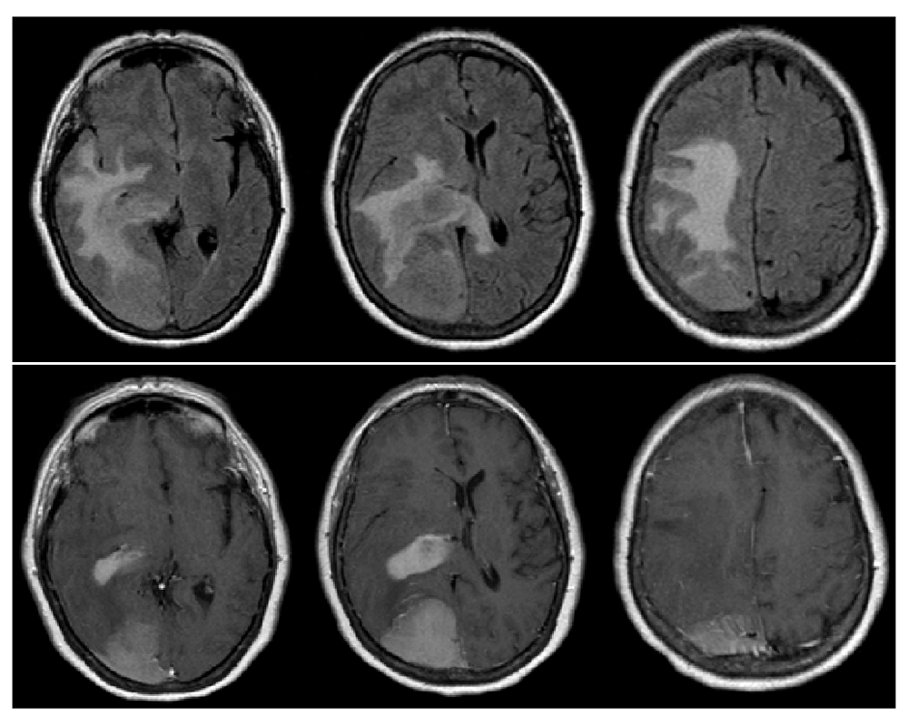
T1-post contrast reveals 2 distinct lesions – a homogenously enhancing extra-axial lesion in the right parietal lobe as well as a homogeneously enhancing periventricular lesion (bottom).
Given the degree of mass effect, presence of neurological deficit and no evidence of metastasis, no further workup was pursued due to the necessity of surgical intervention. After preoperative clearance, a right occipital craniotomy was performed with anticipation for gross total resection of the right parieto-occipital lesion and biopsy with likely subtotal resection and biopsy of the second lesion. Preliminary pathology from intra-operative frozen specimen were consistent with meningioma (extra-axial lesion) and high-grade glioma (periventricular lesion). With the use of intraoperative neuronavigation, gross total resection was performed for the extra-axial lesion and maximal, safe resection of the periventricular lesion was performed. This lesion was accessed via the meningioma cavity after it was removed. She tolerated the procedure well and had an improved neurological exam postoperatively. Her left hemiparesis improved compared with pre-operative exam, however, she did have very minor left visual field deficits. Post-operative MRI demonstrated gross total resection of meningioma and subtotal resection of what was later confirmed to be diffuse large B-cell lymphoma (Figure 3). During this same admission, she also underwent open reduction, internal fixation (ORIF) of her left bimalleolar fracture without complication. She was discharged home in stable condition.
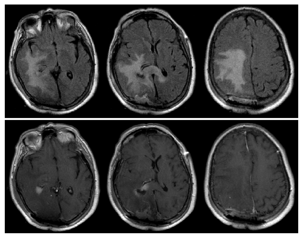
T1-post contrast reveals gross-total resection of the previously seen extra-axial lesion in the right parieto-occipital region as well as subtotal resection of right periventricular lesion (bottom). The midline shift is significantly improved from pre-operative MRI (Figure 2).
Extra-axial lesion: Meningothelial Meningioma
Periventricular lesion: Diffuse Large B-Cell Lymphoma (+CD20, +BCL-6, +BCL-2, +MUM-1, +KI67)
At one month clinic follow-up, she was noted to have an intact motor exam with stable visual field deficits on gross examination. She went into complete remission after a course of methotrexate, cytarabine, and Rituxan and 4 cycles of radiation therapy. She tolerated the treatment relatively well with minor symptoms. At one and two year follow-ups, she continues to be in remission with no signs of recurrence on imaging. Her family reported no evidence of cognitive decline, however, no specific cognitive testing was performed.
We report a rare case of a concurrent meningioma and primary CNS lymphoma (PCNSL), a rare occurrence entity that has only nine reported cases in the literature. The most common concurrent intracranial tumors reported in the literature are meningioma and glioblastoma (Zhang et al., 2018). It is rare to find two or more primary intracranial tumors simultaneously in patients without previous radiation therapy or underlying phacomatosis such as Neurofibromatosis-2 (NF2). The annual incidence of this phenomenon is estimated to be less than one per million (Gordon et al., 2011; Lee et al., 2002).
Accurate diagnosis is essential as the surgical management of these conditions are opposite of one another. One area in which the management in our patient could be improved is a more accurate history and neurological examination. This was likely affected by the fact that the was a non-English speaker and highlights the importance of accurate history taking with a translator to ensure optimal care. Surgical management of PCSNL is typically limited to biopsy if CSF analysis is inconclusive. This is because PCNSL is particularly chemo- and radiosensitive. Conversely, gross total resection is the gold standard in the management of meningiomas and gliomas (Baraniskin & Schroers, 2014; Gordon et al., 2011; Hoang-Xuan et al., 2015; Korfel & Schlegel, 2013; Muñiz et al., 2014). The same principle applies for steroid administration. The administration of glucocorticoids is not recommended in lymphoma as it could affect the diagnostic yield while it is a mainstay in the treatment of vasogenic edema (Hoang-Xuan et al., 2015). Interestingly in our case, the initiation of high-dose dexamethasone did not affect our diagnosis. The typical diagnostic workup for CNS lymphoma consists of CSF analysis for markers such as IL-10, CXCL13, CD19, CD20 or flow cytometry (Baraniskin et al., 2011; Baraniskin & Schroers, 2014; Muñiz et al., 2014; Rubenstein et al., 2013). Due to the mass effect that is exerted by meningiomas, CSF analysis is difficult without a craniotomy as a lumbar puncture would not be recommended in such a setting. MRI is the gold standard diagnostic modality for meningiomas, however, this is complicated by the fact that CNS lymphoma can mimic any and every intracranial pathology, making it difficult to discern whether lymphoma should be considered as a possibility (Bühring et al., 2001; George et al., 2007; Kulkarni et al., 2012).
The most common association of two primary intracranial tumors is that of meningioma and glioma (>40 reported cases), however given that these tumors are two of the most common primary intracranial tumors this is thought by many to be coincidental, however associations between the two pathologies have been proposed (Ruiz et al., 2015; Slowik & Jellinger, 1990; Suzuki et al., 2010; Zhang et al., 2018). In a report of two patients with concurrent meningioma and high grade gliomas, Ruiz et al. reported a mutation in K409Q of the KLF4 gene within the meningiomas (Ruiz et al., 2015). Suzuki et al. reported an oncogenic effect due to overexpression of platelet-derived growth factor (PDGF) receptors (Suzuki et al., 2010). It is also postulated that meningiomas may serve as an oncogenic factor in the development of meningiomas by either inducing a genetic mutation or inciting an inflammatory response in glial cells and B-cell proliferation (Gordon et al., 2011; Slowik & Jellinger, 1990).
Simultaneous presentations tend to affects adults and have a female predominance due to the nature of meningiomas and their apparent relationship with progesterone and estrogen receptors (Pravdenkova et al., 2006). Since meningiomas typically have an indolent course, this is likely why they are often found concurrently with another primary intracranial pathology. In the setting of simultaneous extra-axial and intra-axial lesions, primary CNS lymphoma must remain a consideration to ensure accurate diagnosis and treatment. Prompt treatment in our case contributed to a good outcome. The outcome in our case fares well in comparison to previously reported cases as their average survival was 6 months in those patients whose outcomes were reported.
The patient and her family gave written informed consent for presenting all pertinent clinical information in this case report.
All data underlying the results are available as part of the article and no additional source data are required.
| Views | Downloads | |
|---|---|---|
| F1000Research | - | - |
|
PubMed Central
Data from PMC are received and updated monthly.
|
- | - |
Provide sufficient details of any financial or non-financial competing interests to enable users to assess whether your comments might lead a reasonable person to question your impartiality. Consider the following examples, but note that this is not an exhaustive list:
Sign up for content alerts and receive a weekly or monthly email with all newly published articles
Already registered? Sign in
The email address should be the one you originally registered with F1000.
You registered with F1000 via Google, so we cannot reset your password.
To sign in, please click here.
If you still need help with your Google account password, please click here.
You registered with F1000 via Facebook, so we cannot reset your password.
To sign in, please click here.
If you still need help with your Facebook account password, please click here.
If your email address is registered with us, we will email you instructions to reset your password.
If you think you should have received this email but it has not arrived, please check your spam filters and/or contact for further assistance.
Comments on this article Comments (0)