Keywords
Laminate veneers, Lithium disilicate ceramic, Hybrid ceramic, Fracture resistance, Preparation designs.
Laminate veneers, Lithium disilicate ceramic, Hybrid ceramic, Fracture resistance, Preparation designs.
Laminate veneer restorations have gained popularity and patient contentment owing to their good esthetics and highly conservative tooth preparation designs1. Pressable and machinable ceramics have developed as an option to the conventional porcelain layering manufacturing method2. Ceramics have the benefits of elevated flexural strength and stability of color, while their drawbacks include elevated antagonistic tooth wear and less conservative tooth preparation3. Composite resins can overcome these two drawbacks, although wear of the material itself is higher4. While ceramics have higher stiffness and hardness values than the natural tooth, reduced values are shown by composite resins. Therefore, a material is needed that combines the benefits of ceramics with those of composites, causes minimal wear of both the material itself and the antagonistic tooth and preserves the structure of the sound tooth3.
The success rate of ceramic laminate veneers has been clinically evaluated and has shown range from 18 months up to 15 years; the rate of success varies between 75% and 100%. De-bonding, fracture and micro-leakage failures are seen in ceramic veneers4. Fractures accounted for 67% of the total failures over a 15-year clinical performance period5.
This research aimed to compare the impact of hybrid and lithium disilicate ceramic laminate veneer on their resistance to fracture with two distinct preparation configurations after thermal cycling.
This study was approved by the Research Ethics Committee of the Faculty of Dentistry, Cairo University. Approval number 15531.
A total of 52 human central maxillary incisors were selected for the present study. Teeth scaling and polishing was done to remove any remnants, then they were stored in saline solution at room temperature. To facilitate the handling of teeth, the root of each tooth was mounted vertically with its long axis in Epoxy resin blocks (CMB, Egypt).
Teeth were divided into two equal groups (n=26) according to material: Group A (Cera), Cerasmart laminate veneers, and Group B (e.max), IPS e.max Press laminate veneers). These groups were further subdivided into two subgroups according to preparation design (subgroup I, Featheredge preparation design, and subgroup II, Wraparound preparation design).
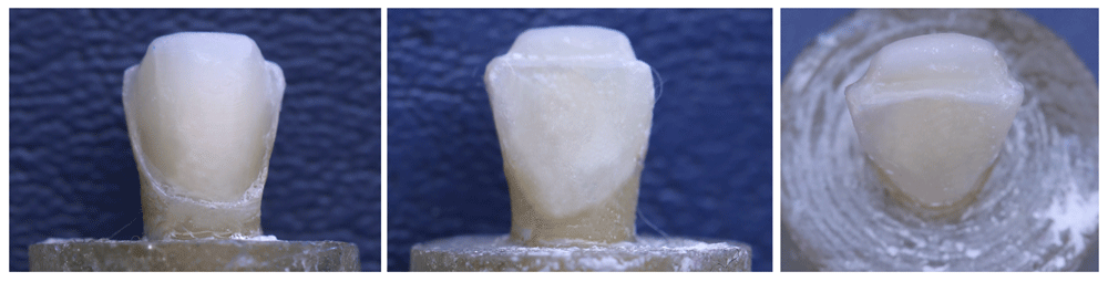
Standardized teeth preparation was done using a five-axis computer numerically controlled (CNC) milling machine (Centroid M400 CNC, USA) with water coolants. The labial surface preparation was performed in mesio-distal direction in two planes (cervical one third and incisal two thirds) (Figure 1). The incisal reduction was only performed in wrap-around design (Figure 2). The parameters are summarized in Table 1.

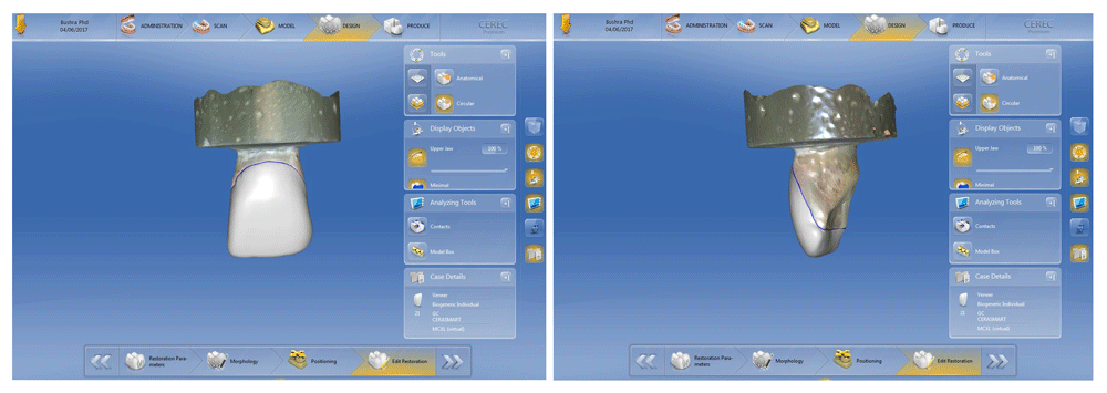
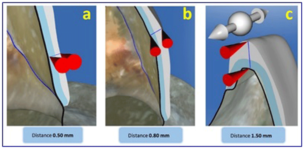
Cerasmart laminate veneers were fabricated using a CAD/CAM system (Omnicam, CEREC MC XL SW 4.0; Sirona Dental Systems GmbH, Germany) using Cerasmart blocks (GC Corporation, Tokyo, Japan). The spacer was adjusted at 30 μm and laminate veneer thickness at 0.5 mm cervically and 0.8 mm incisally (Figure 3, Figure 4).
(a) Cervical, (b) middle, (c) incisal.
IPS e.max Press laminate veneers were fabricated in two steps; first, digital waxing up automated by Exocad CAD software (Exocad GmbH, Germany) to overcome the variation of manually fabricated restorations and to standardize the thickness of all samples. Second, the pressing procedure was done using IPS e.max Press ingots (Ivoclar Vivadent AG, Principality of Liechtenstein) according to the manufacturer’s instructions.
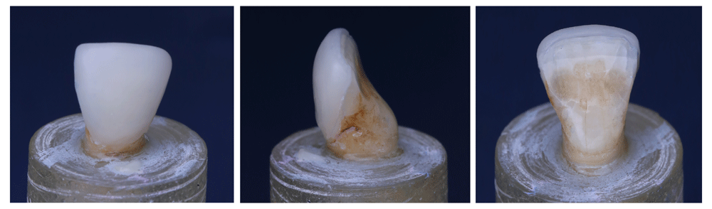
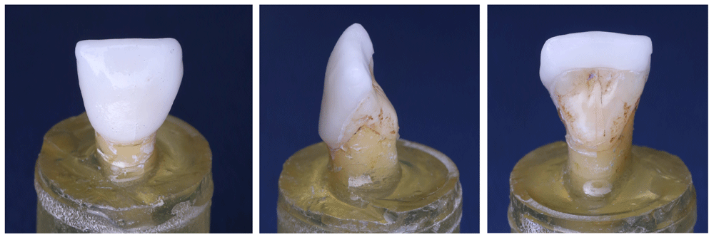
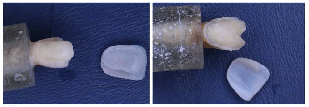
Finally, all laminate veneers were checked on the corresponding prepared tooth for proper seating and marginal adaptation (Figure 5, Figure 6).
All restorations were cleaned using an ultrasonic cleaner (Shenzhen, China) for 180 seconds, the fitting surfaces of the veneers were etched with 9.5% hydrofluoric acid gel IPS Ceramic Refill (Ivoclar Vivadent AG, Principality of Liechtenstein) for 60 seconds for Group A teeth and for 20 seconds for Group B teeth, followed by thorough washing by air/water spray for 30 seconds and drying using air spray. Silane coupling agent Monobond-S (Ivoclar Vivadent AG, Principality of Liechtenstein) was applied for 60 seconds and air-dried before cementation.
The prepared teeth were acid etched using 37% phosphoric acid Scotchbond Universal Etchant (3M ESPE, USA) for 15 seconds and rinsed with air/water spray for another 10 seconds then dried with air spraying. With a fully saturated micro-brush tip two consecutive coats of Single Bond Universal adhesive (3M ESPE, USA) were applied on the prepared enamel surface and thinned gently by air spray for 2–5 seconds. Light cure RelyX Veneer cement (3M ESPE, USA) was used to lute the laminate veneers on their corresponding prepared teeth. The cement was applied on inner surface of the veneers and the veneers were seated with gentle finger pressure on the prepared teeth. Excess cement was removed immediately with an explorer, the exposed margins were covered with glycerin gel to prevent formation of an oxygen-inhibiting layer and ensure the complete polymerization of the cement. Margins were light cured for 20 seconds from the incisal, lingual, mesial and distal directions each respectively.
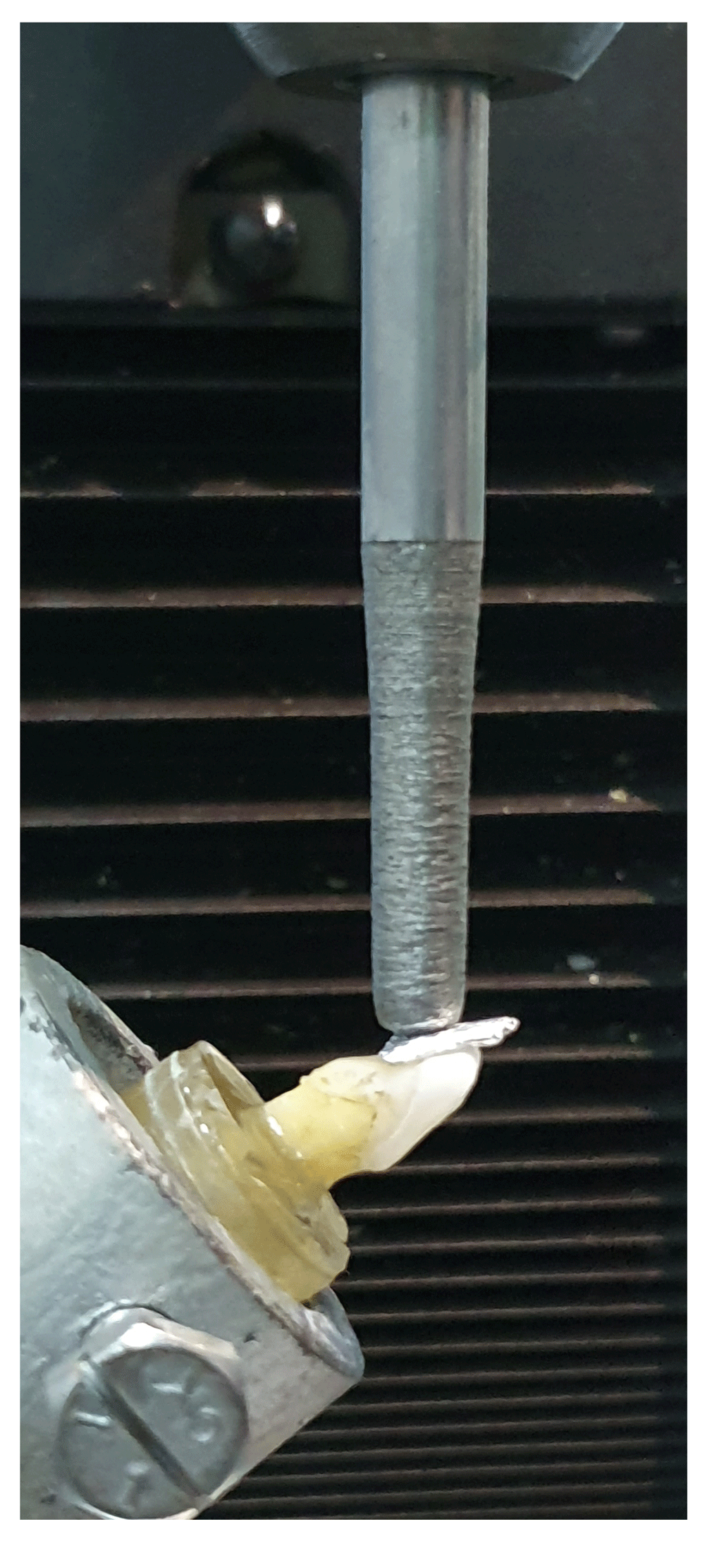
All samples were subjected to thermocycling between 5° and 55°C in a water bath at each temperature for a total of 1750 cycles with a dwell time of 30 seconds at each temperature (water bath) and 10 seconds transport time between the two baths. Fracture resistance test of all samples was performed using a universal testing machine (Instron, Model 3345; Instron industrial, USA) by compressive mode of load applied at 135° angle to the lingual surface of the tooth to simulate the clinical situation as closely as possible6. The load was applied using a metallic rod with flat tip (3.8 mm diameter) attached to the upper movable compartment of testing machine traveling at cross-head speed of 1 mm/min with tin foil sheet in-between to achieve homogenous stress distribution and minimization of the transmission of local force peaks (Figure 7). The load required to fracture was recorded in Newtons.
Data were gathered, tabulated and analyzed statistically using SPSS statistical software (Version 21, Chicago, IL, USA). One-way ANOVA followed by pair-wise Tukey’s post-hoc tests were performed to detect significance between groups. Student t-test was performed to detect significance between paired groups. The level of significance was set at 5% for all statistical analyses and confidence interval at 95%.
E.max wrap-around group recorded statistically non-significant (P>0.05) highest fracture resistance mean value (422.1 N) followed by the Cera wrap-around group (317.23 N), and then E.max feather-edge group (289.6 N), while Cera feather-edge group recorded the lowest fracture resistance mean value (259.3 N) (Table 2 and Figure 8).
| e.max feather- edge, N | e.max wrap- around, N | Cera wrap- around, N | Cera feather- edge, N | P value | ||||
|---|---|---|---|---|---|---|---|---|
| Mean | SD | Mean | SD | Mean | SD | Mean | SD | |
| 289.6 | 88.1 | 422.1 | 203.6 | 317.2 | 108.6 | 259.3 | 109.1 | 0.06 NS |
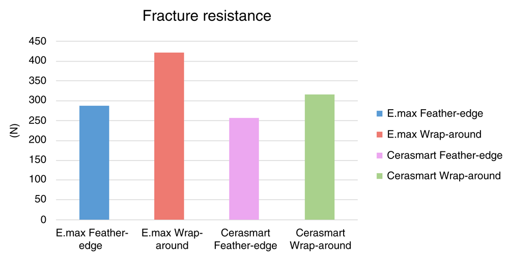
The e.max groups recorded statistically non-significant (P>0.05) higher fracture resistance mean values than Cera groups. Wrap-around groups recorded statistically non-significant higher fracture resistance mean values than feather-edge groups as indicated by the results of Student’s t-test.
The following aspects were considered to assess the failure:
1- Tooth (Intact or fractured)
2- Veneer (Intact or fractured)
3- Tooth-veneer junction (Intact or debonding)
The behavior of samples differed; the results are tabulated in Table 3. None of the restorations tested were fractured. The fracture when occurred was in the tooth structure; root, cervical or incisal edge fracture. The type of failure was either cohesive; fracture in the tooth, adhesive; debonding of the veneer without fracture of the tooth, or adhesive-cohesive; debonding of the veneer with fracture of the tooth (Figure 9, Figure 10). E.max groups showed cohesive type of failure only. Cera groups showed all types of failure.
| Group | Tooth | Veneer | Tooth-veneer junction | |||
|---|---|---|---|---|---|---|
| Intact | Fractured | Intact | Fractured | Bonded | Debonded | |
| E.max feather-edge | 0 | 13 | 13 | 0 | 13 | 0 |
| E.max wrap-around | 0 | 13 | 13 | 0 | 13 | 0 |
| Cera feather-edge | 7 | 6 | 13 | 0 | 6 | 7 |
| Cera wrap-around | 5 | 8 | 13 | 0 | 8 | 5 |
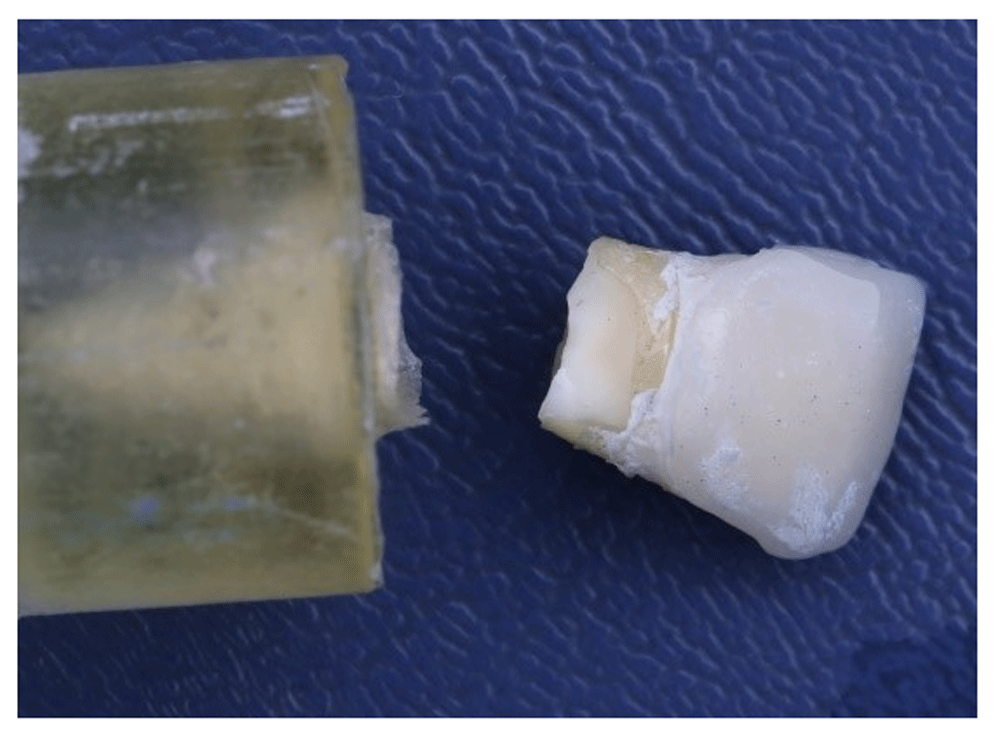
(a) Intact tooth (adhesive failure); (b) fractured incisal edge (adhesive-cohesive failure).
Ceramic laminate veneers are a popular, safe and successful technique to restore discolored, worn, malformed or broken teeth7. The ongoing development of esthetic and functional ceramic adhesive restorations allows the patient’s smile and self-esteem to be improved8.
For this research, maxillary incisors were chosen as more fractures are observed in veneers prepared on maxillary incisors and due to the elevated demand for aesthetics in this area9.
Although some investigators10,11 used periodontal ligament simulation material, it was not used in this research so that the gradual load applied to the coronal part of the embedded tooth would not have been lightened by the interposition of the simulation material between the tooth root and the surrounding epoxy resin12.
Different designs of incisal edge teeth preparations for laminate veneers were suggested by multiple authors6–9. Meijering et al.13 found that the incisal edge preparation design was not linked to the restoration success in a 2.5-year clinical study. Prasanth and Friedman14 found that both feather-edge and butt joint preparations were superior to the palatal chamfer. Moreover, Prasanth et al.15 suggested that the feather-edge offered advantages in tooth reduction, veneer preparation and cementation.
On the other hand, the benefits of the palatal chamfer margin can be explained both mechanically and adhesively. The mechanical advantage of the palatal extension of ceramic veneer is that it serves as a shear key and holds the veneer against the tooth. The adhesive advantage is the increased surface area for adhesive interface bonding on the palatal aspect16. Hence, it is critical to understand whether different preparation designs can affect longevity of ceramic veneers.
In this research, 0.5–0.8 mm labial reduction was performed to assure that the reduction is restricted to enamel, which increases the bonding and the strength without over-contouring17,18.
The materials used in this study were lithium disilicate and Cerasmart. IPS e.max lithium disilicate glass ceramic has a distinctive mixture of strength and optical characteristics and is available as pressable ingots or machinable blocks. The higher flexural strength and fracture toughness of IPS e.max press over e.max CAD made it the material of choice in our study19. The Cerasmart hybrid ceramic was selected as it is less brittle, more flexible and has better stress-absorbing characteristics than standard ceramics20.
In vitro simulation of oral conditions allows better evaluation of performance of dental materials. Thus, thermal cycling was used to simulate oral cavity thermal modifications that can introduce stresses to the bonded interfaces21.
Our fracture resistance results for both IPS e.max lithium disilicate and Cerasmart hybrid materials showed non-significant differences, indicating that Cerasmart is a comparable material to IPS e.max and may represent alternative material for laminate veneers. This result could be related to the favorable association of low flexural modulus and proper flexural strength of the hybrid material, which improves the capacity to resist loading by more elastic deformation before failure22. By contrast, ceramic materials showed comparatively elevated flexural strengths and flexural modules, which reduced the capacity to undergo deformation to absorb the stress of enhanced loading23. Another crucial aspect that illustrates this point is the behavioral synergy between the hybrid and adhesive structure with comparable compositions and elevated bonding ability18.
Regarding the fracture mode, it could be linked to the materials’ flexural strength and modulus of elasticity. Whenever the veneer’s flexural strength cannot offer tooth protection, its fracture will occur to prevent the delivery of the applied force to the tooth24. Additionally, the low modulus of elasticity correlates to increased deformation under load18. Accordingly, Cerasmart veneers were more likely to absorb the stresses which is considered a benefit to endure intra-oral forces and protect the underlying tooth structure against fracture. The e.max veneers showed only teeth fractures (cervical/root) following stresses.
Castelnuovo et al.12 reported the presence of coronal, cervical and root fractures of teeth could be restored with leucite glass ceramic veneers. This is because the enamel not only generates an extremely predictable and stable bond, but also gives the tooth a stiffness that seems appropriate to replicate the tooth’s initial stiffness25.
The findings of this study showed that the feather-edge group had a statistically non-significant reduced mean value of fracture resistance than the wrap-around group in terms of preparation design, regardless of the material.
This was in agreement with the results of Highton et al.26, showing that the incisal wrap-around design reduces the stresses in laminate veneers by distributing the occlusal force to a wider region. Additionally, De Andrade et al.27 found that incisal wrap-around design were three times more resistant to the axial forces than feather-edge design. Moreover, Duzyol et al.28 found that incisal overlap design had the highest values of fracture resistance and justified this by their efficient ability to distribute the applied forces on the teeth.
Conversely, Hui et al.29 showed that the overlap design will transmit maximum stress on the veneer and increase the risk of cohesive fracture. In a systematic review by Albanesi et al.30, ceramic laminate veneers generally had high survival rates regardless of the preparation designs including incisal edge or not.
In the current study, the mean value of the force of fracture was greater than average chewing forces (20–160 N) recorded in the anterior teeth9. Consequently, our findings presented are clinically important.
Finally, our ultimate goal in prosthetic dentistry is to provide our patients with the most conservative and satisfactory results. Therefore, careful selection of the most suitable restorative material and tooth preparation design for each clinical case needs to be done.
The test used in this study is considered a limitation, as the specimens were subjected to static rather than cyclic loading.
1. The material used in this study for fabrication of the laminate veneers restorations has no crucial effect on its performance with regard to fracture resistance. Thus, the Cerasmart hybrid material could be considered a valid alternative to IPS e.max material.
2. The fracture resistance of laminate veneers is not influenced by different preparation designs (feather-edge and wrap-around).
3. Cerasmart veneers are more likely to absorb stresses and protect the underlying tooth structure than e.max veneers.
Open Science Framework: Evaluation of Fracture Resistance of Cerasmart and Lithium Disilicate Ceramic Veneers with Different Incisal Preparation Designs. (In vitro Study). https://doi.org/10.17605/OSF.IO/83N9631.
This project contains the following underlying data:
• Fracture resistance (All groups).xlsx (containing the fracture resistance load in N).
• Fracture resistance (Paired groups).xlsx (containing comparisons of the indicated groups).
Data are available under the terms of the Creative Commons Zero “No rights reserved” data waiver (CC0 1.0 Public domain dedication).
| Views | Downloads | |
|---|---|---|
| F1000Research | - | - |
|
PubMed Central
Data from PMC are received and updated monthly.
|
- | - |
Provide sufficient details of any financial or non-financial competing interests to enable users to assess whether your comments might lead a reasonable person to question your impartiality. Consider the following examples, but note that this is not an exhaustive list:
Sign up for content alerts and receive a weekly or monthly email with all newly published articles
Already registered? Sign in
The email address should be the one you originally registered with F1000.
You registered with F1000 via Google, so we cannot reset your password.
To sign in, please click here.
If you still need help with your Google account password, please click here.
You registered with F1000 via Facebook, so we cannot reset your password.
To sign in, please click here.
If you still need help with your Facebook account password, please click here.
If your email address is registered with us, we will email you instructions to reset your password.
If you think you should have received this email but it has not arrived, please check your spam filters and/or contact for further assistance.
Comments on this article Comments (0)