Keywords
Non-Hodgkins lymphoma, immunodeficiency, candida, disseminated fungal infection, septic shock
Non-Hodgkins lymphoma, immunodeficiency, candida, disseminated fungal infection, septic shock
Immunocompromised patients, those with hematological malignancies, solid organ or bone marrow transplantation, are highly vulnerable to invasive fungal infection, even with administration of systemic antifungal prophylaxis1. Skin damage, which is a natural barrier and considered as one components of innate immunity, may rarely lead to fungal invasion2. Blood stream infection by Candida is very serious and has a mortality rate of 30–60%3. Here, we present a case of fatal mixed fungal infection that started by cutaneous mixed fungal infection that progressed to fatal candidemia.
A male patient 22 months old, 2nd offspring of a consanguineous marriage (1st degree relatives), presented with left lacrimal gland mass that was assumed to be an abscess (a timeline of care is given in Table 1). Empirical antibiotics (amoxicillin/calvulinic: 25 mg/kg/12 hours; for 7 days) were started but with no improvement. A week later another mass appeared at the right submandibular region, firm in consistency, adherent to the underlying tissue, with no skin infiltration. A positron emission tomography (PET)/CT scan was performed, which showed multiple nodal and soft tissue lesions at the left inner canthus infiltrating left nasal bone and lamina propria, and extending to the left orbit, right submandibular lymph nodes, bilateral scattered lung nodules, abdominal, hilar and mediastinal lymph nodes. Biopsy from the lacrimal lesion was consistent with high grade B-cell non-Hodgkin’s Lymphoma.
Full immunological assessment was done as the parents reported a history of recurrent chest infections since birth. Virology PCR for cytomegalovirus, Epstein Barr virus (EBV), herpes simplex virus and HIV was performed (only EBV was positive). The serum level of IgG (226 mg/dl; normal range 330–1260) and CD4 were low (4%; normal range at this age is 32– 51%); however other immunoglobulin levels were normal. Initial bone marrow aspirate and cerebrospinal fluid were normal with no infiltration with atypical cells.
First cycle of chemotherapy (rituximab at a dose of 12.5 mg/kg/d, cyclophosphamide at a dose of 10 mg/kg/d, methylprednisolone at a dose of 12mg/m2/d and vincristine at a dose of 0.33mg/kg/d for one day only) was started; gradual regression was observed in the size of the lesions especially the lacrimal lesion over the next 12 days. Empirical antibiotics (cefepim at a dose of 50 mg/kg/8 hours for 3 days) was added when fever neutropenia occurs at day 4 after chemotherapy.
Central necrosis started to appear at the lacrimal and submandibular lesions. Necrosis increased gradually causing exposure keratitis. Initial culture from the facial lesions showed mixed gram negative growth of Pseudomonas aeruginosa and Escherichia coli. Anti-gram negative antibiotics (meropenem at a dose of 20mg/kg/day for 32 days and amikacin at a dose of 7.5 mg/kg/12 hours for 14 days) were started. Empirical antifungal liposomal amphotericin B at a dose of 3mg/kg/day was stated at the fifth day of fever neutropenia and continued for 32 days. CT paranasal sinus and chest were done as fungal screening at day 6 of chemotherapy and showed stationary pulmonary nodules and other facial and neck lesions. Serum galactomannan was negative. Three days later, a follow-up swab of the wound was taken and showed the first fungal growth of mixed Candida tropicalis and Cryptococcus laurentii. Fluconazole at a dose of 10mg/kg/day; was added to the current liposomal amphotericin B and continued for 7 days.
After 14 days of the first cycle, patient neutrophils were recovered and all culture specimen became negative with no isolated microorganism. So the second cycle chemotherapy (doxorubicin at a dose 0.66 mg/kg/day for 1 day, cyclophosphamide at a dose of 25 mg/kg/day for 2 days, Rituximab at a dose of 12.5 mg/kg/d for 1 day, methylprednisolone at a dose 1.2 mg/kg/day for 5 days, and vincristine at a dose of 0.046 mg/kg/d for only one day) was given.
At day 10 of the second cycle of chemotherapy; the patient became neutropenic again and the facial necrotic lesion had increased in size and a combined surgical consultation (ophthalmology and otorhinolaryngology) decision was made to perform ethmoidectomy and enucleation to remove the source of infection whenever the general condition of the patient permitted.
Two days later, the patient’s general condition markedly deteriorated. The patient was highly febrile, tachycardiac, tachypneic, hypotensive, and shock developed. The patient was transferred immediately to the ICU; initial resuscitative measures were done including IV fluids, mechanical ventilation. Vancomycin at a dose of 15 mg/kg/6 hours was added to broaden the spectrum of antibiotics and continued for 8 days. After 24 hours of ICU admission, when the general condition permitted, follow-up CT maxillofacial and chest showed stationary course regarding the nodal and pulmonary lesions (Figure 1A and B, respectively). Initial blood and tracheal aspirate culture showed heavy growth of Candida glabrata; facial swab wound culture showed Aspergillus flavus (Figure 2A and B, respectively). At this point, caspofungin at a dose of 70mg/m2/day was added to liposomal amphotericin B instead of fluconazole for 8 days. After 2 days, follow-up blood culture gave growth to methicillin resistant Staphylococcus aureus. The patient deteriorated gradually regarding pulmonary condition, ventilator demands increased and follow-up chest imaging revealed newly developed bilateral apical and basal consolidative patches. After 8 days of ICU admission, the patient developed muti-organ dysfunction syndrome secondary to sepsis leading to patient’s cardiac arrest with failed cardio-pulmonary resuscitation.
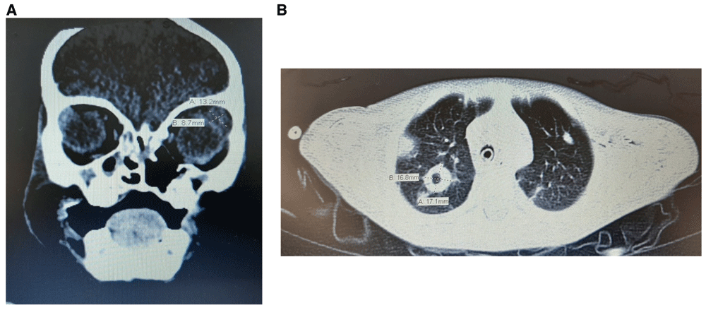
(A) maxillofacial CT scan showing ulcerating lesion in the left inner canthus with erosion of the left nasal bone. Left intra-orbital upper lateral cystic lesion measuring 14 * 12 * 7 mm in its maximal diameters. (B) Well defined right side pulmonary nodule measuring 19*22 mm in maximal diameter.
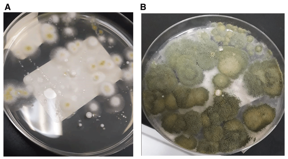
(A) Early fungal after 24 hours, where growth is still not identified. (B) Late fungal growth after 90 hours of incubation 30°C in a humidified environment.
This case describes mixed fungal infection in severely immunocompromised patient after chemotherapy. The infection started local and superficial till ended up with candida septic shock. Early diagnosis of fungal infection is critical to effective treatment that improve the outcome4. Diagnosis of fungal infection can be confirmed by either etiologic agent is isolated from clinical sterile specimens by culture or by histopathological diagnosis of a specimen or biopsy5. In our case we found it difficult to take any tissue biopsy due to rapid deterioration of patient’s general condition which is one of the limitations in our work. Mixed fungal infection is a rare condition, it was reported in 2014 in a case with severe hand injury after corrective surgery6. Other study reported only three mixed fungal infections from 178 immunocompromised pneumonia patients7.
Early and aggressive treatment of invasive fungal infection is very important to improve the outcome. A high index of suspicion is needed, especially with few antifungal agents available and increasing resistance8. Early administration of antifungal combination may salvage the patient’s condition. But in our case the patient died due to his immunocompromised condition.
Neutropenic patients are greatly vulnerable to life threatening fungal infections. Aggressive, early administration of double antifungal drugs may be needed in mixed fungal infection.
All data underlying the results are available as part of the article and no additional source data are required.
Written informed consent for publication of their clinical details and clinical images was obtained from the parents of the patient.
| Views | Downloads | |
|---|---|---|
| F1000Research | - | - |
|
PubMed Central
Data from PMC are received and updated monthly.
|
- | - |
Provide sufficient details of any financial or non-financial competing interests to enable users to assess whether your comments might lead a reasonable person to question your impartiality. Consider the following examples, but note that this is not an exhaustive list:
Sign up for content alerts and receive a weekly or monthly email with all newly published articles
Already registered? Sign in
The email address should be the one you originally registered with F1000.
You registered with F1000 via Google, so we cannot reset your password.
To sign in, please click here.
If you still need help with your Google account password, please click here.
You registered with F1000 via Facebook, so we cannot reset your password.
To sign in, please click here.
If you still need help with your Facebook account password, please click here.
If your email address is registered with us, we will email you instructions to reset your password.
If you think you should have received this email but it has not arrived, please check your spam filters and/or contact for further assistance.
Comments on this article Comments (0)