Keywords
Xanthogranulomatous, Prostatitis, Adenocarcinoma
Xanthogranulomatous, Prostatitis, Adenocarcinoma
A variety of benign conditions of the prostate can clinically mimic prostatic adenocarcinoma, thus raising a diagnostic problem. Xanthogranulomatous prostatitis, a rare form of non-specific granulomatous prostatitis, is considered among those pathologies that can clinically, biochemically and even radiologically behave as a prostate carcinoma1,2. Symmers was the first author to mention this disease in 19503, while Miekoś et al. produced the first case report in Poland in 19864,5. Very few similar cases have since then been described in the literature.
We report an additional case of a patient with initial clinical suspicion of prostatic carcinoma, but whose histopathological report revealed a xanthogranulomatous prostatitis.
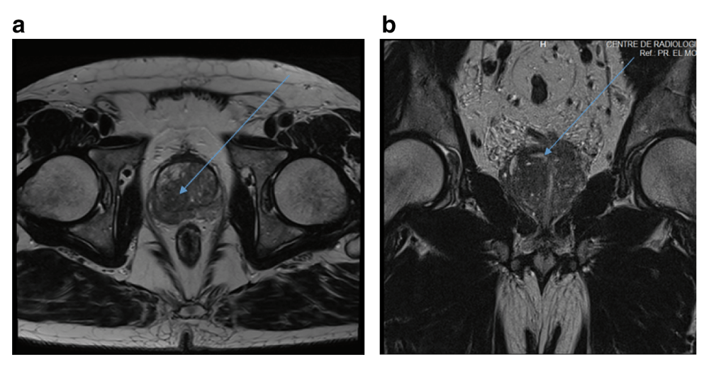
The patient is a 62 year old diabetic male, with no particular medical history, whose main symptoms started 3 months ago with urinary incontinence, increased urinary frequency and urgency, alongside haematuria. The patient also had micturition burning, which came with a recent onset of fever. Digital rectal examination (DRE) was indolent and revealed an asymmetric and enlarged prostate, with a hard fixed nodule on the right lobe. The patient’s general physical examination was normal, and revealed no alterations of the penis, the testicles or the epididymis. A prostate-specific antigen (PSA) test was performed and showed an increase in serum PSA level that reached 43.97 ng/ml (normal level <4 ng/ml). Urine analysis were within normal limits with no growth in urine culture. A transabdominal ultrasonography showed a normal upper renal tract and a normal bladder wall, with a post-micturition residual volume of 48.21 ml. Transrectal ultrasound (TRUS) was not performed, as it was too painful for the patient to sustain. From the clinical, radiological and biochemical data, a locally advanced prostate carcinoma was suspected. A multiparametric magnetic resonance imaging (mpMRI) of the prostate was then performed. The MRI showed an enlarged prostate, with an estimated weight of 51 grams. Two foci were found (Figure 1), which were classified as PI-RADS 4 lesions, thus requiring histological examination. There was no infiltration of the peri-prostatic fat.

(a) The first focus, of 18 mm, was located in the central zone of the prostate, in the right posterolateral region (blue arrow), with a low signal intensity on T2-weighted images. (b) The other focus was a 22 mm nodule located in the transitional zone of the prostate (blue arrow). Both were classified as PIRADS 4 lesions.
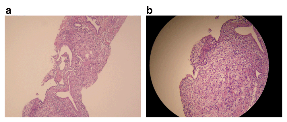
A TRUS guided prostate biopsy was then performed. A systematic 12-core prostate biopsy was realized, alongside three more core samples on zones that were found suspicious on the MRI images. Histological examination of the 15 needle biopsy core samples showed glandular atrophy, fibrosis, and an accumulation of inflammatory cells including polymorphs with eosinophils and neutrophils, with prostatic abscess, as well as the presence of foamy macrophages (lipid-laden histiocytes), also known as xanthoma cells (Figure 2). Tissue cultures and immunohistochemistry tests were not performed. The histopathological examination concluded a suppurated xanthogranulomatous prostatitis, with no evidence of malignancy.

(a) Prostatic parenchyma with abscess and a polymorph inflammatory infiltrate of neutrophils. (b) Xanthomatous infiltrate with foamy histiocytes, with no evidence of malignancy.
Conservative treatment was chosen, and the patient was given Ciprofloxacin for 4 weeks (500mg, twice a day). After three months, a PSA test was performed, and showed a significant decrease in PSA that reached 7.27 ng/ml (compared to the initial 43.97 ng/ml). A transabdominal ultrasonography showed a decrease in prostate volume, with an estimated prostate weight of 31 grams (versus 51 grams previously). A close follow-up will be needed for this case, with a clinical and biochemical check-up every trimester, until PSA levels are within normal limits (<4 ng/ml).
Benign conditions that mimic prostate carcinoma have been divided into six groups5, among them, inflammatory diseases. Granulomatous prostatitis is an unusual prostatic entity that was first described by Tanner and McDonald in 19436 and classified in 1984 by Epstein and Hutchin7 into five groups, based on aetiology and histopathology: idiopathic (non-specific), infectious (specific), malakoplakia, iatrogenic, and cases associated with systematic diseases and allergy3,7,8. Infectious agents that have been encountered in specific granulomatous prostatitis are Mycobacterium tuberculosis, Treponema pallidum, as well as some fungi and viruses3,8. Specific granulomatous prostatitis may also be due to intravesical Bacillus Calmette-Guerin (BCG) therapy for bladder cancer8. Xanthogranulomatous prostatitis is an uncommon form of non-specific granulomatous prostatitis. The aetiology of xanthogranulomatous prostatitis is still unclear; it has been often associated with hyperlipidemia (in our case, the patient had no history of hyperlipidemia) or recurrent urinary tract infection8, although some authors have considered that it might be caused by an autoimmune disease with a HLA-DR15-linked T-cell response against proteins in prostatic secretions4,9,10. Bostwick and Chang brought the theory of ductal obstruction, speculating that blockage of prostate ducts and stasis of secretions cause cellular debris, bacterial toxins, prostatic secretions, sperm, and semen to escape into the stroma through the destroyed epithelium, eliciting a localized inflammatory response4,9,10.
Xanthogranulomatous inflammation occurs very rarely in the prostate. It is vastly known in the kidneys and gallbladder, and has been described in other anatomic sites, such as the mandible, retro peritoneum, third ventricle, choroid plexus, orbit, vagina, lung, stomach, pericardium, and ovary10. Less than 20 cases have been reported in the literature since its first description4,9 with a similar case of suppurated xanthogranulomatous prostatitis recently discovered also in Morocco9.
Histologically, the typical lesion in granulomatous prostatitis is a large inflammatory nodular infiltrate of epithelioid histiocytes, plasma cells, lymphocytes and sometimes polymorphs with eosinophils5,9,11. The specific and distinctive feature of xanthogranulomatous prostatitis is the presence of foamy macrophages (lipid-laden histiocytes), also known as xanthoma cells5,9,11. Immunohistochemistry tests reveal T-lymphocytes in close association with damaged epithelium while B-lymphocytes occur in a more peripheral location or form follicular structures9,11. The presence of xanthoma cells may cause diagnostic confusion with high-grade prostatic carcinoma3,9,12, especially with the hypernephroid pattern of prostate carcinoma (Gleason 4B)3, as well as clear cell carcinoma12. A panel of immunohistochemistry tests, such as cytokeratin, PSA, prostatic acid phosphatase (PAP), leukocyte common antigen (LCA) and CD68 can be useful in differentiating between these two conditions, by showing results more consistent with an inflammatory process3,9,11. Those tests were not performed in our case, as the foamy macrophages were judged sufficient for the histopathological diagnosis of this condition.
In our case, even though all the signs were pointing towards prostate adenocarcinoma, we decided not to rush the diagnosis. Multi-parametric MRI was very helpful, as it allowed us to locate the suspicious foci. We decided then to perform a 15-core needle biopsy, rather than the systematic 12-core biopsy that is usually performed for prostate carcinoma, to increase our chances of finding pathological cells.
Xanthogranulomatous prostatitis causes serious confusion with prostate carcinoma when it comes to diagnosis. It occurs usually in elderly men, usually in the sixth decade3,8. In most cases, xanthogranulomatous prostatitis is diagnosed incidentally on TURP chips or needle biopsy3,8,9. Clinically, the symptoms are either those of urinary obstruction, with low urinary tract symptoms, or severe low urinary tract infection symptoms. Two recent studies have shown that the most encountered symptoms were increased urinary frequency and urgency, sometimes with dysuria, micturition burns, haematuria and urinary incontinence3,8. Few episodes of fever and chills have been reported8. On DRE, it is difficult to distinguish from prostate carcinoma, as the prostate feels hard and nodular3,8. In some cases, DRE finds an asymmetry or an enlargement of the prostate. In addition, xanthogranulomatous prostatitis may cause a transient increase in serum PSA level, which decreases with a resolution of the inflammation4,9,11. In some cases, PSA serum levels reach 150 ng/ml11. In our case, PSA decreased from 43.97 ng/ml to 7.27 ng/ml in the space of 3 months. Furthermore, no imaging (TRUS, MRI) can differentiate between xanthogranulomatous prostatitis and prostate adenocarcinoma, given the radiological similarities, and the absence of a specific feature of xanthogranulomatous prostatitis1,2. In a recent study, it has been shown that 63.6% of patients with PI-RADS (V2) 4 lesions who underwent transrectal biopsy were diagnosed with prostate cancer13. Hence, the only way to differentiate between these two conditions is histopathological examination.
Xanthogranulomatous prostatitis can occur in a normal gland, nodular hyperplastic gland or carcinomatous prostate. It is mostly located in the peripheral or transitional zone3,8. In some cases, xanthogranulomatous prostatitis can co-exist with prostate carcinoma3,8,11.
Unlike prostatic adenocarcinoma, conservative management is the rule for xanthogranulomatous prostatitis8,9. Inflammation is often self-limiting and resolves slowly over time. Surgical management may be considered if there is failure of conservative treatment, and might be needed in case of severe low urinary tract symptoms or due to occurrence of complications that may require radical prostatectomy4,9,10. However, surgical treatment of xanthogranulomatous prostatitis can lead to complications like vesical neck contraction, and might require repeated resections12.
Long-term follow-up is needed for patients with xanthogranulomatous prostatitis, as they require a regular clinical and biochemical check-up. MRI in xanthogranulomatous prostatitis follow-up is not required, but it might be necessary if DRE remains suspicious, or if there is no decrease in PSA serum levels. In addition, benign prostatic hyperplasia and prostatic adenocarcinoma can still occur in patients who previously dealt with xanthogranulomatous prostatitis3,8,11, thus making long-term follow-up inevitable.
Xanthogranulomatous prostatitis is an uncommon inflammatory pathology that can mimic prostatic carcinoma both clinically and/or biochemically. Furthermore, no radiological features can help differentiate between these two conditions. Precise histopathological examination is essential for the final diagnosis of xanthogranulomatous prostatitis.
Given clinical, biochemical and imaging similarities with prostatic carcinoma, as well as the rare nature of this condition, patients with xanthogranulomatous prostatitis raise a major diagnostic issue. Conservative treatment is the rule, with long-term follow-up needed, especially in patients with persisting elevated serum PSA values.
Written informed consent was obtained from the patient for the publication of this case report and accompanying images.
All data underlying the results are available as part of the article and no additional source data are required.
| Views | Downloads | |
|---|---|---|
| F1000Research | - | - |
|
PubMed Central
Data from PMC are received and updated monthly.
|
- | - |
Is the background of the case’s history and progression described in sufficient detail?
Yes
Are enough details provided of any physical examination and diagnostic tests, treatment given and outcomes?
Partly
Is sufficient discussion included of the importance of the findings and their relevance to future understanding of disease processes, diagnosis or treatment?
Yes
Is the case presented with sufficient detail to be useful for other practitioners?
Partly
Competing Interests: No competing interests were disclosed.
Reviewer Expertise: Histopathology
Is the background of the case’s history and progression described in sufficient detail?
Yes
Are enough details provided of any physical examination and diagnostic tests, treatment given and outcomes?
Yes
Is sufficient discussion included of the importance of the findings and their relevance to future understanding of disease processes, diagnosis or treatment?
Yes
Is the case presented with sufficient detail to be useful for other practitioners?
Yes
Competing Interests: No competing interests were disclosed.
Reviewer Expertise: Genitourinary Pathology.
Alongside their report, reviewers assign a status to the article:
| Invited Reviewers | ||
|---|---|---|
| 1 | 2 | |
|
Version 1 22 Oct 19 |
read | read |
Provide sufficient details of any financial or non-financial competing interests to enable users to assess whether your comments might lead a reasonable person to question your impartiality. Consider the following examples, but note that this is not an exhaustive list:
Sign up for content alerts and receive a weekly or monthly email with all newly published articles
Already registered? Sign in
The email address should be the one you originally registered with F1000.
You registered with F1000 via Google, so we cannot reset your password.
To sign in, please click here.
If you still need help with your Google account password, please click here.
You registered with F1000 via Facebook, so we cannot reset your password.
To sign in, please click here.
If you still need help with your Facebook account password, please click here.
If your email address is registered with us, we will email you instructions to reset your password.
If you think you should have received this email but it has not arrived, please check your spam filters and/or contact for further assistance.
Comments on this article Comments (0)