Keywords
ER, PR, HER2neu, CISH, breast carcinoma, FNAC, cell block, IHC
ER, PR, HER2neu, CISH, breast carcinoma, FNAC, cell block, IHC
Breast cancer is a national health problem in Iraq and a serious health concern in all countries1. The early detection of breast cancer represents a major challenge for oncologists, pathologists, and surgical oncologists1. Fine-needle aspiration biopsy (FNAB) is less invasive than a core needle biopsy but may provide insufficient tissue for identifying specific markers and phenotypes1. As an advanced molecular technique, immunohistochemistry (IHC) can be used to assess tumor subtypes based on histo-chemical markers2 and is routinely used in the work-up of breast cancer3. The FNAB method allows the retrieval of small tissue fragments from a fluid specimen that can be embedded in a paraffin block, sectioned, and analyzed4,5. Immunohistochemical detection of hormone receptors (HR) is part of the regular work-up of breast cancer management6. Human EGF receptor 2 (HER2/neu) was one of the first oncogenes identified in samples of invasive cancer and is found in 10%–20% of women with breast cancer. It is an indicator for sensitivity to Herceptin (trastuzumab)7,8. The presence of Her2 can be detected by IHC using either chromogenic in situ hybridization (CISH) or fluorescence in situ hybridization (FISH). CISH resembles FISH in that they are both in situ hybridization techniques used to identify the presence or absence of specific DNA sequences; however, CISH is a much more practical technique in diagnostic laboratories as it requires a bright-field microscope in contrast to the more costly and complex fluorescence microscope required for FISH9.
The present study was conducted to assess the use of FNABs embedded in cell blocks to determine their reliability in assessing HR status and HER2/neu status by IHC compared with standard histological staining and examination of tissue block sections.
We enrolled 67 female participants, of whom 50 completed the study. These were women who attended our clinic for a consultation. They had been diagnosed with breast cancer at our hospital clinic and then visited our institute for a further diagnosis of detected breast masses.
The patients were referred from the Primary Breast Cancer Detection Clinic (PBCDC) screening program in Medical City to our clinic. Although this program is offered at the clinic, it is mainly carried out at our center because we can provide a more accurate diagnosis of breast cancer and offer further assurances about a diagnosis, being oncologists. The primary focus of the screening program is the early diagnosis of breast cancer. Each participant had a file containing records of dates and times of visits, investigations performed, and the date of their next appointment. Detection of breast masses was either due to a patient finding a lump and presenting at the clinic themselves or due to a lump being found by clinic staff during a routine visit. Further assessments included bilateral breast and axilla ultrasonography, mammography, and magnetic resonance imaging (MRI) of the breasts. Following this, FNAC or FNAB was performed on the masses and samples were sent for histopathology. All participants were women diagnosed with breast cancer for the first time. Some patients required further assessment in the form of IHC analysis to determine their eligibility for hormonal or anti-Her2 therapy. Once all diagnoses were complete, we recruited women who consented to inclusion in the study. All patients who presented from 1 April 2017 to 30 November 2017 who met the study criteria and voluntarily gave informed consent were included.
Written, informed consent from all study participants was obtained in accordance with the Helsinki Declaration of 1975, as revised in 2000, at the time of their visit to the clinic. The Medical Ethical Committee of the Iraqi Board for Medical Specializations approved this work (No. 2015; date: 24/03/2017).
Each of the 50 (initially 67) women, aged 42 to 66 years, who participated in the study frequently visited our clinic for either hormonal or chemotherapy treatment. Of the 17 patients lost to follow-up, the reasons included: Patients changed their place of clinical management (n=4), patients refused treatment at our hospital (n=5), patients decided to complete their treatment outside of Iraq (n=4), and patients changed oncologist (n=4).
The inclusion criteria were: Women aged 18-years or more, novel breast cancer diagnosis, provided written, informed consent, and had breast cancer independent of any other primary tumors. (The presence of other primary tumors could lead to the misinterpretation of clinical findings and mismanagement of cases). The exclusion criteria were: Other primary tumor(s), male breast cancer (due to its rarity and its need for special interventions), metastatic breast cancer, and inflammatory breast cancer (due to the need for urgent management).
Following a diagnosis of breast cancer by FNAC by a histopathologist, patients were sent for surgery. This involved either a mastectomy or a lumpectomy based on the stage of the tumor. Women with early-stage breast cancer (stage I or II) they were sent first for surgery, then chemotherapy was planned, and they were given an appointment for radiotherapy. Women with locally advanced breast cancer, or stage III patients, received neoadjuvant chemotherapy before being sent for surgery and then underwent radiotherapy.
Fine-needle aspirate (FNA) samples were obtained from participants upon presentation once the clinical and radiological examinations had been performed. Following the initial diagnosis, participants were sent to our laboratory for FNA sample collection. The samples were analyzed by a histopathologist who also wrote a histopathology report about each patient undergoing FNA. Needle core samples were taken for more accurate staging of breast cancer or if the FNA sample was insufficient. Needle core samples can be obtained either before or after treatment to confirm the diagnosis and staging or after treatment has begun in patients with advanced-stage cancer when they start neoadjuvant chemotherapy. All patients underwent FNA because this analysis process represents the main part of the diagnostic process, and thus happens once a woman has attended our clinic. This protocol was not changed (a patient was not started on any type of treatment if no histopathology analysis had been undertaken). In cases of advanced inflammatory breast cancer neoadjuvant chemotherapy is required immediately, in these cases FNA samples may need to be taken post-treatment. We did not observe any effects of neoadjuvant treatment on the histochemical properties of tumors, since the benefits of neoadjuvant treatment (which is mainly used in patients with locally advanced stages) are to delay the development of local recurrence and to decrease treatment failure rates. Such treatment will also result in the downstaging of a tumor, getting close to the resection negative margin, and making a tumor mass more operable for radical surgery. All histopathology was performed by experienced histopathologists, and we also received written reports from them which contained both patient and tumor data, including a patient’s age, address, stage, grade, number of samples, and the gross and microscopic appearance of the tumor. Cell blocks were prepared using the collodion bag technique5.
This study depended on histopathology reports and/or IHC results and extracted HR and Her/2neu information. The authors did not collect any demographic data. FNAC was performed by routine immune-staining of paraffin block sections for HR and HER2/neu and the results were compared with diagnoses made using stained tissue block sections.
Based on the biopsy results of histopathology findings in reports and the tumor work-up following clinical examination of the breast and axilla of the affected and healthy side, ultrasonography, mammography, and breast magnetic resonance imaging (MRI) were also performed if required to confirm the diagnosis and to evaluate and assess each patient for any locoregional spreading or metastasis elsewhere in the body (this is routine workup that is carried out for each patient), which is a routine steps perform for each patients.
The decision to use either FNAC, core biopsy, or both was influenced by different factors, including the following: lesion size; palpable clinical features of masses; lesion features identified from imaging, such as mass, architectural distortion, asymmetric density, or micro-calcifications; the clinician performing the procedure; the availability of a histopathologist; the need for a hormone receptor assay or tumor marker studies in inoperable cases; consideration for management protocols.
FNA was performed using a 23-gauge needle. After use, the needles and syringes were rinsed with 10 mL 50% (v/v) ethanol (Science Ltd., Catalogue No.: C15K30002-00RE200-L3), and the cell suspension was placed in a 10-mL disposable centrifuge tube and pelleted by centrifugation at 4,000 rpm for 6 min. The supernatant was removed and pellets were fixed and stained with 10% buffered formalin (Sigma-Aldrich, Catalogue No.: F-10NB1G-123456) containing hematoxylin (Sigma-Aldrich, Catalogue No.: 685042) and eosin (Science Ltd., Catalogue No.: E10022661208) for 30–45 minutes at room temperature. The fixed material was filtered, and the fragments were processed for tissue sampling10. Collodion reagent (Sigma-Aldrich, Catalogue No.: E68562K31) was poured into 15-mL glass tubes until they were full. Then, the collodion was left to stand in the tubes for 10–15 min. After that, we poured the collodion back into the reagent bottle, rotating the tubes while pouring. The tubes were placed upside-down in a test-tube rack and left to stand for a further 30 min until the collodion had dried. Once the collodion had dried, the tubes were filled with distilled water. We discarded the distilled water just before use. The collodion bag can be stored for up to one-week5.
Tissue blocks were prepared by embedding samples in paraffin wax (BDH Ltd., Catalogue No.:0008-GFSD), Xyline (BDH Ltd., Catalogue No.:0009-HTCR) and water soluble glycol’s solution (Sigma-Aldrich, Catalogue No.: H-50M1H-01859) with Resin B compound (Sigma-Aldrich, Catalogue No.: H-629110AA-00505) to ensure a good specimen matrix for cryo-state sectioning by CRYOCUT 1800 (YU JIE / Japan/ Model:120ZB BB, Catalogue No.: 11S0B66090ZV). The aims for FNA slide preparation is to get a thin smear that is not subject to crush artifacts, and to allow rapid drying of air-dried slides and quick fixation of wet-fixed slides10. A drop of the aspirated fluid is placed onto two of the pre-labelled glass slides (ATACO / China, Catalogue No.:202441). The slides are then immediately placed into a Coplin jar (JACK Med./ China, Catalogue No.:ME-60166RS) containing 95% (v/v) ethanol (Science Ltd., Catalogue No.: C20K30004-11RE210-L5) for five minutes, to avoid excessive cell shrinkage. The slides were then dried by gently waving them in the air or by using a hair dryer (CROUN/MARTA TRADE INC., Catalogue No.:195220) on a cold or low heat setting. The slide then placed in 25 mL 15% (v/v) methanol (Science Ltd., Catalogue No.: C30K40004-22RE400-L8) for at least 30 seconds. Core biopsy slides were fixed in 10% buffered formalin (Sigma-Aldrich, Catalogue No.: F-10NB1G-123456) for 3–4 hours. Tissue was routinely processed. While practices vary, more common practices include cutting approximately 10 serial ribbons and staining several ribbons at regular intervals, for example, 1, 5 and 10, or staining at least three levels with haematoxylin (Sigma-Aldrich, Catalogue No.: 685042) and eosin (Science Ltd., Catalogue No.: E10022661208) (H&E) stains.
The cell block process was started by pipetting a specimen into a collodion coated test tube, then centrifuging it for 10 min at 2,500 rpm. The supernatant was removed using a pipette, and the edge of the collodion bag was placed on the lip of the tube using forceps. The bag was then lifted from the tube with forceps and tied with cotton string just above the pellet. Excess collodion bag and string was cut away, the remaining bag was placed in a tissue cassette, and then the cassette was placed in a specimen cup filled with 10% neutral buffered formalin (Sigma-Aldrich, Catalogue No.: F-10NB1G-123456). The specimen was then subjected to routine processing in the histopathology laboratory5,10.
Sample analysis was performed by a histopathologist doctor in Lab by comparing to IHC control slides (Cell Signaling Technology, Danvers, USA) by fixing and embedding the tissue, then cutting and mounting the section, followed by de-paraffinizing and rehydrating the section.
Air-dried slides were fixed in methanol (Science Ltd., Catalogue No.: C30K40004-22RE400-L8) for 2 minutes, allowed to dry and then immersed in a stain. Immunohistochemical staining was performed using a rapid Romanovsky stain (Sigma-Aldrich, Catalogue No.: 890601), Papanicolaou stain (Sigma-Aldrich, Catalogue No.: 110399) or haematoxylin (Sigma-Aldrich, Catalogue No.: 685042) and eosin (Science Ltd., Catalogue No.: E10022661208) (H&E) stains. IHC antibodies used in the analysis process were:
1. Anti-estrogen receptor (ER) rabbit monoclonal primary antibody (Roche Medical Systems, Inc., Catalogue No.: 790-4325 SP1) It was diluted as 1:100 in 0.05 M Tris-HCl (GoldBio com., Catalogue No.: T-099) with 0.10% ProClin 300 (Sigma-Aldrich, Catalogue No.: 669400R).
2. Anti-progesterone receptor (PR) rabbit monoclonal primary antibody (Roche Medical Systems, Inc., Catalogue No.: 790-4296 1E2) It was diluted as 1:200 in 0.1M phosphate buffered saline (Sigma-Aldrich, Catalogue No.: 980221M) with 0.05% ProClin 300 (Sigma-Aldrich, Catalogue No.: 659011Y).
3. Anti-HER-2/neu rabbit monoclonal primary antibody (Roche Medical Systems, Inc., Catalogue No.: 790-2991 4B5) It was diluted as 1:100 in 0.05 M Tris buffered saline (Sigma-Aldrich, Catalogue No.: 782330L), 0.01 M EDTA (RPI Ltd., Catalogue No.: E68820-372-2), 0.05% Brij-35 (Sigma-Aldrich, Catalogue No.: 433067H) with 0.05 % sodium azide (Sigma-Aldrich, Catalogue No.: 551108K).
Excess stains were discarded, and the slides rinsed in buffered water (BDH Ltd., Catalogue No.:0011-PODR). The slides were left to drain thoroughly in vertical position and allow them to dry. After that the dehydrating and stabilizing apply. Finally, viewing the staining under the microscope. Reagents:
Peroxidase-blocking reagent (Sigma-Aldrich, Catalogue No.: 559012)
DAB buffered substrate (Sigma-Aldrich, Catalogue No.: 859331)
5% di-amino-benzidine tetra-hydrochloride chromogen solution (Sigma-Aldrich, Catalogue No.: 859221)
Epitope retrieval solution (Bio Mark Pte. Ltd., Catalogue No.: PS003-KD)
Wash buffer (Azure Biosystems crop., Catalogue No.:AC211300)
Ammonium hydroxide (Science co., Catalogue No.: CSA1033-32610051822)
Equipment:
Slides (ATACO / China, Catalogue No.:99111A0350)
Coverslips (ATACO / China, Catalogue No.:98111A8044)
Oven (Memmert/ Germany, Catalogue No.: 1505456300123)
Coplin jars (JACK Med./ China, Catalogue No.:ME-60166RS)
Timer (YU JIE / Japan, Catalogue No.:A1H118DD)
Water bath (Gemmy industrial corp. /Taiwan, Catalogue No.:8996406933110)
Data tabulation and input was performed using IBM© SPSS© V 22. Sensitivity, specificity, and discrepancy rates were analyzed using Spearman’s rank-correlation analysis (ρ) and test of agreement using Cohen’s kappa coefficient (κ).
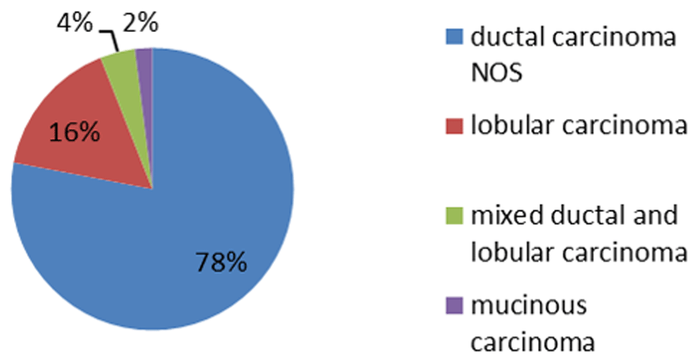
The participants were aged from 42 to 66 years, with women of different ages presenting with different tumor phenotypes (Table 1 and underlying data11). Tissue biopsies, examined either directly or embedded in paraffin blocks, sectioned, and analyzed for breast cancer markers, revealed that the most common type of cancer identified by either technique was ductal carcinoma (Figure 1). Using the cell block method for IHC, 11 cases were diagnosed as HER2/neu-positive, while 39 cases were diagnosed as HER2/neu-negative. Five patient biopsies gave false-negative results when compared with the results from IHC of the tissue block sections (Table 2). In total, 31 cases were diagnosed as ER-positive, and 19 cases were diagnosed as ER-negative, with two false-positive and three false-negative results, when compared with the results of tissue-block histopathology (Table 2). Immunostaining of cell-block sections revealed 12 PR-positive cases and 38 PR-negative cases, with four false-negative results and no false positives. The correlation between cell-block sectioning/IHC and tissue-block staining is shown in Table 3. For HER2/neu, there were 11 patients positive for both cell- and tissue-block, while 34 were negative. For HR, 29 were positive for ER and 16 were negative, whereas PR was positive in 15 and negative in 34 stains. Different H & E stain histopathology images are shown in Figure 2 (raw images are available as underlying data12).
| Mean ± SD | F | p value | |
|---|---|---|---|
| Ductal | 53.13 ± 6.74 | 0.356 | 0.785 |
| Lobular | 51.80 ± 3.23 | ||
| Mixed | 54.00 ± 3.93 | ||
| Mucinous | 54.00 ± 9.64 |
| HER2neu | Cell block | Total | ||
|---|---|---|---|---|
| Positive | Negative | |||
| Tissue block | Positive | 11 | 5 | 16 |
| Negative | 0 | 34 | 34 | |
| Total | 11 | 39 | 50 | |
| ER | ||||
| Tissue block | Positive | 29 | 3 | 32 |
| Negative | 2 | 16 | 18 | |
| Total | 31 | 19 | 50 | |
| PR | ||||
| Tissue block | Positive | 15 | 4 | 16 |
| Negative | 0 | 34 | 34 | |
| Total | 15 | 38 | 50 | |
| ρ | k | Sensitivity | Specificity | Discrepancy | p value | |
|---|---|---|---|---|---|---|
| HER2neu | 0.774 | 0.749 | 68.75% | 100.0% | 10.0% | 0.0419 |
| ER | 0.786 | 0.786 | 90.62% | 88.89% | 10.0% | 0.022 |
| PR | 0.803 | 0.819 | 75.0% | 100.0% | 8.0% | 0.0408 |

Haemotoxylin and Eosin stain histopathology images: IDC on cell block H&E stain A) [10x], B) [20x], C) [20x clear infiltration to the adipose tissue], D) [40x clear infiltration to the adipose tissue], E) [IDC with desmoplasia], F) [Metastatic ductal carcinoma to the axillary LN], G) [ER score 6/8], H) [PR score 6/8], I) [PR score 4/8], J) [ER score 4/8], K) [ER moderate staining 4/8 on 20x], L) [PR weak staining 3/8 on 40x], M) [Her2/Neu score +++ 20x], N) [Her2/Neu score +++ 40x], O) [Her2/Neu score +++ 20x], P) [Her2/Neu score +++ 40x], Q) [Her2/Neu score ++ 20x], R) [Her2/Neu score ++ 40x]. ER - estrogen receptor, PR - progesterone receptor, HER2/neu - human EGF receptor 2.
The cell block results showed a total of 74% of tumors were ductal carcinoma, 16% were lobular, and 4% were mixed, with 6% being mucinous. The tissue block results differed: 64% were ductal, 16% were lobular, and 14% were mixed. This difference in results may be related to the disturbance of morphological features, which has been noted in previous studies such as the one by Makki6. The disparity may also be a result of the disturbance of morphological features and rearrangement of the malignant cells of lobular carcinoma by FNAC. The differentiation of in situ carcinoma from the invasive type cannot be differentiated by cell-block preparation, since the cells and basement membranes of the ducts cannot be differentiated. Our results are consistent with results from studies in various Western countries, which have indicated that invasive ductal carcinoma is the most common variant of invasive breast cancer9,13. The patients’ ages ranged from 42 to 66 years (mean ± SD = 52.94 ± 5.51 years). Patients with lobular breast cancer tended to be younger. These results were different from those obtained in Western countries7, where in situ breast cancer is detected more frequently than the invasive form because of more accurate and reliable screening. Developed countries generally have better screening programs compared with developing countries such as Iraq.
FNA cytology and core biopsy are the two main ways to diagnose and determine the optimal management of breast disease. Accurate procedures depend on an awareness of available techniques, utilization of better tests for a particular clinical setting, and the sensitivity, specificity, and interpretation of results in clinical practice.
These two procedures, FNA cytology and core biopsy, are sufficient for making a diagnosis and deciding whether surgery or other treatments should be applied8.
Some pathologists prefer to histologically evaluate core biopsies because they can be analyzed relatively quickly and easily, and they allow IHC to be applied. Cell blocks of breast FNAB offer these same advantages, while combining FNAB with core biopsies has been found to increase diagnostic accuracy9. Pinder et al.13 were among the first to confirm the value of analyzing multiple prognostic markers for breast cancer. Our data showed good concordance for HER2/neu and PR, with only moderate concordance for ER expression when comparing two blocks. The false-negative rate for HER2/neu amplification did not differ significantly between the two blocks. There was a strong, statistically significant positive correlation (p= 0.0419; ρ = 0.774) between the two blocks regarding HER2/neu status, and with moderate agreement (k = 0.749), 68.75% sensitivity and 100.0% specificity. Five cases were labeled as false-negatives according to the cell-block results.
Similar results were obtained by Bueno Angela et al.14 and Vohra et al.15. There was a strong, positive, statistically significant correlation (p value = 0.022; ρ = 0.786) between both blocks for the detection of ER status with moderate agreement (k = 0.786), 90.62% sensitivity and 88.89% specificity. Our findings differed slightly from the Bueno Angela study11, in which it was concluded that cell blocks provide a useful method for assessing HR and HER-2/neu mainly in inoperable and recurrent cases, but consideration should be given to carrying out FISH analysis on 1+ as well as 2+ HER-2/neu findings14; our results were similar, however, to those of Vohra et al12. There was a strong, positive, statistically significant correlation (p= 0.0408; ρ = 0.803) and strong agreement (k = 0.819) between the two blocks with respect to the detection of PR status, with 75.0% sensitivity and 100.0% specificity. These results were similar to those reported by Bueno Angela et al.14 but differed slightly from Vohra et al.15. Vohra et al.15 reported ER, PR, and HER2 determination on FNA-acquired cell blocks which showed excellent agreement with ER and HER2, and moderate agreement for PR with the corresponding tissue block. These findings support the equivalency of ER and HER2 evaluation performed on FNA cell blocks compared with surgical tissue blocks15.
It is possible that a small number of ER-positive, PR-negative cases could be missed by IHC when using a cell block, and therefore further research is needed to evaluate the significance of this possibility. One of the limitations of this study was its small sample size.
Other limitations included the time consumed in performing IHC analyses at the histopathology laboratory due to a lack of materials necessary for completing the procedures (most of the time these materials are lacking in our clinic and we send samples to a private clinic for analysis, which is more expensive and patients often cannot afford it). This causes delays in the completion of the IHC analysis we require, and sometimes the private laboratory loses samples, so we suggest that they are done outside of the country. Other challenges include the small number of pathologists available, poor facilities for the accurate recording of data, and ineffective screening programs. All of the above points may cause changes in the sequence of obtaining optimal work-up data for our patients, which is necessary if they are to have better outcomes from the management of newly diagnosed breast cancer. All above points may cause alteration in sequences of obtain optimal work-up for our patients to have a better results from management of newly diagnosed breast cancer.
The combined use of FNAC smears and cell blocks can be useful for establishing a more definitive diagnosis of breast cancer. HR and HER2/neu determination by IHC on sections from cell blocks was in close agreement with HER2/neu and PR, and in partial agreement with ER. These findings support the equivalency of these markers on cell blocks compared with tissue blocks.
Zenodo: Cell block vs tissue block. http://doi.org/10.5281/zenodo.350735611
This project contains the following underlying data:
Dr. Mazin Data (2).xlsx (Excel file of Patients age, breast cancer types, and results of cell and tissue block for ER, PR, and Her2neu)
Zenodo: Histological staining of samples [Clinico-pathology on newly diagnosed breast cancer with expression of ER, PR and HER 2neu in the cell block: observational study]. http://doi.org/10.5281/zenodo.333814612
This project contains the following underlying data:
10X.png (Raw microscope image, Invasive ductal carcinoma (IDC) on cell block H&E stains)
20 x clear infiltration to the adipose tissue.png (Raw microscope image, IDC cells infiltrated the adipose tissue on H&E stains)
20X.png (Raw microscope image, IDC on cell block H&E stains)
40 x clear infiltration to the adipose tissue.png (Raw microscope image, IDC cells infiltrated the adipose tissue on H&E stains)
ER moderate staining 4–8 on 20 x.png (Raw microscope image, IHC staining using the Anti-ER antibody showed distinct nuclear positivity in glandular cells in tumor cells in breast cancer)
ER score 4–8.png (Raw microscope image, IHC staining using the Anti-ER antibody showed distinct nuclear positivity in glandular cells in tumor cells in breast cancer)
ER score 6–8.png (Raw microscope image, IHC staining using the Anti-ER antibody showed strong nuclear positivity in glandular and stromal cells in tumor cells in breast cancer)
Her2score ++.png (Raw microscope image, IHC staining using the Anti-Her-2/neu antibody showed HER2-negative ductal carcinoma with no membranous positivity)
Her2Neu score +++ 20x.png (Raw microscope image, IHC staining using the Anti-Her-2/neu antibody showed strong membranous (combined with moderate cytoplasmic) positivity in tumour cells in HER2-positive ductal carcinoma)
Her2Neu score +++ 20x.png (Raw microscope image, IHC staining using the Anti-Her-2/neu antibody showed strong membranous positivity in tumour cells in HER2-positive ductal carcinoma)
Her2Neu score +++ 40x.png (Raw microscope image, IHC staining using the Anti-Her-2/neu antibody showed strong membranous positivity in tumour cells in HER2-positive ductal carcinoma)
Her2neu score ++.png (Raw microscope image, IHC staining using the Anti-Her-2/neu antibody showed HER2-negative ductal carcinoma)
IDC with desmoplasia.png (Raw microscope image, IDC cells grouped as desmoplasic fashion on H&E stains)
Metastatic ductal carcinoma to the axillary LN.png (IDC cells spreading to lymphatic tissue)
PR score 4–8.png (Raw microscope image, IHC staining using the Anti-PR antibody showed weak nuclear positivity in glandular cells in breast cancer)
PR score 6–8.png (Raw microscope image, IHC staining using the Anti-PR antibody showed strong nuclear positivity in glandular cells in breast cancer)
PR weak staining 3–8 on 40 x.png (Raw microscope image, IHC staining using the Anti-PGR antibody showed very weak nuclear positivity in glandular cells in breast cancer)
| Views | Downloads | |
|---|---|---|
| F1000Research | - | - |
|
PubMed Central
Data from PMC are received and updated monthly.
|
- | - |
Provide sufficient details of any financial or non-financial competing interests to enable users to assess whether your comments might lead a reasonable person to question your impartiality. Consider the following examples, but note that this is not an exhaustive list:
Sign up for content alerts and receive a weekly or monthly email with all newly published articles
Already registered? Sign in
The email address should be the one you originally registered with F1000.
You registered with F1000 via Google, so we cannot reset your password.
To sign in, please click here.
If you still need help with your Google account password, please click here.
You registered with F1000 via Facebook, so we cannot reset your password.
To sign in, please click here.
If you still need help with your Facebook account password, please click here.
If your email address is registered with us, we will email you instructions to reset your password.
If you think you should have received this email but it has not arrived, please check your spam filters and/or contact for further assistance.
Comments on this article Comments (0)