Keywords
Nodular colloid degeneration, Colloid milium, Nodular amyloidosis
Nodular colloid degeneration, Colloid milium, Nodular amyloidosis
Nodular colloid degeneration (NCD), also named paracolloid, is a rare subtype of colloid deposition disorders. It presents as a single or multiple soft to rubbery nodules or plaques usually on chronically sun-exposed skin. Histologically, there are masses of amorphous, eosinophilic material expanding the papillary dermis and extending into the deep dermis1. Nodular colloid degeneration is a rare disease. Only 12 cases have been previously reported in the literature. Here, we present a case of NCD that occurred on the face and discuss the topic of how to discriminate between NCD and other similar entities such as nodular amyloidosis.
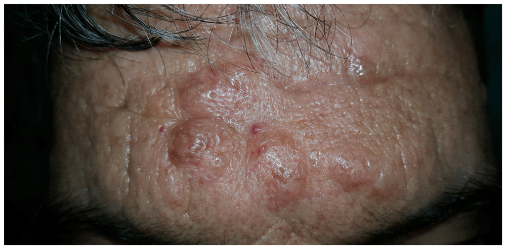
A previously healthy 58-year-old farmer from Hamedan province, Iran, presented to the dermatology clinic with a three year history of multiple papular and nodular lesions on his face. The lesions had been gradually enlarging during last six months. An examination of the skin carried out by a dermatologist revealed multiple skin-colored to yellow papules and nodules of different sizes (10–25mm) located on the forehead, cheeks, and nares (Figure 1 and Figure 2). The nodules were painless and mobile, they were rubbery-soft in consistency and were attached to the overlying skin. Findings of the systemic physical examination were otherwise unremarkable.

After local anesthesia using 2–3ml of intralesional lidocaine (2%), an excisional biopsy was obtained from forehead lesions. The specimen was put in formalin and was sent to the lab for routine hematoxylin-eosin stain and special stains (periodic acid-Schiff (PAS) staining, crystal-violet and Congo red staining).
Histopathologic examination revealed hyperkeratosis, flattened epidermis, and amorphous eosinophilic material deposits in the upper dermis, which were separated from the epidermis by a Grenz zone of collagen fibers and extended into the deep dermis. The hair follicles and sebaceous glands were well preserved. There were some irregular fissures and clefts in the hyaline material dividing this material into smaller islands. Scattered nuclei of fibroblast were aligned along the line of fissuring (Figure 3).

The amorphous colloid material shows numerous irregular clefts. Scattered nuclei of fibroblasts are observed in the colloid material (haemotoxylin and eosin staining, ×40).
The special stains revealed positive reactivity of materials with periodic acid-Schiff (PAS) staining but negative reactivity with crystal-violet and Congo red staining. According to clinical and histopathological findings, nodular lesions on sunexposed areas with colloid material deposition in the dermis, a diagnosis of nodular colloid degeneration was made.
For treatment, forehead lesions were excised and for the lesions on the nose, resurfacing using a Co2 laser (Jeisys Medical Korea) was applied with a frequency of 50kHz and pulse duration of 500μs. Unfortunately, the patient did not return for follow-up and we could not obtain data on treatment efficacy, cosmetic results or recurrence.
Colloid milium and colloid degeneration are rare degenerative cutaneous disorders, with at least four distinct clinicopathological variants. The four variants of this disease are as follows: adult colloid milium, juvenile colloid milium, pigmented colloid milium, and nodular colloid degeneration (para colloid)2.
In both the adult and juvenile forms, numerous yellow-brown, semitranslucent dome-shaped papules are developed in areas of chronic sun exposure; for example, the cheeks, ears, neck, and dorsum of the hands. In pigmented colloid milium, gray to black grouped or confluent papules are present on the face, following the excessive use of topical hydroquinone for skin bleaching2.
NCD clinically presents with solitary or multiple asymptomatic nodules or plaque-like lesions usually in chronically sun-exposed areas, particularly the face. The lesions have been rarely reported on sun protected areas such as penile skin3. Although the exact etiologic role of chronic sun exposure remains uncertain, it is suggested that photo-induced damage to dermal elastic fibers, as in solar elastosis, produce the colloid4.
Clinically, the differential diagnosis of NCD includes nodular amyloidosis, nodular mastocytosis, nodular histiocytosis, steatocystoma multiplex, sarcoidosis, lepromatous leprosy, leishmaniasis, cutaneous lymphoma, and pseudolymphoma1,2. A histopathologic workup is a helpful tool in establishing the appropriate diagnosis. Upon histopathologic examination, an amorphous eosinophilic material with small fissure and clefts is observed in the dermis, separated from the epidermis by a Grenz zone of narrow collagen tissue2,5.
Histologically, NCD must be distinguished from lipoid proteinosis, different types of colloid milium, and amyloidosis. In cutaneous lesions of lipoid proteinosis, there are masses of amorphous or laminated hyaline-like PAS-positive deposits in the dermis that have the striking perivascular pattern and also can surround the eccrine glands as well as the hair follicles and sebaceous glands6.
In juvenile colloid milium, amorphous eosinophilic deposits are found in the dermis next to basal layer without Grenz zone. In adult colloid milium, nodules of homogenous eosinophilic colloid material expand the papillary dermis and extend into the mid dermis. Fissures and clefts divide this material into smaller islands and evidence of solar elastosis are commonly seen beneath the nodules. The Grenz zone is often present in the dermis. A pigmented colloid milium is similar to adult colloid milium but contains an area of yellow-brown islands of colloid material in the upper dermis. Nodular colloid degeneration is distinguished from adult colloid milium and juvenile colloid milium by more deeply extended and less conspicuous clefting of amorphous materials within the dermis2,7.
The main histologic differential diagnosis of nodular colloid degeneration includes nodular amyloidosis. In nodular amyloidosis, there are large masses of amyloid in the dermis and subcutaneous fat, accentuate around deep vascular channels, adnexal structures, and infiltrate blood vessel walls8. Infiltration of plasma cells are usually present in and around the amyloid deposition7.
There is a considerable resemblance between colloid milium and amyloidosis, as both may stain positively for Congo red, PAS and crystal violet, and produce fluorescence with Thioflavin-T. In contrast to colloid, amyloid reacts with Pagoda-red and other cotton dyes8. Moreover, infiltrate of plasma cells and positive immunostaining for light chain deposition can aid in the differentiation of amyloidosis from NCD8.
To improve the appearance of the lesions, cryotherapy, dermabrasion, erbium:YAG or carbon dioxide laser resurfacing have been reported as effective methods, although lesions may recur. Considering the pathogenesis of NCD, sun protection can be effective in preventing NCD2. In this case, we used excision and Co2 laser resurfacing to flatten the lesions. Unfortunately, the patient did not return for follow-up and we could not obtain data on treatment efficacy, cosmetic results or recurrence.
In summary, we encountered a rare case of nodular colloid degeneration arising on the face, which was diagnosed by histopathological examination. Although it is rare, the clinical diagnosis of nodular colloid degeneration should be considered in differential diagnosis of any cases presenting with facial nodules.
All data underlying the results are available as part of the article and no additional source data are required.
Written informed consent for publication of their clinical details and clinical images was obtained from the patient.
| Views | Downloads | |
|---|---|---|
| F1000Research | - | - |
|
PubMed Central
Data from PMC are received and updated monthly.
|
- | - |
Provide sufficient details of any financial or non-financial competing interests to enable users to assess whether your comments might lead a reasonable person to question your impartiality. Consider the following examples, but note that this is not an exhaustive list:
Sign up for content alerts and receive a weekly or monthly email with all newly published articles
Already registered? Sign in
The email address should be the one you originally registered with F1000.
You registered with F1000 via Google, so we cannot reset your password.
To sign in, please click here.
If you still need help with your Google account password, please click here.
You registered with F1000 via Facebook, so we cannot reset your password.
To sign in, please click here.
If you still need help with your Facebook account password, please click here.
If your email address is registered with us, we will email you instructions to reset your password.
If you think you should have received this email but it has not arrived, please check your spam filters and/or contact for further assistance.
Comments on this article Comments (0)