Keywords
breast cancer, sarcoidosis, mediastinal lymph adenopathy, positron emission tomography
breast cancer, sarcoidosis, mediastinal lymph adenopathy, positron emission tomography
Breast cancer is the most common cancer in women1. It is a highly curable cancer, and with proper treatment protocols, the five-year survival of Stage IV disease is 22%2. The overall survival of a breast cancer patient depends on the stage of disease with visceral or bony metastasis; therefore at the time of initial treatment planning, it is highly important to determine the intent, which is either curative or palliative. The oncological team exerts the utmost effort to properly stage cancer by giving attention to history and clinical examination for judicious use of staging workup. Usually, on examination of breast cancer patients, the axillary lymph nodes are palpable, but internal mammary lymph node involvement is less commonly seen. We present herein a rare case of breast carcinoma with sarcoidosis. Sarcoidosis is a multisystem disease that has different grades. Most patients remain asymptomatic, and very few require treatment. It is very important to differentiate and diagnose breast cancer with metastasis or breast cancer with sarcoidosis, as there is limited literature available on the coexistence of breast cancer with sarcoidosis.
A 36-year-old woman presented with concerns of a lump in her left breast along with pain and discharge from the nipple. Her age of menarche was 12 years. She had no family history of breast or ovarian cancer.
We noted a 3-cm hard and tender mass in the upper medial quadrant of the left breast. There were no palpable axillary lymph nodes. The breast ultrasound showed a solid lesion with ill-defined margins in the upper medial quadrant of the left breast. The bilateral mammography demonstrated left Breast Imaging Reporting and Data System (BI-RADS) category IV and right breast BI-RADS category I.
The ultrasound-guided Tru-Cut® biopsy of the mass in the left breast showed Grade 2 infiltrating ductal cell carcinoma. The preoperative chest x-ray showed bilateral hilar lymphadenopathy. The preoperative chest computed tomography (CT) scan showed a small (2.5 cm × 1.6 cm) soft tissue density mass with speculated margins in the upper quadrant of the left breast with possible focal infiltration of the underlying chest wall muscle (Figure 1).

Small (2.5 cm × 1.6 cm) soft tissue density mass with speculated margins in the upper quadrant of the left breast with possible focal infiltration of the underlying chest wall muscle is observed (shown by yellow arrows).
The results of the axillary lymphadenopathy were negative bilaterally. Multiple enlarged discrete and confluent lymph nodes were seen in the mediastinum and both hilar regions, which could have been malignant.
Positron emission tomography (PET) scan showed multiple enlarged hypermetabolic lymph nodes in the mediastinum, right paratracheal, carinal, bilateral hilar region, and aortopulmonary window (Figure 1).
The largest lymph node in the subcranial region was 1.5 cm × 4.0 cm with a maximum standardized uptake value (SUV max) of 8.3 (Figure 2). One of the lymph nodes in the right paratracheal region measured 2.5 cm × 2.1 cm with an SUV max of 9.9, likely representing metastasis.
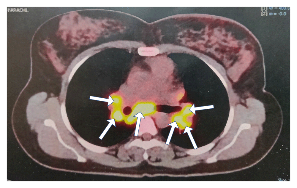
Multiple enlarged hypermetabolic lymph nodes in the mediastinum, right paratracheal, carinal, bilateral hilar region, and aortopulmonary window are observed (shown by yellow arrows).
The bronchoscopic biopsy of mediastinal lymph node showed non-caseous necrotic granulomatous inflammation, suggestive of sarcoidosis. This allowed us to correctly stage the disease as T2N0M0 stage IIA. The patient underwent lumpectomy followed by adjuvant chemotherapy, radiotherapy and hormonal therapy for breast cancer, while corticosteroids (prednisone, 1mg/kg for 9 months then tapered off over period of 3 months) were given for sarcoidosis for 1 year. The patient is being followed up with clinical examination every 3 months and breast mammogram done initially at 6 months following completion of radiotherapy (post mastectomy radiation therapy, total dose of 60 Gy: 50 Gy in 25 fractions given to chest wall with scar boost 10 Gy in 5 fractions) then at 1 year. At 18 months, there is no evidence of recurrence of disease and the patient is well.
Sarcoidosis is a multisystem granulomatous disease of unknown aetiology that manifests as non-caseating granulomas predominantly in the lungs, intrathoracic lymph nodes, and skin. The less commonly affected organs are the eyes, liver, heart, and brain. Sarcoidosis is more common in women; it occurs in patients aged 20 to 50 years3. The incidence rate is higher among African Americans. Many features of sarcoidosis are suggestive of infectious origin4. The diagnosis of sarcoidosis requires radiographic signs (e.g., bilateral hilar lymphadenopathy and pulmonary infiltrations)3 and non-caseating granulomas on histopathology. Chronic inflammation is associated with an increased risk for malignant lymphoma and cancer in the affected tissue. The association between sarcoidosis and cancer is indistinct; although it has been proposed that patients with sarcoidosis are at increased risk for developing cancer of the lung, small intestine, stomach, liver, and skin4. Cancer can produce sarcoid-like reactions in the lymph nodes, and sarcoidosis can occur in patients with cancer. The radiological features of neoplasms can be mistaken for sarcoidosis. Therefore, histologic confirmation is required before making a diagnosis3,5.
There is increased risk of breast cancer occurrence in patients with sarcoidosis than other inflammatory diseases5. Lungs are common sites for breast cancer metastasis and are detected on chest radiographs in 25% of patients4,5.
Cavitation of metastatic nodule is a rare feature on radiography. However, cavitation in metastatic adenocarcinoma including breast cancer is frequently seen on CT5. The axillary, mediastinal, and hilar lymph nodes are commonly involved. PET/CT imaging is superior in detecting distant metastasis in patients with stage II and stage III breast cancer, but false positive 18F-fluorodeoxyglucose (FDG) uptake and negative PET/CT are frequently seen6. The conditions that cause false positive FDG uptake for malignancy include tuberculosis, fungal infection, and sarcoidosis. Initial staging with PET/CT is not recommended in preoperative early stage breast cancer because of high suboptimal sensitivity of the axillary lymph nodes, but specificity is high, and it has excellent positive predictive value confirming immediate axillary dissection instead of sentinel lymph node biopsy in the presence of high FDG uptake. However, sentinel lymph node biopsy is necessary if there is normal or slightly increased FDG uptake6.
Sarcoidosis has been reported to increase levels of CA 15-3 in some cases, so the presence of this biomarker might be misleading7.
If abnormal FDG uptake is detected on FDG PET in isolated mediastinal lymph nodes, in the absence of axillary lymph node involvement in patients with breast cancer, a thorough preoperative evaluation is warranted with histopathologic confirmation, thereby allowing the choice of correct staging and curative strategy.
Written informed consent was obtained from the patient for the publication of this case report and any associated images.
All data underlying the results are available as part of the article and no additional source data are required.
| Views | Downloads | |
|---|---|---|
| F1000Research | - | - |
|
PubMed Central
Data from PMC are received and updated monthly.
|
- | - |
Provide sufficient details of any financial or non-financial competing interests to enable users to assess whether your comments might lead a reasonable person to question your impartiality. Consider the following examples, but note that this is not an exhaustive list:
Sign up for content alerts and receive a weekly or monthly email with all newly published articles
Already registered? Sign in
The email address should be the one you originally registered with F1000.
You registered with F1000 via Google, so we cannot reset your password.
To sign in, please click here.
If you still need help with your Google account password, please click here.
You registered with F1000 via Facebook, so we cannot reset your password.
To sign in, please click here.
If you still need help with your Facebook account password, please click here.
If your email address is registered with us, we will email you instructions to reset your password.
If you think you should have received this email but it has not arrived, please check your spam filters and/or contact for further assistance.
Comments on this article Comments (0)