Keywords
Urine cytology, Urinary abnormalities, Urothelial carcinoma, Khartoum, Sudan
This article is included in the Neglected Tropical Diseases collection.
Urine cytology, Urinary abnormalities, Urothelial carcinoma, Khartoum, Sudan
Urine cytology is a safe and inexpensive method to diagnose urinary tract abnormalities. It is similarly used for the detection of primary and recurrent urothelial carcinoma1,2; as well as in many other diseases related to the urinary system such as urinary schistosomiasis3 and bacterial infections4. Urine cytology has variable sensitivity and specificity depending on the sample collection method and tumor grade in the case of malignancy5–7. Although the determination of cell morphology through microscopy remains the gold standard8, false positive and negative results have been reported. The false-positive results from urine cytology may be attributed to the presence of viral infections, such as polyomavirus. False negative results have been linked to the sampling method used, the number of the samples obtained from the patient and the volume of the urine being processed1,6–10. Urothelial carcinoma is one of the most commonly reported cancers11. The major clinical concern for the management of bladder cancer patients is that most of the patients relapse12,13. Therefore, regular follow-up is required for patients who are at risk of urothelial carcinoma14. Despite many attempts to develop tests with higher sensitivity and specificity, cytology remains one of the best ways to diagnose a variety of bladder lesions15.
Hematuria is one of the common indications for urine cytology examination. It is also known to be caused by an underlying malignancy in 5% to 10% of patients16. Urine cytology is also used for the detection of abnormalities related to diseases like schistosomiasis3, candidiasis17, polyomavirus (BK virus (BKV)) infection18,19, Human Papillomavirus (HPV)20,21 and cryptococcosis22. In this study, we aimed at investigating the frequency of urine abnormalities among Sudanese patients attending the outpatient clinic at Omdurman Teaching Hospital, Khartoum, Sudan using urine cytology.
A descriptive cross-sectional study conducted from November 2016 to April 2017, an organized mass screening programme for urine cytology was introduced during the study period. The target screening population consists of the entire patients attending the outpatient’s clinic and agreed to participate in the study. A total of 1238 Patients were recruited from the outpatient clinic at Omdurman Teaching Hospital in Khartoum state, Sudan. All the patients attending the outpatients clinic were assessed by our research team and we discussed with all the patients the objective of our research. After obtaining written informed consent from the patients whom were willing to participate our research team gathered the patients information and demographic data including name, age, and any clinical signs or symptoms using a questionnaire, the process of filling and collecting the patients clinical data were conducted by our research team and it took place in the patients waiting area in the clinic, focusing on patients with HIV, diabetes mellitus and transplant recipients, and any patients with other conditions mentioned in the questionnaire.
Early in the morning, first pass urine sample was rejected as the cells may be abnormal, with features such as enlarged nuclei and washing out of the chromatin, features that mimic malignant cells; this is due to the pH of the urine23–25. Accordingly, the patients were asked if they passed urine before and if they drank water? Then each patient was provided with a sterile screw cap urine container and asked to pass some urine into the toilet at first, then without stopping the flow of the urine catch some in the provided container.
The entire volume of the urine samples were centrifuged (using Hettich® EBA 20 centrifuge, Germany) for five minutes at 3000rpm then the supernatant was discarded leaving the sediment, this was followed the addition of suspending media (74 ml saline, 6 ml Glacial acetic acid and 20 ml of 100% ethanol) to the sediment. The quantity of the suspending media was proportional to the quantity of the obtained sediment this is done by measuring the sediment quantity using automated pipette and the same quantity of the sediment was added the suspending media (after centrifugation we measured the quantity with a pipette). It was left for 30 minutes, followed by centrifugation for 5 mins at 3000 rpm. The supernatant was discarded and 1% acid alcohol (1 ml of Hydrochloric acid added to 99 ml of 70% alcohol) was added to the sediment and left for 30 minutes, followed by centrifugation at 3000 rpm for 10 mins, and discarding of the supernatant again. The resulting sediment was then used for smears by spreading the entire obtained volume on a microscopical slide, and then the slide were left to concentrate. Then the smears were fixed in 95% ethyl alcohol (V/V) for 15 mins.
The smears were stained using Papanicolaou staining procedures as follow: the smears were immersed in a descending gradient of alcohol from 80%, 70% to 50% (V/V) and then immersed the smear in tap water for 2 mins. Then the smears were stained using Harris Hematoxylin for 2 mins, after that the smears were washed in tap water and differentiated in 1% acid alcohol for seconds. This was followed by bluing by leaving it in a bath of running water for 10 mins, then immersed in 80% and 95% alcohol and stained with Orange G solution (O.G.6) for 2 mins. This is followed by washing in 95% alcohol, and then staining with Eosin Azure (EA.50) for 4 mins and washed again in 95% alcohol. Then rinsed quickly in 100% alcohol and blot using a sterile filter paper to dry. Clear in Xylene and mounting in distyrene (a polystyrene), a plasticiser (tricresyl phosphate), and xylene (D.P.X) (Catalog No. 100579; sigma – Aldrich, USA)14.
For reporting the cytological smears, two cytopathologist (EES, MAM) examined the entire slides blindly using Olympus light microscope (Model No. CX21FS1, Olympus, Tokyo, Japan). The cytological diagnosis was grouped into 5 categories according to The Paris System for Reporting Urinary Cytology (PSRUC)23, categories included nondiagnostic/unsatisfactory, negative for high-grade urothelial carcinoma (NHGUC), atypical urothelial cells, suspicious for high-grade urothelial carcinoma (SHGUC), high-grade urothelial carcinoma (HGUC) and Low-grade urothelial neoplasm (LGUN). The presence of koilocytosis, decoy cells, and pseudo-hyphae, as well as detecting the variable sizes of cells with clear hollow and narrow base with single budge were used as a diagnostic remark for the presence of other urinary abnormalities such as HPV and BKV. Urine cytology results obtained by the cytologists were sent to the physician who made the final decision on the patients’ diagnosis for proper patients management according to their findings of the cytological smears.
Statistical analysis was performed using the Statistical Package for Social Sciences (SPSS. Version 16). Chi-square test was used to compare urine cytology results with age, gender and to test the odds ratios of patients showing abnormalities in relation to their medical condition (A p value of <0.05 was considered to be statistically significant).
A total of 1238 urine samples were examined, of them, 832 (67.2%) were males (mean age 41.7±12.67), and 406 (32.8%) were females (mean age 43.8±10.94). The age range of the patients was 10 to 77 years, with a median age of 43 years. Patients were grouped according to their age into 4 groups; 46 (3.7%) were ≤ 20 years, 539 (43.5%) were 21 to 40 years, 522 (42.2%) were 41 to 60 years and the last age group were those aged more than 60 years; 131 (10.6%). Of the 1238 patients, 1091 (88.1%) reported no significant medical history, the remaining 147 (11.9%) patients reported an underlying medical condition which included HIV/AIDS, diabetes mellitus type 2 (DM2), and renal transplantation history. A total of 13 (1.1%) had HIV, of which 12 were male (92.3%) and one was a female (7.7%). Regarding the gender distribution among the DM2 and renal transplantation, 64 (92.8%) and 57 (87.7%) were males, respectively; and 5 (7.2%) and 8 (12.3%) were females, respectively.
Urine cytology showed a total of 1113 (89.9%) with normal urine findings. The remaining 125 (10.1%) patients showed a variety of findings; 43 (3.5%) cases of candidiasis, 36 (2.9%) with reactive urothelial cells, 11 (0.9%) with cryptococcosis, 9 (0.7%) with urothelial carcinoma, 9 (0.7%) with HPV, 8 (0.6%) with BKV, 6 (0.5%) with schistosomiasis and 3 (0.2%) with low grade urothelial cells. Frequency of cytological findings from urine analysis was higher in males than females and the results were found to be statistically significant (p value= 0.0001). (Table 1).
Regarding the relationship between age and cytological findings, our study showed that candida infection, Schistosomiasis, Polyoma virus and HPV were highest among the age group 41 – 60 years; while Cryptococcus infection was highest among 21 – 40 years old patients.
A representative microscopic result of urine cytology diagnosis is shown in Figure 1, and the distribution of gender, presence of hematuria, medical condition and urine cytology diagnosis based on age groups is described in Table 2.
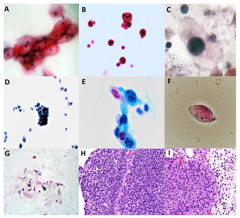
A: Cytopathic effect of human papilloma virus (HPV) with the eosinophilic intranuclear inclusion and a perinuclear halo and koilocytosis. B: Cryptococcus infection showed the organism with double contours. C: Polyomavirus cytopathic effect, the presence of decoy cells and pseudo hyphae and cells exhibit threads and small, round clumps of degenerating chromatin. D: Papillary cluster and single urothelial cells from low-grade urothelial cells. E: Cytological smears showed a monolayer sheet of cells with hyperchromasia and irregular distribution of chromatin and prominent nucleoli features of high-grade urothelial carcinoma. F: Schistosoma haematobium egg. G: Candida infection with bacterial. H and I: The counterpart diagnosis of D and E for the detected low-grade urothelial neoplasm and high-grade urothelial carcinoma using Hematoxylin and Eosin (H&E) histopathology staining (x40). H: Atypical urothelial cells. I: High-grade urothelial carcinoma showing very large, irregular and hyperchromatic nuclei. A–G: Staining using Papanicolaou stain (x100).
Of the 1238 patients, a relative risk estimation was conducted for both male and female with any of abnormal cytological finding and the results showed a significant proportion of females to be infected with cryptococcosis and HPV; p value = 0.001, odds ratio was 0.106 [95% CI, 0.023-0.494] for cryptococcosis, and p value = 0.001, odds ratio was 0.060 [95% CI, 0.007-0.480] for HPV. Risk estimates of HGUC, atypical urothelial cells, presence of hematuria, polyomavirus, and schistosomiasis are presented in Table 3.
| Male | Female | Total | P value | Odds ratio | 95% Confidence Interval | ||
|---|---|---|---|---|---|---|---|
| Lower | Upper | ||||||
| HGUC* | |||||||
| Yes | 9 (75.0%) | 3 (25.0%) | 12 (1.0%) | 0.408 | 1.469 | 0.396 | 5.456 |
| No | 823 (67.1%) | 403 (32.9%) | 1226 (99.0%) | ||||
| AUC* | |||||||
| Yes | 23 (63.9%) | 13 (36.1%) | 36 (2.9%) | 0.394 | 0.859 | 0.431 | 1.715 |
| No | 809 (67.3%) | 393 (32.7%) | 1202 (97.1%) | ||||
| Hematuria | |||||||
| Yes | 100 (67.0%) | 45 (31.0%) | 145 (11.7%) | 0.352 | 1.096 | 0.754 | 1.593 |
| No | 732 (67.0%) | 361 (33.0%) | 1093 (88.3%) | ||||
| Candidiasis | |||||||
| Yes | 33 (76.7%) | 10 (23.3%) | 43 (3.5%) | 0.190 | 1.636 | 0.798 | 3.352 |
| No | 799 (66.9%) | 396 (33.1%) | 1195 (96.5%) | ||||
| Cryptococcosis | |||||||
| Yes | 2 (18.2%) | 9 (81.8%) | 11 (0.9%) | 0.001 | 0.106 | 0.023 | 0.494 |
| No | 830 (67.6%) | 397 (32.4%) | 1227 (99.1%) | ||||
| Human papilloma virus | |||||||
| Yes | 1 (11.1%) | 8 (88.9%) | 9 (0.7%) | 0.001 | 0.060 | 0.007 | 0.480 |
| No | 831 (67.6%) | 398 (32.4%) | 1229 (99.3%) | ||||
| Polyomavirus | |||||||
| Yes | 7 (87.5%) | 1 (12.5%) | 8 (0.6%) | 0.285 | 3.436 | 0.421 | 28.024 |
| No | 825 (67.1%) | 405 (32.9%) | 1230 (99.4%) | ||||
| Schistosomiasis | |||||||
| Yes | 3 (50.0%) | 3 (50.0%) | 6 (0.5%) | 0.309 | 0.486 | 0.098 | 2.419 |
| No | 829 (67.3%) | 403 (32.7%) | 1232 (99.5%) | ||||
The results of risk estimation for patients having a current or history of a medical condition showed that patients with AIDs are particularly vulnerable to candidiasis and HPV; p value= 0.009 and 0.005, respectively. While those with DM2 are more at risk of candidiasis and atypical urothelial cells, p values = 0.000 and 0.003, respectively. On the other hand, patients with a medical history of renal transplant showed increased risk for having BKV; p value= 0.000, and candidiasis, p value= 0.022. Results of risk estimation based on current medical condition are shown in Table 4.
| Urine cytology diagnosis | Medical Condition | P value* | Odds ratio | 95% Confidence Interval | ||
|---|---|---|---|---|---|---|
| Yes | No | Lower | Upper | |||
| AIDs | ||||||
| HGUC | 0 | 13 | 0.881 | 1.010 | 1.004 | 1.016 |
| AUC | 2 | 11 | 0.052 | 6.369 | 1.359 | 29.849 |
| Candidiasis | 3 | 10 | 0.009 | 8.888 | 2.355 | 33.541 |
| Cryptococcosis | 2 | 11 | 0.005 | 24.566 | 4.750 | 127.040 |
| HPV | 0 | 13 | 0.909 | 1.007 | 1.003 | 1.012 |
| BKV | 0 | 13 | 0.919 | 1.007 | 1.002 | 1.011 |
| Schistosomiasis | 0 | 13 | 0.939 | 1.005 | 1.001 | 1.009 |
| Diabetes Mellitus Type-2 | ||||||
| HGUC | 0 | 69 | 0.501 | 1.010 | 1.004 | 1.016 |
| AUC | 7 | 62 | 0.003 | 4.438 | 1.871 | 10.531 |
| Candidiasis | 10 | 59 | 0.000 | 5.835 | 2.744 | 12.406 |
| Cryptococcosis | 0 | 69 | 0.531 | 1.009 | 1.004 | 1.015 |
| HPV | 0 | 69 | 0.596 | 1.008 | 1.003 | 1.013 |
| BKV | 2 | 67 | 0.690 | 5.786 | 1.146 | 29.212 |
| Schistosomiasis | 0 | 69 | 0.708 | 1.005 | 1.001 | 1.009 |
| Renal Transplant | ||||||
| HGUC | 0 | 65 | 0.522 | 1.010 | 1.004 | 1.016 |
| AUC | 3 | 62 | 0.292 | 1.672 | 0.499 | 5.601 |
| Candidiasis | 6 | 59 | 0.022 | 3.122 | 1.268 | 7.691 |
| Cryptococcosis | 2 | 63 | 0.110 | 4.106 | 0.869 | 19.403 |
| HPV | 1 | 64 | 0.386 | 2.275 | 0.280 | 18.471 |
| BKV | 6 | 59 | 0.0001 | 59.542 | 11.765 | 301.334 |
| Schistosomiasis | 0 | 65 | 0.723 | 1.005 | 1.001 | 1.009 |
Urine cytology is considered a noninvasive, cost effective technique that is able to diagnose a wide range of abnormalities including infections, atypical cellular changes and malignancy26,27. However, in this study, we investigated a varied array of urine abnormalities among Sudanese patients attending the outpatient clinic at Omdurman Teaching Hospital, Khartoum-Sudan using urine cytology.
In this study, we observed that urine cytology could easily differentiate benign from malignant cells showing high specificity for detecting HGUC; this result agrees with previous reports8,28–30. HGUC are aneuploid and are detected with high sensitivity, as compared to atypical urothelial cells29–31. The cytological features of HGUC identified in this study showed that the smears exhibited high cellularity and dehiscence cellular pattern. Malignant cells were larger than normal cells and showed pleomorphic nuclei with prominent nucleoli. Also, the cytoplasm ranged from homogenous to scant vacuolated one. Depending on these features, previous studies have indicated that detection of malignant cells using urine cytology is the gold-standard method for diagnosing urothelial cancer.2,29,32.
In this report we are also able to identify Candida spp with great confidence by the presence of the budding yeast and pseudohyphae which is considered as the cytomorphological characteristic of this fungus, however if the organism was not identified, cytological changes such as stain anomaly, nuclear degeneration and bacterial background may pinpoint the presence of candida.
Cryptococcus infection was detected in 2 out of 13 HIV patients; 0 out of 69 DM2 patients and 2 out of 65 renal transplant recipients. The organism was correctly identified by the presence of a semitransparent fungal organism with double contours in an inflammatory background22,31,33.
Polyoma virus (BKV) was detected in 0 out of 13 (0%) HIV positive patients; 2 out of 69 (2.89%) DM2 patients and 6 out of 65 (9.2%) renal transplant recipients. The key feature for the existence of polyomavirus is depending on the presence of the unique cytomorphological features, i.e., presence of the typical decoy cells, the nuclei of this cell appeared as uniform with washed chromatin34–42. The results obtained by this study demonstrated that urine cytology can be used as a simple monitoring test for the renal transplant recipient for the presence of polyoma virus, since polyoma virus infection in renal transplant recipient is considered as one of the factors that can lead to graft rejection42–48. Our results are in concordance to that conducted by Kapila and his associates; they investigated the diagnostic efficacy of urine cytology in detecting Polyoma virus among renal transplant recipients comparing urine cytology with molecular diagnosis and their results showed that urine cytology can be used as an easy tool for monitoring the patients49. Furthermore, our results showed that no polyoma virus was been detected among HIV patients, which is in contrast to the findings of Boldorini and his associates as they screened 78 patients with HIV for the presence of polyoma virus using urine cytological examination and molecular and immunohistochemistry, there results demonstrated that out of 78 patients, only 17 patients were positive for Polyoma virus using cytology50.
Furthermore, Human papilloma virus was been detected among 9 out of 1238 (0.7%) of whom 8 were females and one was a male; the results obtained here should be evaluated with care especially in the settings of female individuals as it is very difficult if not impossible to differentiate whether these koilocytic cells originated from the urinary tract or they belong to the uterine cervix and therefore in such circumstances we highly recommend that these females should go for pap smear to locate the exact site of HPV infections.
Urine cytology is a safe, noninvasive, and reliable diagnostic tool for identifying viral cytopathic effects in urothelial cells, and deserves more widespread use in the monitoring of patients, especially those with renal transplants. It now seems that urine cytology needs to be implemented in clinical settings for the benefit of patients and urologists. Polyoma virus infection detection among transplant patients helps urologists to identify and manage renal graft rejection early which is of invaluable benefit to patients, physicians and economically. Due to the risk of the presence of polyoma virus among renal transplant recipient and based on the fact that 50% of the cases with Polyoma virus was associated with the graft rejection we highly recommended to use urine cytology as an early screening tool for routine checks among renal transplant patients. Furthermore, Pap staining was considered as an excellent cytological stain that provided excellent morphological quality for the visualization of the nuclear changes.
The study was approved by the Faculty of Medical Laboratory Sciences Research Ethics Committee – University of Khartoum, Sudan (Nov/2016/102). Written informed consent was obtained from each participant prior to participation.
Harvard Dataverse: Replication Data for: Urine cytology in our daily practice", https://doi.org/10.7910/DVN/JHZCCM51.
Harvard Dataverse: Replication Data for: Urine cytology in our daily practice", https://doi.org/10.7910/DVN/JHZCCM51.
This project contains the following extended data:
- Supplementary Table 1: Paris Reporting System for Urinary Cytology based on age groups and gender of patients.
- Supplementary Table 2: Gender risk estimation for having urine abnormalities
- Supplementary Table 3: Patients medical condition or medical history and risk estimation for having urine abnormalities
Data are available under the terms of the Creative Commons Zero "No rights reserved" data waiver (CC0 1.0 Public domain dedication).
| Views | Downloads | |
|---|---|---|
| F1000Research | - | - |
|
PubMed Central
Data from PMC are received and updated monthly.
|
- | - |
Provide sufficient details of any financial or non-financial competing interests to enable users to assess whether your comments might lead a reasonable person to question your impartiality. Consider the following examples, but note that this is not an exhaustive list:
Sign up for content alerts and receive a weekly or monthly email with all newly published articles
Already registered? Sign in
The email address should be the one you originally registered with F1000.
You registered with F1000 via Google, so we cannot reset your password.
To sign in, please click here.
If you still need help with your Google account password, please click here.
You registered with F1000 via Facebook, so we cannot reset your password.
To sign in, please click here.
If you still need help with your Facebook account password, please click here.
If your email address is registered with us, we will email you instructions to reset your password.
If you think you should have received this email but it has not arrived, please check your spam filters and/or contact for further assistance.
Comments on this article Comments (0)