Keywords
Seronegative myasthenia gravis, malignant thymoma, tomotherapy, paraneoplastic disorder.
Seronegative myasthenia gravis, malignant thymoma, tomotherapy, paraneoplastic disorder.
Malignant thymoma is a rare epithelial neoplasm and accounts for 30% of anterior mediastinal tumors. The highest incidence is in the 7th decade of life and is more common in men. About one-third of patients are asymptomatic at diagnosis, which is commonly found as incidentaloma. Of the symptomatic patients, 60% presents with a parathymic syndrome manifestation and 40% has symptoms relating to impingement by intrathoracic mass1–3. Myasthenia Gravis (MG) has the highest incidence among the parathymic syndromes and occurs in 50% of patients with thymoma. Of patients with MG, 15% have a thymoma and 60% will have thymic lymphoid hyperplasia1,4. As an autoimmune disorder, 85% of these patients have autoantibodies directed against postsynaptic nicotinic acetylcholine receptor (AChR). Thymectomy is the main treatment modality and complete resection is an important prognostic factor, with locoregional relapse reduction and possible resolution of MG symptoms. If high risk factors are present, radiotherapy should be considered as adjuvant treatment1.
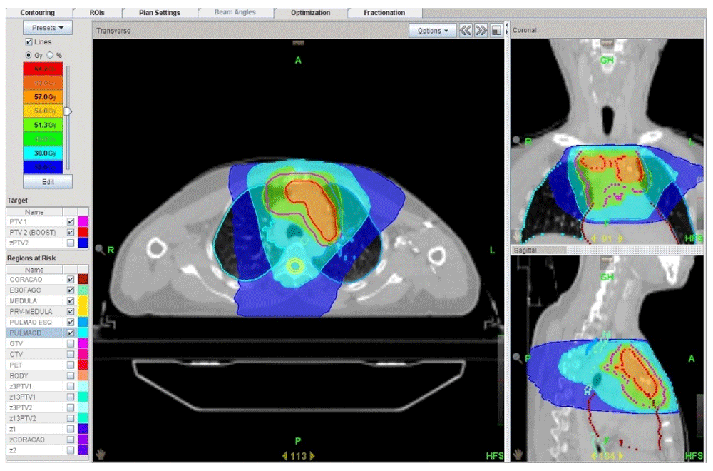
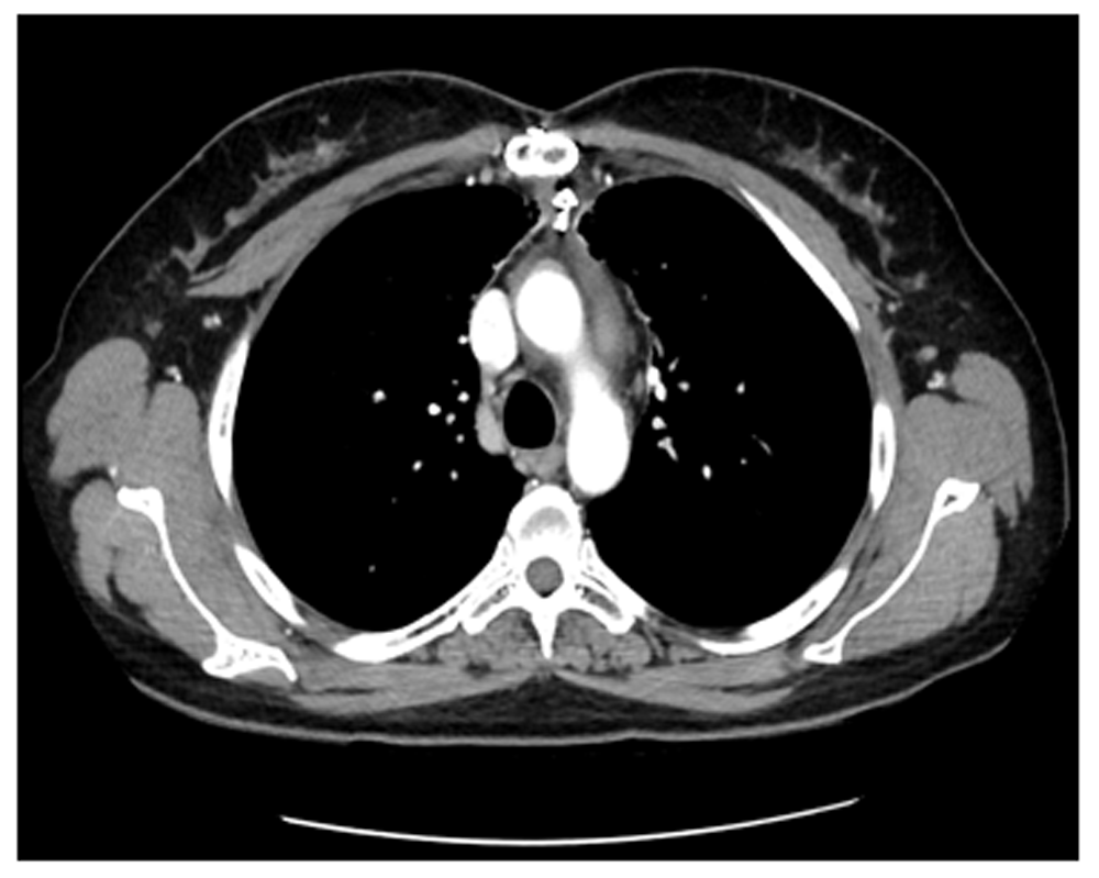
A 47-year-old caucasian woman, businesswoman, without important medical history, presented with fatigue and occasional dyspnea in May 2016. A computed tomography (CT) scan was performed and showed a pre-vascular mass in the anterior mediastinum with 7 centimeters of axial axis with features of a malignant thymoma. A 18F-fluorodeoxyglucose positron emission tomography integrated with computer tomography (18F-FDG PET/CT) showed a hypermetabolic mediastinal mass, suggestive of high-grade neoplasia. The remaining diagnostic investigation was normal, without alterations of autoantibodies directed against postsynaptic nicotinic acetylcholine receptor (AChR), thyroid hormones, β-human chorionic gonadotropin (β-HCG) and α-fetoprotein. The patient underwent incomplete surgical resection due to tumor adherence to the brachiocephalic venous trunk. The histological study revealed a WHO type B1 in IIA stage of Modified Masaoka. The patient was referred for adjuvant radiotherapy and received 54Gy (1.8Gy/fraction) over the tumor bed and a total of 60 Gy (2Gy/fraction) in 30 fractions over residual tumor, by tomotherapy (Figure 1). During treatment the patient maintained an excellent general status, without relevant toxicity. One year after radiotherapy, the patient revealed severe worsening of fatigue with muscle weakness in the upper limbs, dysphagia and right diplopia. Since AChR antibodies were negative, fibromyalgia was considered. Due to maintenance of suspected MG, she started pyridostigmine 180 mg per day and presented fatigue improvement, but poor tolerance. Currently in the 33rd month of clinical control, the patient is medicated with deflazacort 6 mg, fluoxetine 20 mg, alprazolam 0.5 mg, trazodone 100 mg and maintains a stable symptomatic condition with no signs of disease recurrence (Figure 2). Surveillance continues through chest CT every 6 months and regular evaluation in radiation oncology, pneumology, psychiatry, neurology and neuro-ophthalmology.
Paraneoplastic neurological degenerations (PND’s) affects 1 in 10,000 patients with cancer4. MG is included and is often associated with pathological abnormalities of the thymus. Given the incidence, when a diagnosis of thymoma is suspected, further investigation should be undertaken to exclude MG1. A problem of seronegative MG has been the lack of a gold standard in its diagnosis and, it is known that, the proportion of these patients varies from 5% to 30% among studies5. Seronegative MG has been reported in a few cases of benign thymoma and in only one case of malignant thymoma6. It should be noted that autoantibodies against muscle-specific kinase (MuSK) are present in about 10% of the all cases of MG and in 30% of AChR seronegative MG7. In our case, the diagnosis of MG was supported on clinical and pharmacological basis. We should be aware that, besides autoantibodies against AChR, other antibodies can be associated with this disease, including autoantibodies against MuSK and lipoprotein-related protein 4 (LRP4), however these were not tested. Additionally, new assays could improve the sensitivity in antibody detection7.
Generally, the highest incidence of malignant thymoma is in the 7th decade of life, but for patients with MG, the peak of incidence is in the 4th decade, as observed in this case1.
The differential diagnosis allowed the exclusion of neuroendocrine, germinative, hematologic and pulmonary tumors, which is crucial for the treatment plan. Surgery is the main treatment for malignant thymoma and 85% of stage II tumors are resectable. Complete resection is an important prognostic factor. In these patients, thymectomy can also be considered as an effective treatment for MG with symptomatic improvement in 50% of cases. This response seems to be associated with high AChR activity1. The efficacy of thymectomy in the treatment of MG is noted in the MGTX trial, showing that local control allows less dependence on immunosuppressive medication and less exacerbations requiring hospitalization8.
Adjuvant RT is associated with better disease free-survival (DFS) with an impact on the overall survival of stage II and III tumors with a positive margin1. In this case, with incomplete resection, RT is considered to be mandatory for satisfactory local control. Advanced RT techniques, such as tomotherapy, that allows the combination of intensity-modulated radiotherapy (IMRT) and image-guided radiotherapy (IGRT), with helical radiation deposition, can provide high tumor control rates and a satisfactory toxicity profile1.
In our case, the patient showed worsening of neurological symptoms after the thymectomy and adjuvant RT. At that moment, fibromyalgia was considered in the absence of autoantibodies against AChR. Characterized by widespread musculoskeletal pain, fatigue and sleep disorder, fibromyalgia is more frequent in women, especially if other autoimmune disorders are present. MG is a differential diagnosis of fibromyalgia, as it is associated with post-exercise and generalized fatigue but not coupled with widespread pain, a symptom that our patient did not present9,10.
Some studies report the importance of assuring clinical stability with immunosuppression prior to surgery in order to prevent rapid perioperative deterioration7. Given the late diagnosis, our patient only started corticotherapy after thymectomy and the systemic progression of the immunological disease.
It is possible that, in this case, the early diagnosis with initiation of immunosuppressive therapy, especially before surgery, could have changed the course of the disease1,8.
In conclusion, given the impact of an earlier diagnosis of MG on quality of life, when thymoma is suspected, seronegativity should prompt further investigated rather than result in MG exclusion.
Obtained.
Data are available under the terms of the Creative Commons Zero “No rights reserved” data waiver (CC0 1.0 Public domain dedication).
| Views | Downloads | |
|---|---|---|
| F1000Research | - | - |
|
PubMed Central
Data from PMC are received and updated monthly.
|
- | - |
Provide sufficient details of any financial or non-financial competing interests to enable users to assess whether your comments might lead a reasonable person to question your impartiality. Consider the following examples, but note that this is not an exhaustive list:
Sign up for content alerts and receive a weekly or monthly email with all newly published articles
Already registered? Sign in
The email address should be the one you originally registered with F1000.
You registered with F1000 via Google, so we cannot reset your password.
To sign in, please click here.
If you still need help with your Google account password, please click here.
You registered with F1000 via Facebook, so we cannot reset your password.
To sign in, please click here.
If you still need help with your Facebook account password, please click here.
If your email address is registered with us, we will email you instructions to reset your password.
If you think you should have received this email but it has not arrived, please check your spam filters and/or contact for further assistance.
Comments on this article Comments (0)