Keywords
peripartum cardiomyopathy, female, stroke, anticoagulation
peripartum cardiomyopathy, female, stroke, anticoagulation
Peripartum cardiomyopathy (PPCM) is a rare cause of heart failure affecting women in late pregnancy or in the puerperium. The overall incidence of PPCM ranges from 1 in 1300 to 1 in 15,000 pregnancies1. However, the incidence fluctuates globally, and is higher in developing countries2. PPCM is often recognized when the patient has severe myocardial dysfunction, less severe forms of PPCM often go unrecognized3. The 2010 European Society of Cardiology (ESC) Working Group defined PPCM as an idiopathic cardiomyopathy with following characteristics3:
1. The development of heart failure (HF) towards the end of pregnancy or within five months following delivery.
2. The absence of an identifiable cause of HF.
3. Left ventricular (LV) systolic dysfunction with an LV ejection fraction (LVEF) of less than 45 percent. The LV may or may not be dilated.
Despite many attempts to establish the exact etiology of PPCM, the cause remains unknown. Viral, autoimmune, dietary deficiencies, and idiopathic causes may contribute1. Familial occurrence of PPCM suggests a possible role of genetic predisposition1. Altered prolactin processing, and elevated soluble Fms-like tyrosine kinase 1 (Flt 1) have also been associated with the pathogenesis of PPCM4,5. During pregnancy, increased oxidative stress leads to cleavage of prolactin by cathepsin D into abnormal 16 kDa protein. This protein damages the heart, and blood vessels4. Soluble Flt 1 is secreted by placenta, that inhibits vascular endothelial growth factor signaling, which leads to angiogenic imbalance, and endothelial dysfunction5. Relaxin-2, a hormone produced by ovaries, breast, and placenta has a potential beneficial effect in PPCM. It increases cardiac output, and decreases vascular resistance6. Risk factors of PPCM are increased maternal age, increased parity, multiple pregnancy, malnutrition, use of tocolytics, and preeclampsia or eclampsia1. Patients usually present with shortness of breath, orthopnea, cough, hemoptysis, paroxysmal nocturnal dyspnea, and ankle edema3. Tachycardia, elevated jugular venous pressure, third heart sound (S3), and displaced apex beat are common, however; basal crackles are less common3. A high index of suspicion is required for diagnosis, as these features are common in advanced pregnancy and the peripartum period3.
About 6 % of patients of PPCM present with thromboembolic complications such as deep vein thrombosis, pulmonary thromboembolism, stroke, acute limb ischemia etc7. Morbidity and mortality is high in PPCM, especially when associated with cardioembolic phenomenon3. Fortunately, despite the high morbidity, mortality, and a high risk of relapse in succeeding pregnancies, many patients with peripartum cardiomyopathy recover within three to six months of disease onset2. Here, we report a rare case of a young female with peripartum cardiomyopathy complicated by cardioembolic stroke.
A 20-year-old female farmer from Nepal presented with shortness of breath and cough which had lasted 10 days in November, 2019. The shortness of breath had a gradual onset and was progressive. Initially patient had shortness of breath on exertion only, but with time she had shortness of breath even at rest, and it was associated with orthopnea. The patients cough was productive with mucoid sputum lasting for 10 days. There was no history of fever, chest pain, and hemoptysis. The patient was 45 days post-partum, and was breastfeeding. She didn’t indicate history of illicit drug abuse and use of hormonal contraceptives. The family history was negative for premature coronary artery disease, young stroke, and premature death. With the above symptoms the patient present at her local hospital, where she received injectable and oral antibiotics (intravenous ceftriaxone 1 gram twice a day and oral azithromycin 500 mg daily) thinking that it could be a chest infection. However, her symptoms were not relieved and, she visited our center.
On examination the patient had a temperature of 36.7°C (normal = 36.5°C to 37.5°C), heart rate of 109 beats per minute (normal = 60 to 100 beats per minute), blood pressure was 100/60 mm Hg (< 120/80 mm Hg), respiratory rate of 20 breaths per minute (normal = 14 to 16 breaths per minute), and oxygen saturation of 92% (normal = 95% to 100%)while he was breathing ambient air. There was no peripheral edema. On chest examination there was dullness in the right infra-scapular and infra-axillary region and the intensity of breath sound was decreased, however her trachea was in the midline. Examinations of other systems were unremarkable.
On investigation total leukocyte count was 8,300/uL, hemoglobin was 11.4 gm/dL, platelet count was 4,35,00/uL, and random blood sugar was 100 mg/dL. Details are shown in Table 1.
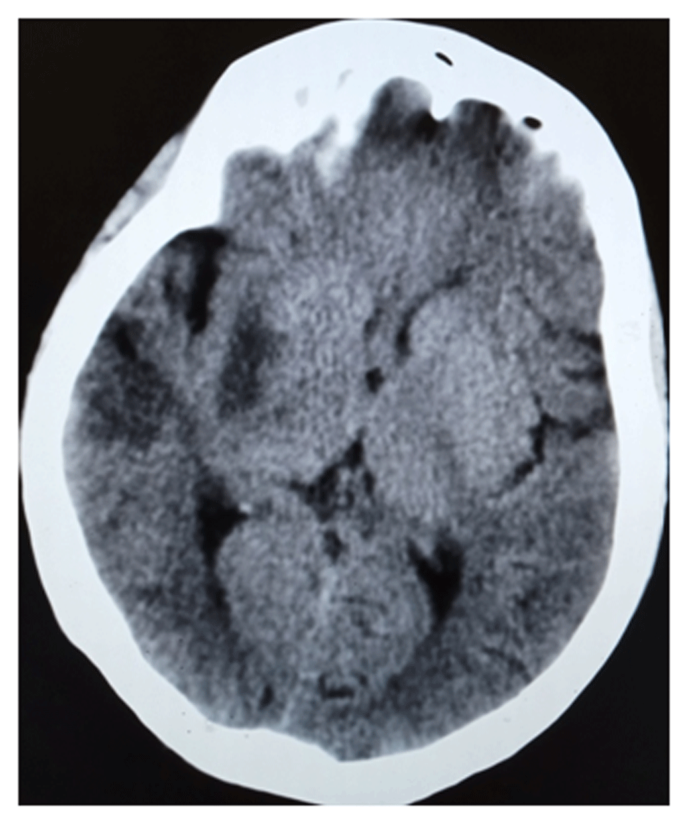
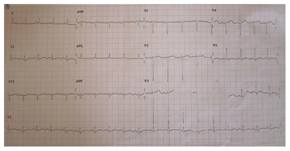
A provisional diagnosis of right sided pleural effusion was made and the patient was admitted for further evaluation. Antibiotic treatment (intravenous ceftriaxone 1 gram twice a day) was initiated. One day after hospital admission the patient developed weakness of left half of the body. The weakness was more in the upper limb compared to the lower limb. It was associated with slurring of speech, and deviation of face towards the right side. On neurological examination, the patient had left-sided upper motor neuron type facial nerve palsy, muscle strengths in the left upper and lower limbs were 0/5 and 1/5 respectively on the Medical Research Council (MRC) scale, and there was an ipsilateral Babinski sign. Fundoscopy findings were unremarkable. A computed tomography scan of the head was done immediately which was normal. A repeat CT scan head at 48 hours showed a wedged shaped hypodensity involving the gray and white matter of the right anterior temporal lobe, a hypodense area in the posterior portion of lentiform nucleus, the adjacent internal capsule region, and in the head of caudate nucleus (features suggestive of right sided acute ischemic stroke) [Figure 1]. Electrocardiogram showed sinus tachycardia, and poor progression of the R-wave [Figure 2].
Transthoracic echocardiography (TTE) showed global hypokinesia of the left ventricular wall with an LVEF of 25%, moderate mitral regurgitation, mild tricuspid regurgitation, and minimal pericardial effusion. On transesophageal echocardiography (TEE) there was no left atrial or ventricle clot.
A diagnosis of right sided ischemic stroke (cardioembolic) with peripartum cardiomyopathy was formulated. The patient was treated with aspirin 150 mg daily, frusemide 20 mg twice a day, spironolactone 25 mg daily, metoprolol 25 mg daily, and prophylactic unfractionated heparin (UFH) 2500 units subcutaneously twice a day. Limb physiotherapy was initiated. Two weeks after the incident of stroke, anticoagulation with warfarin 5 mg daily bridged with low molecular weight heparin 40 mg subcutaneously twice a day for an initial five days was administered. Aspirin and UFH were stopped. On discharge, her HF medications were optimized (frusemide 20 mg twice a day, spironolactone 50 mg daily, and metoprolol 50 mg daily), and anticoagulation with warfarin 5 mg daily was continued with provision for regular monitoring of prothrombin time (PT), and international normalization ratio (INR). At the time of discharge, her power was 3/5 and 4/5 in the left upper limb, and lower limb respectively. The patient was counselled about avoiding subsequent pregnancies.
Strokes in young adults are uncommon, comprising 10–15% of all stroke patients8,9. Though uncommon, stroke in young adults has a large economic burden, often leaving the victims disabled during their most productive years.
Previously published articles have defined the cut-off age for strokes in young adults as those younger than 45 or 49 years10. The etiologies and nature of strokes in young adults are different from the older patients. In young adults, congenital and acquired heart diseases, hematological conditions (such as sickle cell disease), vasculopathy (such as arterial dissection and vasculitis), pregnancy (cortical venous sinus thrombosis, preeclampsia/eclampsia), other hypercoagulable states, smoking, illicit drugs, premature atherosclerosis, hypertension, metabolic disorders (such as Fabry disease, homocystinuria etc), and possibly migraine are common causes The etiology of stroke in young patients remains a diagnostic challenge. In our patient, the etiology was cardioembolic secondary to hypokinesia of the left ventricle (EF=25%) due to peripartum cardiomyopathy. In a case report from Kumbham et al.11 the patient had multifocal infarct involving the left frontoparietal lobe, both occipital lobes, and the right insular cortex as well as thrombi in the left ventricle, which is in contrast with our case. In our case, we performed TEE to rule out a thrombus in the left atria or ventricle, the results of which were negative. The case report from Kumbham et al.11 had a multifocal infarct involving both hemispheres, but in our case the infarct was unilateral. Maternal age > 30 years is one of the risk factors for PPCM12, however our patient was 20 years old. Other conventional risk factors for PPCM such as increased parity, multiple pregnancy, use of tocolytics, and preeclampsia or eclampsia were not present in our patient.
Management of PPCM with cardio-embolic stroke requires a multidisciplinary approach that involves cardiologist, neurologist, obstetrics, and physiotherapist. Components of HF therapy in PPMC are similar to that of other types of HF, with attention to avoiding particular medications that have an effect on the fetus2. Angiotensin converting enzyme (ACE) inhibitors, angiotensin II receptor blockers (ARBs), angiotensin receptor-neprilysin inhibitors (ARNI), and aldosterone antagonists are teratogenic, and shouldn’t be used during pregnancy. In our patient we have avoided all these medications, but we used aldosterone antagonist (spironolactone) as our patient was 45 days post-partum, and is relatively safe during lactation13. Anticoagulation in PPMC is administered when LVEF is < 30 %14. In our patient (LVEF=25 %), anticoagulation was started at two weeks following the ischemic stroke to avoid the risk of bleeding, as the infarct was involving more than one-third of the middle cerebral artery region.
The risk of recurrent HF in patients with PPCM is around 25 % when LV function has recovered, and 50 % when LV function has not recovered15. We therefore, strongly counselled our patient to avoid subsequent pregnancies.
Limitations: Patient developed acute ischemic stroke during hospital stay, however; we were not able to thrombolyse the patient with intravenous recombinant tissue plasminogen activator (rtPA) therapy due to financial constraints for the patient. Work-up for thrombophilias, and vasculitis disorders were not feasible for the same reason. At presentation to our center, we missed the diagnosis of PPCM initially, and didn’t consider that pleural effusion could be due to HF in PPCM.
PPCM is one of the rare causes of HF in puerperium. One should consider PPCM as a differential diagnosis in any patient presenting with shortness of breath, and cough during puerperium. Stroke in PPCM is rare, but has a devastating effect on mother, infant, and other family members. Early diagnosis, optimization of HF medication, and anticoagulation therapy prevents the further complications. Patient counselling to avoid subsequent pregnancies is the cornerstone to prevent recurrent HF.
Written informed consent for publication of their clinical details and clinical images was obtained from the patient.
| Views | Downloads | |
|---|---|---|
| F1000Research | - | - |
|
PubMed Central
Data from PMC are received and updated monthly.
|
- | - |
Provide sufficient details of any financial or non-financial competing interests to enable users to assess whether your comments might lead a reasonable person to question your impartiality. Consider the following examples, but note that this is not an exhaustive list:
Sign up for content alerts and receive a weekly or monthly email with all newly published articles
Already registered? Sign in
The email address should be the one you originally registered with F1000.
You registered with F1000 via Google, so we cannot reset your password.
To sign in, please click here.
If you still need help with your Google account password, please click here.
You registered with F1000 via Facebook, so we cannot reset your password.
To sign in, please click here.
If you still need help with your Facebook account password, please click here.
If your email address is registered with us, we will email you instructions to reset your password.
If you think you should have received this email but it has not arrived, please check your spam filters and/or contact for further assistance.
Comments on this article Comments (0)