Keywords
Head Neck Cancer , Sarcoidosis, Lymph Nodes
Head Neck Cancer , Sarcoidosis, Lymph Nodes
Sarcoidosis is a systemic disease of unknown etiology that is characterized by the development of noncaseating epithelioid granulomas in various organs, mainly the lungs and lymphatic system1.
Several cases have been published describing the coexistence of sarcoidosis and cancer. It has been reported at diagnosis, during treatment, and in the surveillance of cancer patients2,3. In head and neck cancer, there are few cases reporting the simultaneous occurrence of sarcoidosis4,5.
We present a case of a patient with a squamous cell carcinoma of the right alveolar ridge of the mandible, that during treatment developed mediastinal lymphadenopathies, causing a diagnostic and therapeutic dilemma between disease progression and benign lesions.
A 42-year-old male, Caucasian, presented to his stomatologist in May 2013 with a painless lump in the right jaw with 3 months of evolution. His Eastern Cooperative Oncology Group Performance Status was 0. The patient was an active smoker (24 pack-year) and denied drinking alcohol. He didn´t have other comorbidities. Oral cavity inspection revealed an ulcero-infiltrative lesion on the gingival margin of tooth 45 with extension to the tongue. In the lymph node assessment, an enlarged right submandibular node was palpable. The remainder of the physical examination was unremarkable. He had a normal complete blood count and biochemical profile. A biopsy of the lesion was performed and revealed a squamous cell carcinoma of the oral cavity. The patient was then referred to our institution.
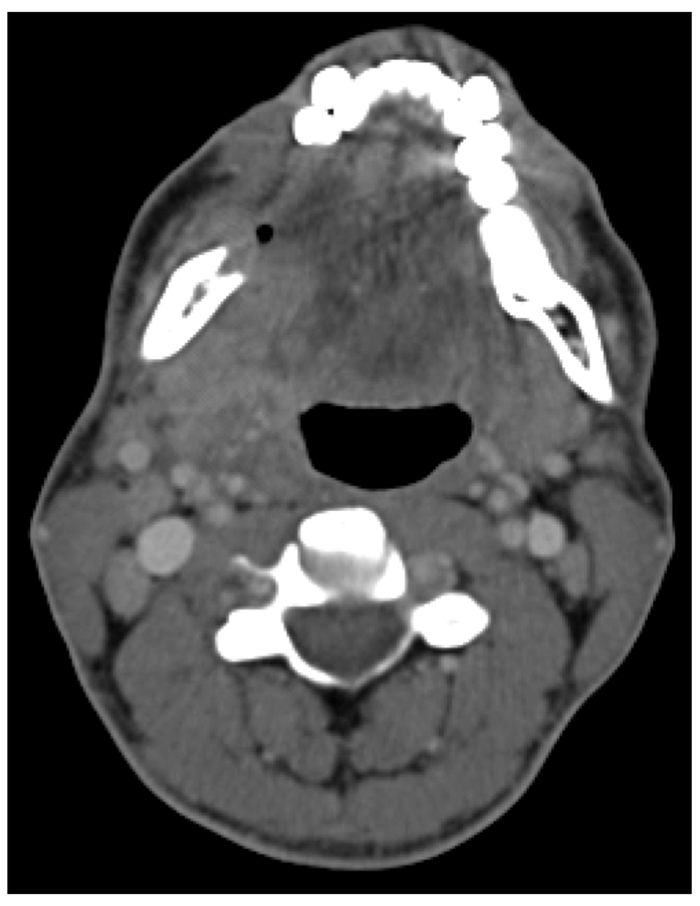
A cervical and thoracic computed tomography (CT) scan revealed a lesion in the right alveolar ridge of the mandible with bone reabsorption, and multiple bilateral enlarged cervical lymph nodes with high contrast enhancement (Figure 1). No other anatomical changes were reported. The tumor was clinically classified in T4 N2c M0.

The case was discussed at the Head and Neck multidisciplinary team meeting (MDT) and a surgical approach was decided.
In August 2013, the patient was submitted to a partial glossectomy, right hemimandibulectomy and bilateral neck dissection. Histological analysis confirmed a squamous cell carcinoma of the oral cavity with cervical lymph node metastasis in 2 of 18 lymph nodes of the right lymph node dissection; the 12 lymph nodes dissected from the left side of the neck were clear from metastasis. The surgical margins were negative although the tumor was 1mm from the deep margin (pT4 N2b M0).
Due to the presence of positive lymph nodes, high T stage and close margins, the patient underwent adjuvant radiation therapy (60 Gy/30 fr over 6 weeks) from October to November. During the treatment he developed a grade 3 mucositis requiring a nasogastric feeding tube.
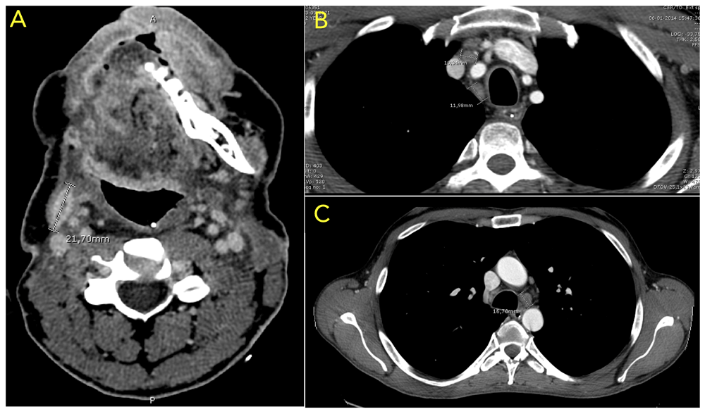
In January 2014, the cervical and thoracic CT scan revealed local and distant relapse with an area of heterogenous contrast enhancement in the right side of the floor of mouth and tongue as well as presence of mediastinal lymphadenopathies (Figure 2). Due to this fact the patient started palliative chemotherapy with the EXTREME regimen: Cisplatin (at a dose of 100 mg/m2) and cetuximab [at a dose of 400 mg/m2 initially (loading dose), then 250 mg/m2 on day 1 and 5-FU (at a dose of 1000 mg/m2 per day for 4 days)] every three weeks. As major toxicities he developed a grade 3 skin rash and grade 2 hypomagnesemia.

(A) Area of heterogenous contrast enhancement in right side of the floor of mouth and tongue as well as area of peripheral contrast enhancement with central necrosis (21,7×8 mm); (B) and (C) presence of enlarged mediastinal lymph nodes (16,78mm, 10,34mm and 11,98mm).
After 6 cycles with clinical and imagological complete response, Cetuximab was maintained in monotherapy.
In November 2014, the control CT scan revealed tumor progression in mediastinal lymph nodes without local tumor relapse. An 18F-fluorodeoxyglucose positron emission tomography (FDG-PET) scan was obtained, showing hypermetabolic mediastinal and hilar lymph nodes (Figure 3).

(A) Enlarged lymph node (12,8 mm) visible in the CT scan with correspondent abnormal in FDG uptake in the PET (B); (C) Focus of increased uptake in multiple mediastinal and hilar lymph nodes.
The Head and Neck MDT planned to enroll him in a clinical trial with immunotherapy. To meet the inclusion criteria, it was necessary to confirm histologically the mediastinal lymph node metastasis. Therefore, the patient underwent mediastinoscopy with mediastinal lymph node dissection in March 2015. Histopathological findings of the surgical specimens revealed granulomatous inflammation consistent with sarcoidosis, no metastatic disease was identified in the lymph nodes and special stains for fungi and acid-fast bacilli were negative.
The patient was referred to the Pneumology Department. Since he had no symptoms and the levels of serum angiotensin converting enzyme were normal no further pharmacological intervention was done.
In September 2017 he was submitted to a Facial Reconstructive Surgery and until now, the patient remains in follow-up without signs of local recurrence or metastasis.
Clinical and imagiological evaluation (CT and FDG-PET scan) are widely used to evaluate tumor response. Positive CT and FDG-PET findings in patients with suspected cancer recurrence are often used to guide therapeutic approach6. However false-positives results can occur7.
We reported a rare case of a patient with a locally advanced squamous cell carcinoma of the right alveolar ridge of the mandible, that during the systemic treatment developed sarcoidosis of mediastinal lymph nodes. In our clinical case the diagnosis of sarcoidosis was incidental. The positivity in the CT scan and FDG-PET scan led us to the assumption of tumor progression. At that time, we considered the enrollment of the patient in a clinical trial because the therapeutic options were limited. The obligation of a histological confirmation of recurrent disease (inclusion criteria) led to the diagnosis of sarcoidosis and exclusion of cancer recurrence, providing a major shift in our therapeutic approach. Since then the patient remains exclusively in follow-up.
In conclusion sarcoidosis is an uncommon, but critical disease in the setting of Head and Neck cancer because it might mimic metastatic disease as the literature8 and our case shows. In order to avoid incorrect treatment decisions a biopsy of the suspected metastatic site should always be performed for confirmatory diagnosis.
Informed written consent for the publication of clinical details and images was obtained from the patient
All data underlying the results are available as part of the article and no additional source data are required.
| Views | Downloads | |
|---|---|---|
| F1000Research | - | - |
|
PubMed Central
Data from PMC are received and updated monthly.
|
- | - |
Is the background of the case’s history and progression described in sufficient detail?
Yes
Are enough details provided of any physical examination and diagnostic tests, treatment given and outcomes?
Yes
Is sufficient discussion included of the importance of the findings and their relevance to future understanding of disease processes, diagnosis or treatment?
Partly
Is the case presented with sufficient detail to be useful for other practitioners?
Yes
Competing Interests: No competing interests were disclosed.
Reviewer Expertise: Immunology, Biomarker, Calprotectin, Tonsil, HNSCC, Sarcoidosis, Quality of Life, PROM Research
Is the background of the case’s history and progression described in sufficient detail?
Yes
Are enough details provided of any physical examination and diagnostic tests, treatment given and outcomes?
Yes
Is sufficient discussion included of the importance of the findings and their relevance to future understanding of disease processes, diagnosis or treatment?
Yes
Is the case presented with sufficient detail to be useful for other practitioners?
Yes
Competing Interests: No competing interests were disclosed.
Reviewer Expertise: Radiotherapy and chemotherapy, head and neck cancer
Alongside their report, reviewers assign a status to the article:
| Invited Reviewers | ||
|---|---|---|
| 1 | 2 | |
|
Version 1 25 Feb 19 |
read | read |
Provide sufficient details of any financial or non-financial competing interests to enable users to assess whether your comments might lead a reasonable person to question your impartiality. Consider the following examples, but note that this is not an exhaustive list:
Sign up for content alerts and receive a weekly or monthly email with all newly published articles
Already registered? Sign in
The email address should be the one you originally registered with F1000.
You registered with F1000 via Google, so we cannot reset your password.
To sign in, please click here.
If you still need help with your Google account password, please click here.
You registered with F1000 via Facebook, so we cannot reset your password.
To sign in, please click here.
If you still need help with your Facebook account password, please click here.
If your email address is registered with us, we will email you instructions to reset your password.
If you think you should have received this email but it has not arrived, please check your spam filters and/or contact for further assistance.
Comments on this article Comments (0)