Keywords
Cone Beam CT, Linear Measurements, Low Dose.
Cone Beam CT, Linear Measurements, Low Dose.
Two-dimensional (2D) imaging techniques have been used in dentistry since 1896. Despite its long clinical success, 2D imaging possesses a number of problems, including superimposition and magnification, which may result in interpretation problems of the images, whether it actually represents the anatomical structures and/or pathological conditions1.
Lately, dental imaging techniques have advanced with the introduction of tomography. Cone beam computed tomography (CBCT) is the most recently introduced tomography that greatly approximates the accepted standard for three-dimensional (3D) maxillofacial imaging that can guide diagnosis, treatment planning, and follow-up2,3. CBCT produces accurate images, leading it to be utilized for many dental fields, such as surgical, endodontics, prosthodontics, and orthodontics4,5.
The radiation dose imparted by a CBCT examination varies as it depends on many variables, such as the type of the CBCT machine, the chosen field of view (FOV), the number of basis images, the mode of exposure (continuous or pulsed), and the exposure parameters used for scanning6. Varying the machine’s exposure parameters will result in considerable reductions in radiation dose, which is considered advantageous from a biological point of view. However, theoretically reductions in radiation dose may possibly lead to under sampling artifacts or quantum noise that could adversely affect the diagnostic quality of the images, thus affecting the accuracy of measurements obtained from these images6.
This study aimed to compare the accuracy of linear measurements conducted using a low dose CBCT protocol in comparison with direct skull linear measurements.
The current study was conducted on ten dry human skulls that were obtained from the Anatomy Department, Faculty of Medicine, Cairo University.
The study was approved by the Research Ethics committee of Faculty of Dentistry, Cairo University.
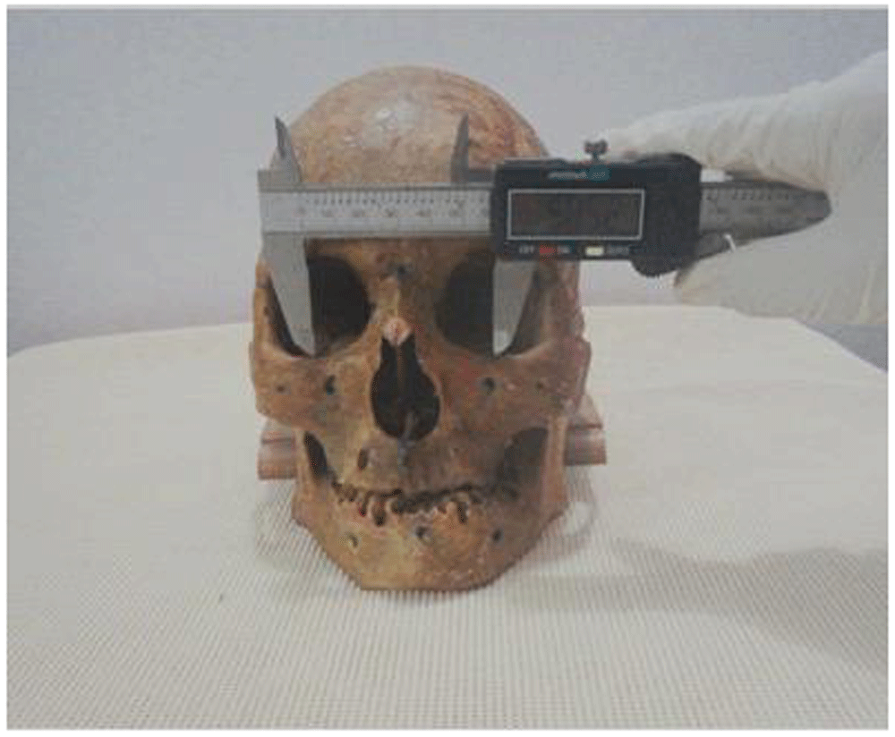
Using a blue permanent marker, 23 anatomical landmarks were identified on each skull by marking small points representing each landmark, then 12 linear measurements were conducted directly on the skull between these anatomical landmarks using an electronic digital calliper (Allendale Electronics Ltd, Hertfordshire, UK) (Figure 1). These linear measurements are shown in Table 1.
Gutta-percha cones, size 80, were cut into 2 mm rod and were glued over the drawn anatomical landmarks on the skull, to be used as radiopaque radiographic markers. After gutta-percha application, the skulls were covered with block of pink wax of 10-12 mm thickness, which was adapted carefully on the facial surface of the skull from the inferior border of the mandible till above the frontonasal suture for soft tissue simulation7,8.
CBCT examinations were performed using Planmeca ProMax 3D Mid CBCT unit (Planmeca, Helsinki, Finland).
The skulls were mounted on the machine, and the laser beams were adjusted to centralize the skull within the scanning field. The skulls were then scanned with a low dose protocol of 90 kVp, 7.1 mA, 9 sec, 600 µm voxel size and 20×20 cm FOV.
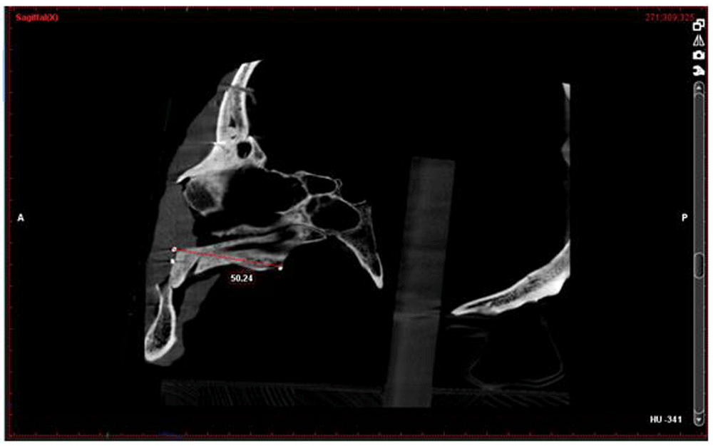
After scanning each skull, the reconstructed images were viewed on the computer screen using Romexis Viewer 4.4.O.R software. The same linear measurements conducted on the skulls were conducted on CBCT orthogonal images (Figure 2; Table 1).
The CBCT and linear measurements were conducted by two observers; the first observer repeated the reading two times with a time interval of one month between each reading.
Statistical analysis was performed using SPSS (version 17), and Microsoft office Excel was used for data handling and graphical presentation. For assessment of the agreement between all measurements Dahlberg error (DE) and Relative Dahlberg Error (RDE) were used together with Intra-class Correlation Coefficients (ICC), including the 95% confidence limits of the coefficient calculated assuming analysis of variance two-way mixed model ANOVA with absolute agreement on SPSS. For both inter and intra observer reliability analysis, DE and RDE were used with ICC, including the 95% confidence limits of the coefficient. Significance level was set at P < 0.05 and two tailed test assumption was applied.
There was no statistically significant difference between the low dose CBCT measurements when compared to direct skull measurements. Using the low dose protocol, mean DE was recorded as 0.83. RDE ranged from 0.8% to 1.9% for almost all measurements (Table 2).
SD, standard deviation; DE, Dahlberg error; RDE, Relative Dahlberg error; ICC, Intraclass Correlation Coefficient.
For many CBCT machines, it is possible to optimize one or more of the investigated exposure parameters and therefore reduce the patient's radiation dose, while maintaining diagnostic image quality and accuracy for some diagnostic tasks9,10. Accordingly, the current study aimed to investigate the effect of reducing the mA and exposure time on the accuracy of CBCT linear measurements as compared with a digital calliper.
The results of the present study revealed that the RDE of almost all the measurements ranged from 0.8% to 1.9%, which didn't exceed 5%. This percentage has been considered as clinically acceptable and permissible relative error in the medical field, as reported by Tarazona-Álvarez et al.11 and Rokn et al.12.
The highest accepted RDE was recorded on the MORw-MORw horizontal linear measurements, 3.4%. The inverse relation between the RDE and the mean of the gold standard of MORw-MORw among all conducted measurements clearly explained its high RDE relative to its low mean of gold standard, which was 21.37%.
The results of Hidalgo et al.13 was in accordance with this study as it showed that the coefficient of variation for measurements was between 1.0% and 1.3% using different tube voltage and tube current. They concluded that a low dose protocol of 80 kV and 3 mA could be used for clinical practice, which represented as much as a 50% dose reduction compared with manufacturer’s recommendations, while giving the operator the freedom to adjust the mA by +0.5 mA on the basis of their judgments of the patient’s size.
Further confirmation was obtained from Vasconcelos et al.14. They concluded that there was no association between the increase in milliamperage and the reliability of the measurements, and recommended the use of low dose protocols when the purpose of the examination is to obtain linear measurements. They added that the 2 mA and 4 mA should be avoided because they could cause degradation to the image and could affect the visualization of the mandibular cortical bone.
In accordance with the current study, Al-Ekrish6 results revealed that on decreasing exposure time the reliability and dimensional accuracy of linear measurements for implant site evaluation were not affected.
In conclusion, the results of this study support the idea that decreasing mA and/or exposure time will not affect the accuracy of linear measurements when craniofacial imaging tasks is required.
Open Science Framework: Dataset 1. Measurements for all 12 linear measurements for both methods: digital calliper and CBCT for both repeats of the experiment, and inter/intra observer measurements, https://doi.org/10.17605/OSF.IO/NH8FE15.
Open Science Framework: Dataset 1. Additional images of the process of linear measurements using both the digital calliper and CBCT, https://doi.org/10.17605/OSF.IO/NH8FE15.
Data are available under the terms of the Creative Commons Zero "No rights reserved" data waiver (CC0 1.0 Public domain dedication).
| Views | Downloads | |
|---|---|---|
| F1000Research | - | - |
|
PubMed Central
Data from PMC are received and updated monthly.
|
- | - |
Provide sufficient details of any financial or non-financial competing interests to enable users to assess whether your comments might lead a reasonable person to question your impartiality. Consider the following examples, but note that this is not an exhaustive list:
Sign up for content alerts and receive a weekly or monthly email with all newly published articles
Already registered? Sign in
The email address should be the one you originally registered with F1000.
You registered with F1000 via Google, so we cannot reset your password.
To sign in, please click here.
If you still need help with your Google account password, please click here.
You registered with F1000 via Facebook, so we cannot reset your password.
To sign in, please click here.
If you still need help with your Facebook account password, please click here.
If your email address is registered with us, we will email you instructions to reset your password.
If you think you should have received this email but it has not arrived, please check your spam filters and/or contact for further assistance.
I recommend publishing a further work on this article in a forensic journal and change the title of “low dose CBCT”. Imitation of soft tissue is a ... Continue reading Dear authors,
I recommend publishing a further work on this article in a forensic journal and change the title of “low dose CBCT”. Imitation of soft tissue is a good idea. You may employ the suggestions of the second reviewer or use clay for example. This type of research is important and lacking in forensic medicine.
The high dose vs low dose CT is not an issue in autopsy cases albeit the image quality and precision of measurements are paramount. Nevertheless, the relatively elevated dose due to the high kV and mAs settings when combined with a large FOV attains a dose range which is comparable with low-dose MSCT protocols (Pauwels et al. 2011; Loubele et al., 2006).
References
Pauwels R, et al. Effective dose range for dental cone beam computed tomography scanners. Eur J Radiol (2011), doi:10.1016/j.ejrad.2010.11.028
LoubeleM,JacobsR,MaesF,etal.Radiationdosevs.imagequalityforlow-dose CT protocols of the head for maxillofacial surgery and oral implant planning. Radiat Prot Dosimetry 2006;117(1–3):211–6.