Keywords
surgery, neuropathy, arteriovenous, congenital, malformation, interventional
surgery, neuropathy, arteriovenous, congenital, malformation, interventional
Carpal tunnel syndrome (CTS) is a condition commonly encountered by physicians and surgeons alike, with the prevalence in the general population ranging from 3–5%1 and accounting for up to 90% of all entrapment neuropathies2. The finding of a congenital arteriovenous malformation (AVM) is a much less common occurrence in clinical practice, with some studies suggesting a prevalence of 1 in 100,000, with the majority of these occurring in the head and neck, with peripheral limb AVMs rarer still3. AVMs have a tendency to grow progressively larger with age, only becoming symptomatic when associated with haemodynamic instability or compression of surrounding structures4.
Medline, PubMed, Ovid, WorldCat, ProQuest and Access Medicine databases were accessed with the input of the following search terms: arteriovenous malformation; arteriovenous malformation upper limb; arteriovenous malformation flexor retinaculum; compressive lesion upper limb. This initial search yielded 1290 results to which the following filters were applied: Peer reviewed; English Language; date within 25 years, which narrowed the results to 675. To further refine the search, of these articles, only those containing the following terms were included: carpal tunnel syndrome; carpal tunnel; median neuropathy; flexor retinaculum, which resulted in a total of 57 articles all of which were reviewed. Of these articles, 8 were identified as being similar.
The most similar case published in the literature was presented by Krishnamoorthy and colleagues who described a case of an 18-year-old man with persistent median artery and causing compression of the carpal tunnel with the median artery itself behaving as the main feeding vessel to the AVM5. Although the pathology described was similar, the lesion was less complex than the one reported in our case and accordingly, attracted a different approach to the management. The remaining 7 cases shared some similarity with respect to the aetiology of symptoms6–10 or the underlying pathology11,12; however, in each of these cases, the lesions described were not as progressive or involved as that which was observed in our patient.
Our patient was a 58-year-old man who had migrated to Australia from South Sudan. He was left hand dominant with no known medical comorbidities or past surgical history. He had been referred to our general surgical outpatients clinic by his general practitioner with a nerve conduction test that had confirmed the presence of a severe left sided CTS.
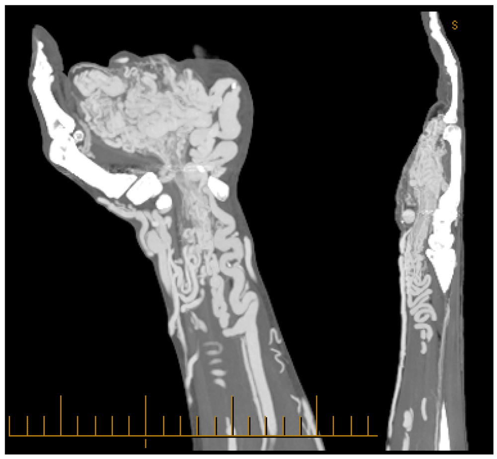
The patient provided us with a 12 month history of transient paraesthesia in the distribution of the median nerve with sparing of the palmar cutaneous region. He reported that his left hand had been pulsatile his whole life and despite being ungainly was, until recently, asymptomatic. On inspection, his symptomatic left hand was 50% larger in comparison to his right. On palpation, thrills were appreciated in association with the multiple aneurysmal bulges. We hypothesised that an AVM was present deep to the flexor retinaculum and had caused some degree of compression to the median nerve. We subsequently arranged for him to undergo CT angiography, which confirmed our suspicions. (Figure 1)

In an attempt to relieve his symptoms we arranged for the patient to undergo angiography with endovascular embolisation. Multiple large aneurysmal sacs were coiled and feeding vessels embolised; however, the extent of the abnormal vasculature in this instance was considerable and definitive treatment was unable to be established without incurring a significant risk of ischaemia to the patient’s hand.
Immediately post-intervention, the patient did not report any difference in his symptoms and this remained to be the case until 6 months post-procedure. At this visit it was observed that his AVM had significantly reduced in size macroscopically and the patient reported improvement in his symptoms. This change correlated with a volumetric reduction on repeat CT angiogram; it was postulated that this could be attributed to a delayed fibrosis following embolisation (Figure 2).
Unfortunately, this improvement was only transient and at 9 months post procedure his AVM had again spontaneously reverted to its original pre-intervention dimensions.
The patient’s case was subsequently discussed at several multidisciplinary meetings, eventually a high-risk surgical procedure involving resection of the AVM with multiple vascular graft reconstruction was offered to the patient. However, he ultimately decided to continue living with his symptoms rather than risk compromising his hand function or viability.
Direct compression of the median nerve by a congenital AVM within the flexor retinaculum is a particularly unlikely cause of carpal tunnel syndrome, made even more remarkable by the fact that the patient in this case only became symptomatic in the fifth decade of life.
Low-flow AVMs may be clinically less concerning as they are more amenable to surgical or endovascular intervention and appear to have lower rates of recurrence when compared to high-flow AVMs.
Most AVMs involing the extremities feature low-resistance niduses between the arteries and veins. Rosen and colleagues suggest that in the management of high-flow vascular malformations if these niduses are not adequately controlled, then attempts at embolising or coiling of the proximal feeding vessels are likely to fail as new collateral feeders will invariably develop to replace them13, such as was the experience with our patient.
Peripheral limb high-flow AVM can be a rare and therapeutically challenging cause of carpal tunnel syndrome. It is a complex issue which warrants multidisciplinary team discussion, as complications of treatment and recurrence must be carefully weighed against the likelihood of improvement on a case-by-case basis.
Formal written consent for publication was obtained from the patient prior to the writing of the case report.
All data underlying the results are available as part of the article and no additional source data are required.
| Views | Downloads | |
|---|---|---|
| F1000Research | - | - |
|
PubMed Central
Data from PMC are received and updated monthly.
|
- | - |
Provide sufficient details of any financial or non-financial competing interests to enable users to assess whether your comments might lead a reasonable person to question your impartiality. Consider the following examples, but note that this is not an exhaustive list:
Sign up for content alerts and receive a weekly or monthly email with all newly published articles
Already registered? Sign in
The email address should be the one you originally registered with F1000.
You registered with F1000 via Google, so we cannot reset your password.
To sign in, please click here.
If you still need help with your Google account password, please click here.
You registered with F1000 via Facebook, so we cannot reset your password.
To sign in, please click here.
If you still need help with your Facebook account password, please click here.
If your email address is registered with us, we will email you instructions to reset your password.
If you think you should have received this email but it has not arrived, please check your spam filters and/or contact for further assistance.
Comments on this article Comments (0)