Keywords
Tuberculosis, Scrofuloderma, Cutaneous Tuberculosis, Cold Abscesses
This article is included in the World TB Day collection.
Tuberculosis, Scrofuloderma, Cutaneous Tuberculosis, Cold Abscesses
Tuberculosis (TB) is a systemic illness caused by a rod-shaped bacillus, Mycobacterium tuberculosis. It is usually pulmonary. Cutaneous TB constitutes 1–2% of extrapulmonary TB and 0.15% of skin diseases; this is significant, considering the overall prevalence of TB is 2% per year1–4.
Cutaneous TB can present in a number of ways clinically, during histopathology as well as in treatment response, making it difficult to diagnose. These cases should be recognized early for timely and accurate management4,5. Cutaneous TB most commonly presents as Tuberculosis cutis colliquativa (also known as scrofuloderma), a type of cutaneous TB presenting with cold abscesses and commonly affecting the supraclavicular region, axilla, and the cervical region6. Lupus vulgaris (LV) is another less common manifestation of TB7. Some rarer ones include inguinal scrofuloderma, ulcerative type of LV, and acute military cutaneous TB3. Cutaneous TB is usually confined to the skin but can be multifocal8.
Here, we describe a case of scrofuloderma presenting with different cutaneous lesions at the same time, which were culture negative for TB.
A 23-year-old male laborer, with no known co-morbidities was admitted to Jinnah Postgraduate Medical Centre, Karachi, Pakistan in March 2018, with complaints of fever for 18 months and multiple swellings on different parts of the body for 12 months.
According to the patient, he had developed a fever, which was gradual in onset, low grade (100oF), intermittent, occurring mostly in the evening and relieved by taking Paracetamol. The fever was not associated with rigors or chills. After six months of fever, the patient noticed multiple swellings of variable size on different parts of body, which included the right lower back, left lower base of neck, upper part of middle chest, front of right ear and upper surface of right foot. The largest swelling was over the back. Swelling was gradual in onset, increasing in size, ranging from lemon size (upper surface of foot) to melon size (abdomen), associated with discharge from left lower neck and chest swelling. Discharge was yellowish in color with no blood in it and was not associated with itching or pain.
The patient had a history of undocumented weight loss for one year and using antipyretics and proton pump inhibitors (Omeprazole, 20mg once daily). The rest of the history was unremarkable.
On general physical examination, the patient was average height and thinly built, cooperative, with visible parotid swelling on the right side, and lying comfortably on the bed. His vitals were all normal. There was no pallor, icterus, cyanosis, clubbing, koilonychias, splinter hemorrhages, edema, or ear discharge. Oral and thyroid examination was normal.
Bilateral anterior cervical and inguinal lymph nodes were palpable, firm in consistency, non-tender, matted, mobile, with no underlying erythema or discharging sinus, not adherent to overlying skin or any underlying structure. Size ranged from 2cm to 4.5cm. Parotid swelling was noted, non-tender, soft in consistency, size ranging from 5cm to 7cm, mobile over underlying structure, with no overlying erythema, sinus or discharge. There was a round, well demarcated lesion on the chest, ulcerated, 3 inches in size, reddish color with a yellowish crust. Right lumbar swelling measured ~3×16cm, soft, non-tender, non-discharging and without any gibbus. There was a scar noted over the left supra-clavicular region, above and parallel to the clavicle. Figure 1 provides images of the patient’s swellings. There was no hyperpigmentation or petechiae noted. Bone tenderness was absent. Systemic examination was found to be unremarkable.
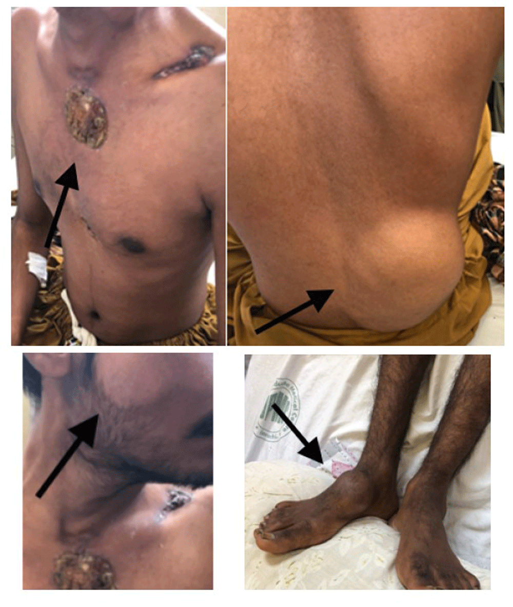
A round, well demarcated lesion on the chest, ulcerated, 3 inches in size, reddish color and with yellowish crust. Right lumbar swelling measured about 13×16cm, soft, non-tender, non-discharging and without any gibbus. A swelling was also noted on the right foot.
The patient’s laboratory test results are presented in Table 1. Viral markers and HIV testing were negative, while pus for gram staining and Acid-Fast Bacillus staining was also negative. Chest X-ray was unremarkable.
On ultrasound of the abdomen, the liver was 14cm and spleen was 11.7cm in size with normal borders. A psoas abscess was found measuring 11×11.9cm with septations and calcifications. Fine-needle aspiration cytology of the cheek swelling showed necrotizing inflammation. Fungal stain was negative and skin split test for Leishmania donovani bodies was also negative. Mycobacterium tuberculosis was detected after GeneXpert analysis of the pus from the psoas abscess, which had previously shown no growth on culture. Blood culture was also negative.
Computed tomography of chest with contrast showed multiple fluid collections at right supraclavicular region, anterior chest wall and along with bilateral psoas abscess formation predominantly on right side.
Initially, a differential diagnosis of deep fungal infection (actinomycosis), cutaneous leishmaniasis, lymphoma and TB was made. After GeneXpert, HIV serology, and fungal stain were performed, cutaneous tuberculosis (scrofuloderma) was the final diagnosis and anti-tuberculous therapy was started. After consulting the Dermatological Department, Lupus vulgaris was also ruled out. A standard regimen of four drugs, i.e. Isoniazid (5 mg/kg), Rifampin (10 mg/kg), Ethambutol (20 mg/kg), and Pyrazinamide (24 mg/kg), in combined form, i.e. Tablet Myrin-P-Forte 4 × daily, was given for two months and converted to the combination of Isoniazid and Rifampin in combined form, i.e. Tablet Rifna 4 × daily, for a further 8 months. The patient was discharged after 8 days and kept on regular follow-up.
The incidence of TB is increasing worldwide, which may be due to the growing number of HIV cases and resistance to antituberculous drugs4. Immunocompromised individuals have a greater propensity to get this disease. Cutaneous TB is either transmitted via a hematogenous route or direct extension from a primary focus, which is elsewhere in the body, usually pulmonary. However, primary infection can also occur by direct introduction of the microbe into the skin or mucosa of a susceptible individual by trauma or injury4.
In our case, no primary source was found in any organs and there was no history of TB contact. Moreover, there was no reason to identify the patient as immunocompromised.
Cutaneous TB rarely involves the region of head and neck, especially lymph nodes, larynx, oropharynx, salivary glands, nose, ear, skin, and paranasal sinuses9. Our patient had bilateral cervical and inguinal lymphadenopathy, parotid swelling, an ulcerated lesion on the chest, a right lumbar swelling, and a scar on the left supra-clavicular region. This distribution also favored our final diagnosis of scrofuloderma.
Due to the different type of lesions, correct diagnosis may be delayed because investigations fail to detect TB, as in our case where cultures were negative and TB was only detected by GeneXpert10.
Our case illustrates that scrofuloderma, although a rare disease with a wide spectrum of cutaneous lesions and higher rates of false negative investigations, should still be considered in differential diagnosis of cold abscesses and nodules, especially of the head and neck region.
No data are associated with this article.
Written informed consent for publication of their clinical details was obtained from the patient
| Views | Downloads | |
|---|---|---|
| F1000Research | - | - |
|
PubMed Central
Data from PMC are received and updated monthly.
|
- | - |
Provide sufficient details of any financial or non-financial competing interests to enable users to assess whether your comments might lead a reasonable person to question your impartiality. Consider the following examples, but note that this is not an exhaustive list:
Sign up for content alerts and receive a weekly or monthly email with all newly published articles
Already registered? Sign in
The email address should be the one you originally registered with F1000.
You registered with F1000 via Google, so we cannot reset your password.
To sign in, please click here.
If you still need help with your Google account password, please click here.
You registered with F1000 via Facebook, so we cannot reset your password.
To sign in, please click here.
If you still need help with your Facebook account password, please click here.
If your email address is registered with us, we will email you instructions to reset your password.
If you think you should have received this email but it has not arrived, please check your spam filters and/or contact for further assistance.
Comments on this article Comments (0)