Keywords
basal cell adenoma, rare case, case report
basal cell adenoma, rare case, case report
The National Cancer Institute, USA, describes adenoma as “a tumor that is not cancer. It starts in gland-like cells of the epithelial tissue (thin layer of tissue that covers organs, glands, and other structures within the body)”1. Basal cell adenomas are a rare type of benign glandular tumors, salivary in origin. Accounting for less than 1-2% of salivary gland tumors2, they are located usually superficial within the glandular body, and often a brownish appearance is observed3. First described by Kleinasser and Klein in 1967, they are termed as rare benign tumors with a high recurrence rate and, in general, good prognosis and recognized as an independent entity in the Second Edition of the Salivary Gland Tumors Classification of the World Health Organization4. Normally indigenous to (parotid) salivary glands, basal cell adenomas have also been reported in the buccal mucosa, palate and nasal septum. They are often seen in patients in their fifth-seventh decade of life, a contrast to other benign tumors5. Basal cell adenoma can be divided into four subtypes: solid, trabecular, tubular and membranous6. Histologically characterized by the presence of uniform and regular basaloid cells having two differenced morphologies, the tumor cells are intermingled. Among the two groups, one has characteristic constitution of small cells with little cytoplasm and intense basaloid nuclei located near the tumoral nests while another is made up of large cells with abundant cytoplasm and pale nuclei in the centre of tumoral nests. The tumoral nests are separated from the surrounding connective tissues by a basal membrane like structure, which surrounds them5. Immunologically speaking, they strongly express PAN CK and CK 5/6, along with Calponin, P-63 and weak staining of Ki-67. Vimentin, actin also stain positive in basal cell adenomas, along with S-100 (alpha subunit) in ductal cells and S-100 (beta subunit) in basaloid cells.
A 63-year-old woman of Chinese (Han) origin was referred to Taihe Hospital, Department of Digestive Medicine after esophagogastroduodenoscopy (EGD) in her county hospital due to a protuberant mass in the esophagus, 35 cm from the incisors. The patient had initially complained of sensation of regurgitation and was a non-smoker and non-drinker. There was no history of heartburn, chest pain, nausea, vomiting or diarrhea. She also had no complaints of hematemesis, hematochezia or melena. The patient was non-diabetic, non-hypertensive, with no history of renal disorders, cardiac diseases or tuberculosis. She had no remarkable family history (medically relevant) and no signs of genetic disorders. The patient had undergone appendectomy 13 years back and had been diagnosed and treated for intestinal obstruction 4 years previously. Laboratory investigation reports were normal. Upper abdominal and chest contrast-enhanced computer tomography (CECT) showed: lower esophagus and cardiac region of stomach wall hypertrophy (highly indicates presence of a tumor); right hepatic lobe diffuse (indicates small cyst present); B/L lung field showed some scattered fibrotic and calcified lesions.
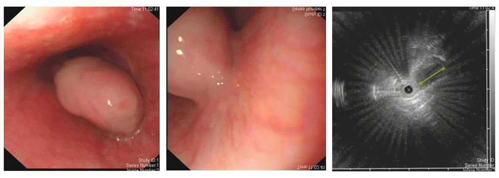
EGD was repeated and showed a protuberant lesion in the esophagus, 3cm in size, 35 cm from the incisors (Figure 1). It also showed another protuberant lesion 0.7 cm in size in the gastric cardia. Multiple yellow spots (gastric xanthoma) were seen in the stomach. Pylorus, pyloric sphincter and duodenum were normal. Endoscopic ultrasound (EUS) of the protuberance in the esophagus showed that the lesion as hypoechoic, an originated from and limited to the submucosal layer with an area of more than 3 cm (Figure 1). No lymphadenopathy was found around the lesion. Radiofrequency ablation was done on the yellow spots.

(EGD by OLYMPUS EVIS LUCERA CV-260SL; EUS by OLYMPUS MAJ-935.)
With the working diagnosis of esophageal leiomyoma/polyp, the patient was scheduled for surgery. Endoscopic submucosal dissection (ESD) was done under general anesthesia (Figure 2). The surgery went well without complications. The protuberant mass was then sent for histopathological examination and the patient was kept for observation.

Right panel: Closure of the defect by hemoclips. (ESD by single-use Olympus DualknifeTM, tip thickness-0.3mm.)
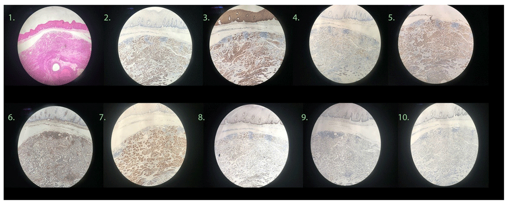
Immunohistology of the sample showed Calponin (+), CD117 (+), CD 56 partially (+), CKP (+), GATA-3(-), CK5/6 (+), CK7 (+), SOX 10(+), P63 (+), INI-1 (+), and Vimentin (+) (Figure 3). It also showed P63 (+), S-100(+), KI-67 about 2%. Light microscopy revealed the submucosal layer of esophagus showing glandular duct like structure also with double layer epithelium. Around the gland duct there was transparent substance partially solid in nature. The sample also showed two types of epithelium-muscular epithelium and glandular epithelium, which corresponds with the diagnosis of basal cell adenoma, a type of salivary gland tumor. The patient was then discharged seven days after surgery with PPI (Cap. Lansoprazole 30mg BD for 45 days), Itopride (50mg TDS for 45 days) and Hydrotalcite (1gm TDS for 7 days) as discharge medication. Follow up was scheduled two months later.

Follow-up EGD of the patient was performed two months later and showed no recurrence of the tumor. The patient has had no fresh complaints since the ESD procedure (Figure 4).
Basal cell adenomas are uncommon variants of benign salivary gland tumor with varying recurrence rates. There have been very few reported cases of basal cell adenomas in ectopic sites. Its rarity makes comparison between two similar cases very difficult7. Basal cell adenomas are generally characterized by no regional node involvement, no calcification and no cystic component within the tumor8.
This case posed a challenge to our team since ectopic occurrences of basal cell adenoma have been reported very rarely in literature and is not a very common occurrence in itself. The prognosis of basal cell adenoma, when found at ectopic sites is not clear and thus poses a challenge to predict the outcome even after intervention.
Differentiation between benign and malignant basal cell adenoma is vital since they share the same site and characteristics. However, the mechanism and rate of progression is vastly different. Histology is not alone enough to differentiate or predict the progression, hence immunohistology plays a vital role in differentiation. Immunohistological staining with CD5/6, Keratin, Alpha-1 antichymotrypsin, CEA, S-100, Vimentin, CD117, Actin must be done to differentiate it from other benign and malignant tumors.
Whenever encountered and suspected, differentials must be established in favor of basal cell adenoma. Pleomorphic adenoma, adenoid cystic carcinoma, basaloid squamous carcinoma must be excluded first with the help of gross microscopic examination and immunohistological staining.
Surgical excision remains the first and foremost approach of choice for treatment of basal cell adenoma. However, during removal, it is mandatory not to disturb the tumor capsule to prevent recurrence. Moreover, the patient must be kept under regular follow-up to monitor malignant transformation as well as recurrence.
Written informed consent was obtained from the patient for the publication of this case report and any associated images.
All data underlying the results are available as part of the article and no additional source data are required.
| Views | Downloads | |
|---|---|---|
| F1000Research | - | - |
|
PubMed Central
Data from PMC are received and updated monthly.
|
- | - |
Is the background of the case’s history and progression described in sufficient detail?
Yes
Are enough details provided of any physical examination and diagnostic tests, treatment given and outcomes?
Partly
Is sufficient discussion included of the importance of the findings and their relevance to future understanding of disease processes, diagnosis or treatment?
Yes
Is the case presented with sufficient detail to be useful for other practitioners?
Yes
Competing Interests: No competing interests were disclosed.
Reviewer Expertise: Gastrointestinal and Liver Pathology
Is the background of the case’s history and progression described in sufficient detail?
Yes
Are enough details provided of any physical examination and diagnostic tests, treatment given and outcomes?
Yes
Is sufficient discussion included of the importance of the findings and their relevance to future understanding of disease processes, diagnosis or treatment?
Yes
Is the case presented with sufficient detail to be useful for other practitioners?
Partly
Competing Interests: No competing interests were disclosed.
Alongside their report, reviewers assign a status to the article:
| Invited Reviewers | ||
|---|---|---|
| 1 | 2 | |
|
Version 1 16 Jan 19 |
read | read |
Provide sufficient details of any financial or non-financial competing interests to enable users to assess whether your comments might lead a reasonable person to question your impartiality. Consider the following examples, but note that this is not an exhaustive list:
Sign up for content alerts and receive a weekly or monthly email with all newly published articles
Already registered? Sign in
The email address should be the one you originally registered with F1000.
You registered with F1000 via Google, so we cannot reset your password.
To sign in, please click here.
If you still need help with your Google account password, please click here.
You registered with F1000 via Facebook, so we cannot reset your password.
To sign in, please click here.
If you still need help with your Facebook account password, please click here.
If your email address is registered with us, we will email you instructions to reset your password.
If you think you should have received this email but it has not arrived, please check your spam filters and/or contact for further assistance.
Comments on this article Comments (0)