Keywords
Key words: heparin; thrombocytopenia; thrombosis; coronary artery; stent; myocardial infarction; femoral artery; coronary angioplasty; thrombectomy
Key words: heparin; thrombocytopenia; thrombosis; coronary artery; stent; myocardial infarction; femoral artery; coronary angioplasty; thrombectomy
Heparin is a commonly used anticoagulant for hospitalized patients, but its use can lead to devastating complications, such as heparin-induced thrombocytopenia (HIT)1. We report an unusual case of concomitant subacute coronary stent and femoral artery thrombosis in the setting of HIT. This case highlights the importance of considering HIT as a cause of coronary or arterial thrombosis few days after heparin exposure.
A 66-year-old male patient, with a history of smoking (30 pack-years) and no known medical or surgical history, was admitted in our department for a spontaneously resolved inferior ST elevation myocardial infarction (STEMI). The intra-hospital treatment included enoxaparin 0.6 ml twice a day, clopidogrel 75 mg once a day, aspirin 100 mg once a day, bisoprolol 2.5 mg once a day and atorvastatin 40 mg once a day. The coronary angiogram (performed at day 3 through the right radial artery) showed a severe thrombotic lesion of the distal circumflex. The patient underwent an ad-hoc successful angioplasty of the circumflex with a drug eluting (everolimus) stent. Initial laboratory tests at admission were normal except elevated troponin. Echocardiography showed a 65% left ventricular ejection fraction. The patient was discharged after 5 days of anticoagulation by low molecular weight heparin (enoxaparin). Laboratory tests were not controlled during the hospitalization. The discharge treatment included clopidogrel 75 mg once a day, aspirin 100 mg once a day, bisoprolol 2.5 mg once a day and atorvastatin 40 mg once a day.
One week later, the patient was referred again to our department for both chest and right lower limb pain. The electrocardiogram showed an inferior STEMI and the physical exam of the right lower limb found ischemic signs with absence of the femoral pulse. There was no history of aspirin or clopidogrel discontinuation. An urgent coronary angiogram (performed through the left femoral artery) showed total thrombosis of the circumflex stent (Figure 1A). The patient underwent a successful primary angioplasty of the circumflex by simple balloon (Figure 1B). Urgent lower limb contrast-enhanced computed tomography was performed immediately after the angioplasty, revealing total acute thrombosis of the right common femoral artery (Figure 2A, B). The patient underwent an urgent successful thrombectomy with Fogarty catheter. Immediate evolution was favorable with total regression of coronary and right lower limb ischemic signs. Laboratory tests showed a marked fall in the platelet count (68,000/L) which was normal (364,000/L) in the previous hospitalization. A diagnosis of concomitant coronary stent and femoral artery thrombosis due to HIT was strongly suspected (4T score = 8). Our therapeutic strategy was immediate discontinuation of low molecular weight heparin (enoxaparin), aspirin and clopidogrel with strict daily control of platelet count. During this period, no alternative anticoagulation was initiated because of the unavailability of direct thrombin inhibitors in our center. Anticoagulation with a vitamin K antagonist (acenocoumarol 4 mg once a day) and dual antiplatelet therapy with aspirin 100 mg once a day and clopidogrel 75 mg once a day were initiated at day 3 once platelet count had recovered. The in-hospital outcome was favorable and the patient was discharged after 15 days on acenocoumarol 4 mg once a day, aspirin 100 mg once a day and clopidogrel 75 mg once a day. The 3-month follow-up, with controlled blood tests and lower limb contrast-enhanced computed tomography showing total reperfusion of the right femoral artery (Figure 2C), was unremarkable.
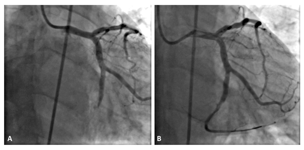
(A) The intra-stent thrombosis of the circumflex coronary artery. (B) The final result after successful angioplasty of the circumflex coronary artery.
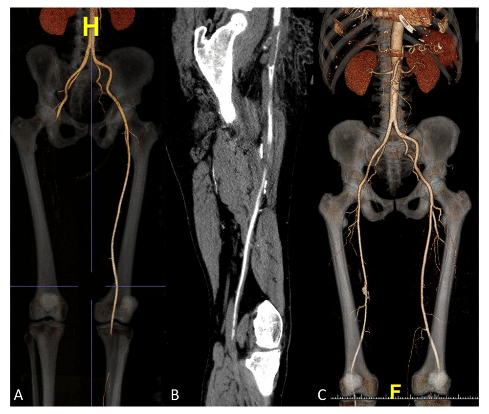
(A, B) show the thrombotic occlusion of the right common femoral artery. (C) Reperfusion of the right femoral artery after thrombectomy.
HIT is defined as a sudden fall in the platelet count (e.g. <100,000 per L or >50% drop from baseline) a few days after heparin use. Its incidence ranges from <1% to 7% depending on the heparin type (more than 10 times higher with unfractionated heparin compared to low-molecular-weight heparin), duration of heparin exposure and patient population1. There are two types of HIT, with type I HIT the most common form. Type 1 HIT is due to the direct effect of heparin on platelets and may manifest as only a slight decrease in platelet count, mostly within 2 days of commencing heparin. Type II HIT is secondary to the formation of antibodies against the heparin-platelet factor 4 complex, resulting in 50% of cases in thrombotic events, mostly in veins, within 5 to 14 days. Coronary artery thrombosis secondary to HIT is very rare and usually occurs in the setting of coronary stents or bypass grafts2. However, the concomitant occurrence of coronary stent and other arterial site thrombosis secondary to HIT is very rare and few cases have been reported in the literature3. The use of scoring systems such as the “4T score” is helpful in assessing the pretest probability of HIT4. Platelet factor 4–heparin antibody tests should be ordered only if the diagnosis of HIT is strongly suggested by clinical features5.
Treatment of HIT requires immediate discontinuation of all heparin products and initiation of alternative therapeutic dose anticoagulation, including direct thrombin inhibitors (argotraban, bivalirudin, fondaparinux, danaparoid) or direct oral anticoagulants (apixaban or rivaroxaban or dabigatran)6. The decision to continue antiplatelet therapy during treatment with a non-heparin anticoagulant may be influenced by the risk of vascular events and bleeding. Routine platelet transfusion is not recommended for patients with acute HIT and thrombosis or average bleeding risk, but it may be an option for patients with active bleeding or at high bleeding risk. In the acute phase of HIT with life-threatening thrombosis, bivalirudin (or argatroban if bivalirudin is unavailable) is the best option for alternative anticoagulation therapy7. Primary angioplasty and thrombectomy with Fogarty catheter should be recommended in the setting of life-threatening thrombosis with STEMI or acute limb thromboembolism8. Oral anticoagulation is required for at least 3 months, preferably with direct oral anticoagulant, which can be initiated at the first day. Warfarin is the most highly recommended vitamin K antagonist when indicated, and should not be given until platelets have substantially recovered (e.g. usually to at least 150 000 per L)7. In our case, acenocoumarol was the sole available alternative anticoagulation therapy. It was initiated at day 3 after platelet count recovery and was continued for 3 months.
Concomitant coronary and femoral artery thrombosis due to HIT is a rare life-threatening complication of heparin therapy. The present case highlights the importance of considering such diagnosis among patients with prior heparin exposure. Prompt identification and management of this disorder is critically important to avoid devastating complications. To prevent such events, strict control of platelet count during heparin therapy is of paramount importance.
All data underlying the results are available as part of the article and no additional source data are required.
Written informed consent for publication of their clinical details was obtained from the patient.
| Views | Downloads | |
|---|---|---|
| F1000Research | - | - |
|
PubMed Central
Data from PMC are received and updated monthly.
|
- | - |
Provide sufficient details of any financial or non-financial competing interests to enable users to assess whether your comments might lead a reasonable person to question your impartiality. Consider the following examples, but note that this is not an exhaustive list:
Sign up for content alerts and receive a weekly or monthly email with all newly published articles
Already registered? Sign in
The email address should be the one you originally registered with F1000.
You registered with F1000 via Google, so we cannot reset your password.
To sign in, please click here.
If you still need help with your Google account password, please click here.
You registered with F1000 via Facebook, so we cannot reset your password.
To sign in, please click here.
If you still need help with your Facebook account password, please click here.
If your email address is registered with us, we will email you instructions to reset your password.
If you think you should have received this email but it has not arrived, please check your spam filters and/or contact for further assistance.
Comments on this article Comments (0)