Keywords
3D conformal radiotherapy, Breast cancer, Progression free survival, Luminal subtypes, HER2 neu receptors
3D conformal radiotherapy, Breast cancer, Progression free survival, Luminal subtypes, HER2 neu receptors
Breast cancer is a common cancer in women, and it is estimated that one in eight women in the US will develop it in their life1. It is the leading cancer among women in both Europe and the US and has become an emerging disease in developing countries2,3. In Iraq, 3845 patients were estimated to have breast cancer in 20114. Therapy requires a multidisciplinary team involving surgeons, medical oncologists, radiation oncologists, pathologists, radiologists, and supportive care personnel1. Adjuvant radiotherapy decreases locoregional relapse, increases survival and palliates symptoms; after breast-conserving surgery (BCS), it is an essential component of treatment1. In oncology, progression free survival (PFS) referred to the length of time during and after the treatment of a cancer, that a patient lives with it but it does not get worse and refers to situations in which a tumor is present, as demonstrated by laboratory, radiology, and/or clinical evidence5. In developing countries, survival rates are poorer than those in developed countries6. Approximately 30% patients with early-stage cancer develop metastases, whereas metastatic breast cancer occurs in 6 – 7% of newly diagnosed patients7. The 5-year survival rate with metastasis is 25%, and the overall survival (OS) is reported to be 24 months8. Adjuvant radiotherapy reduces the incidence of locoregional recurrence from 30% to 10.5% at 20 years and decreases deaths by 5.4% at 20 years9.
This study aimed to assess PFS in patients treated by hypofractionated three-dimensional conformal radiotherapy (3DCRT) and correlate PFS with patients' clinical and pathological profiles.
We retrospectively reviewed 299 women who were consecutively treated at Baghdad Radiation Oncology Center between October 2017 and May 2018. Patients were included if they planned to undergo adjuvant 3DCRT. Medical records were reviewed in detail after approval was obtained from our institution. All women were treated after completing chemotherapy cycles in a period not less than 13 weeks from the last cycle.
1. T3, T4 stages.
2. All patients with node positive status.
3. All patients after breast conservative surgery.
4. Multifocal breast tumors, extensive DCIS, central tumors in a small breast and incomplete excision.
1. Significant pre-existing cardiac or lung disease.
2. Scleroderma.
3. Limited shoulder mobility.
4. M1 breast cancer (metastasis).
Individual data and information on primary and advanced disease were collected. The following variables were studied as possible prognostic factors for specific clinical outcomes: age, tumor-node-metastasis staging, lymph nodes (LN), histopathology, grades, estrogen receptors, progesterone receptors, human epidermal growth factor receptor 2 (HER2) neu, surgery types, body mass index (BMI), period between the last cycle of chemotherapy and radiotherapy, date of radiotherapy, follow-up time, and metastatic or recurrent events (brain, supraclavicular LN, axillary LN, liver, chest wall scars and bones).
1. 4005 cGy in 15 fractions over 3 weeks adopted in mastectomy, the indications are10–13:
a. Involvement of one or more axillary lymph nodes.
b. T3 (>5 cm tumor), T4 (skin/chest wall invasion) or stage III tumor.
c. Positive surgical margin.
d. Gross residual disease.
e. Multiple primary tumors (multi-centricity).
2. 4005 cGy / 15 fractions / 3 weeks performed for breast conserving surgery BCS plus 1000 cGy / 5 fractions / 1 week as booster10,11,13.
Data were first entered into an Excel file, then transferred for statistical analysis into SPSS v22. For categorical variables, such as age, histo type, ER, PR, HER2, the frequency distribution calculated and analyzed with Fisher’s test and chi-squared. Correlation coefficient (r) was used to detect significance relations. Kaplan-Meier survival curves for PFS were used, which is a way of graphically displaying the time until the study developed an endpoint, often death, or an event such as recurrence of cancer, which was obtained during follow-up.
Written informed consent was obtained from the patients, for the use of their data in this study, and the study was conducted according to the ethical standards established by the 1964 Declaration of Helsinki. The Medical Ethical Committee of College of Medicine, Baghdad University approved this study (code: 6/2018), which covers the Baghdad Radiation Oncology Center.
The majority of patients were in the age group 46–55 years (115, 38.5%). BMI revealed that the majority of the patients were either overweight (60, 27.2%) or had moderate obesity (73, 33%) (Table 1).
| Variables | N (%) | Mean ± SD | |
|---|---|---|---|
| Age (years) Range: 25–27 | 25–35 | 36 (12) | 49.9±11 |
| 36–45 | 70 (23.4) | ||
| 46–55 | 115 (38.5) | ||
| 56–65 | 61 (20.4) | ||
| 66–75 | 17 (5.7) | ||
| Total | 299 | ||
| BMI (m2/Kg) Range: 18–43 | Underweight (<18.5) | 2 (0.9) | 29±5.2 |
| Normal (18.6–24.9) | 51 (23.1) | ||
| Overweight (25–29.9) | 60 (27.2) | ||
| Moderate obesity (30–34.9) | 73 (33) | ||
| Severe obesity (35–39.9) | 29 (13.1) | ||
| Morbid obesity (>40) | 6 (2.7) | ||
| Total | 221 (78 missing)* | ||
| Molecular subtypes (n=293) | |||
| HER2 enriched | 29 (9.9) | ||
| Triple negative | 33 (11.3) | ||
| Luminal B | 34 (11.6) | ||
| Luminal A | 197 (67.2) | ||
| Total | 293 (6 missing)* | ||
| Events | |||
| Non-relapse | 264 (88.3) | ||
| Relapse** | 35 (11.7) | ||
| Total | 299 (100) | ||
Luminal A breast cancer type present in 197 patients (67.2%), while non-luminal A phenotypes were recorded as luminal B subtype (34, 11.6%), triple negative (33, 11.3%) and HER2 enriched (29, 9.9%) (Table 1).
The T2 stage was predominant (156, 53.9%), which mostly comprised luminal A (105, 36.3%). Other stages and subtypes presented in different proportions (Table 2). The results showed a high frequency of N1 in 109 patients (37.2%), related to molecular luminal A (69, 23.4%) (Table 2).
| T stages | Molecular subtypes | Total | |||
|---|---|---|---|---|---|
| Luminal A (%) | Luminal B (%) | HER2 enriched (%) | Triple negative (%) | ||
| T1 | 36 (12.4) | 10 (3.4) | 6 (2.0) | 2 (0.7) | 54 (18.6) |
| T2 | 105 (36.3) | 18 (6.2) | 15 (5.1) | 18 (6.2) | 156 (53.9) |
| T3 | 40 (13.8) | 5 (1.7) | 6 (2.0) | 7 (2.4) | 58 (20.0) |
| T4 | 13 (4.5) | 1 (0.3) | 2 (0.7) | 5 (1.7) | 21 (7.2) |
| Total | 194 (67.1) | 34 (11.7) | 29 (10.0) | 32 (11.0) | 289 (10 missing)* |
| Fisher's Exact test=9.15, Chi-square=9.5, P = 0.031 | |||||
| N stages | |||||
| N0 | 43 (14.6) | 6 (2.0) | 7 (2.3) | 5 (1.7) | 61 (20.8) |
| N1 | 69 (23.5) | 16 (5.4) | 12 (4.1) | 12 (4.1) | 109 (37.2) |
| N2 | 56 (19.1) | 10 (3.4) | 5 (1.7) | 12 (4.1) | 83 (28.3) |
| N3 | 29 (9.9) | 2 (0.6) | 5 (1.7) | 4 (1.3) | 40 (13.6) |
| Total | 197 (67.2) | 34 (11.6) | 29 (9.9) | 33 (11.2) | 293 (6 missing)* |
| Fisher's Exact Test=6.2, Chi-square=6.23, P = 0.004 | |||||
A significant result was obtained between age, BMI, and T stage when correlated to molecular subtypes, ER and HER2 neu receptors. Age correlated to molecular subtypes (r = +1); there was good correlation (2-tailed = 0.46 and P = 0.043). BMI was significantly correlated to estrogen receptors (ER) (r = +1, 2-tailed = 0.5 and P = 0.046). Furthermore the correlation between T stages and HER2 neu receptors showed a significant value (r = +1, 2-tailed = 0.99 and P = 0.001).
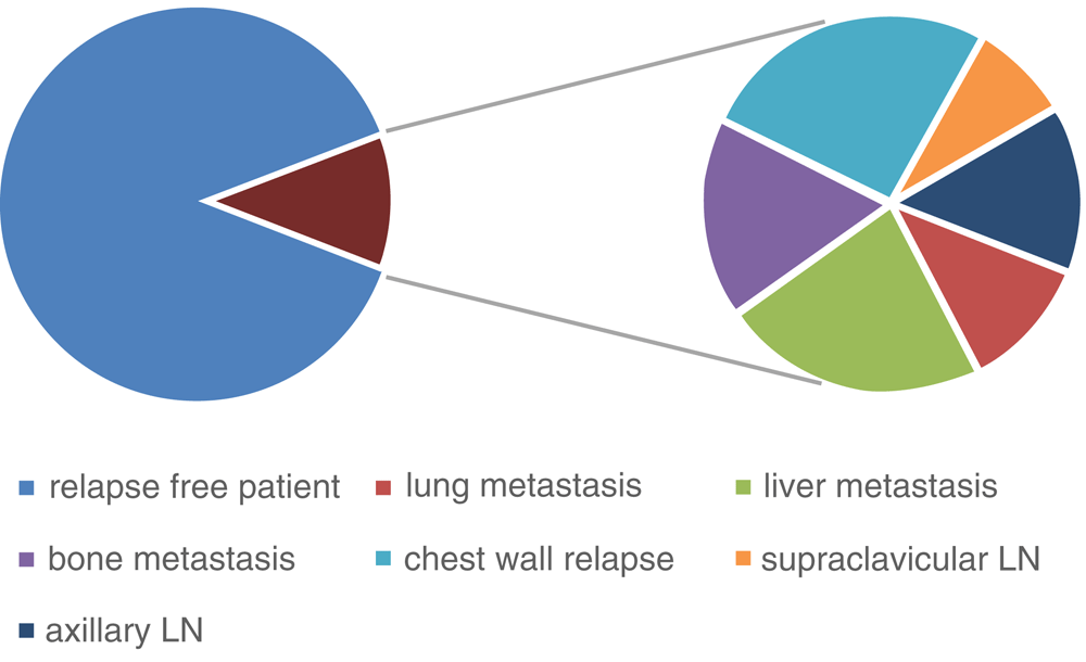
Relapse occurred in 35 (11.7%) women, while the reminder (264, 88.3%) were non-relapsed (Table 1). The waiting period from last cycle of chemotherapy to the date of starting radiotherapy in weeks of this study had median 17 weeks and mode about 9 weeks, which ranged from 4 to 30 weeks (Figure 1).

PFS in this study presented in 264 women, while relapsing was evident in 35 cases. Chest wall relapse occurred in 9 patients (25.7%). Other relapses included liver metastasis, bone secondaries, axillary LN relapse, lung metastasis and supraclavicular LN in 8 (22.9%), 6 (17.1%), 5 (14.3%), 4 (11.4%), 3 (8.6%) patients, respectively (Figure 2).
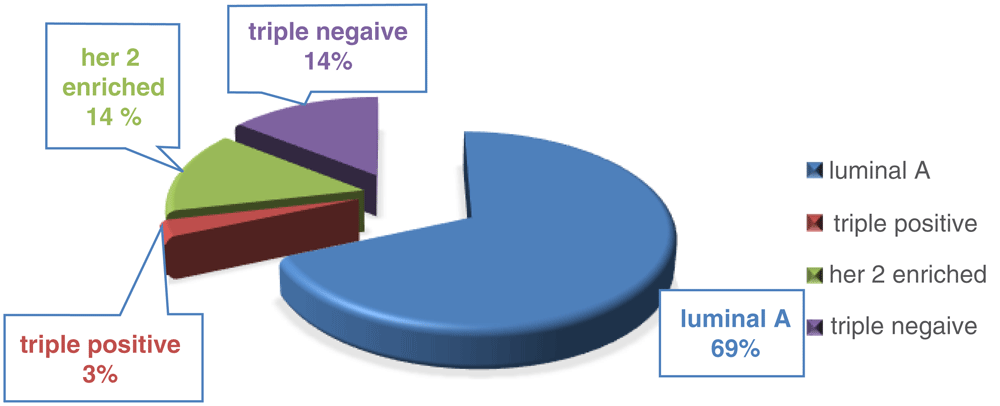
The luminal A subtype relapsed in 69% of patients which represented major subtypes reliable for relapse, while HER2 enriched and triple negative subtypes occurred in 14% patients for both and 3% for luminal B (Figure 3).
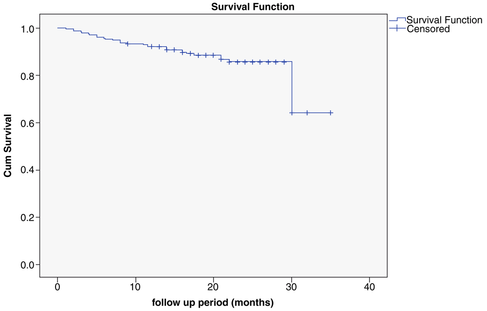
Kaplan Meier survival curve estimation of PFS is shown in Figure 4. The PFS rates for relapse were 35 (11.7%) versus 264 (88.3%) for non-relapse, estimated using this curve, which indicated that when a woman received adjuvant radiotherapy, she was about nine times more likely to survive beyond the time than someone who did not receive adjuvant radiotherapy. The follow-up period was 12 months; the Kaplan Meier estimates of PFS for all the patients was censored at 9 months and the last was at 35 months. There was a trend for better PFS in patients receiving adjuvant radiotherapy, so radiotherapy was significantly associated with prolonged PFS.
In the present study, the age of patients was 25–75 years with a mean± standard deviation of 49.9±10.99 years, with the majority of patients in the group of 46–55 years (115, 38.5%). This is similar to the results of previous studies conducted in Iraq: Al-Rawaq et al., 201614, Al-Khafaji et al., 201015, and Al-Naqqash et al., 200916. Age represents an important factor both for occurrence and management of breast cancer17. In our study, the median age of onset was > 10 years younger than that shown in studies in Europe and North America18. It has been proposed that these differences are due to differences in exposure to hormones, diet, physical activities, and other risk factors17. In most Arabian countries, breast cancer is commonly diagnosed in women younger than 50 years, which is consistent with the findings of our study, unlike in the US, where women aged 50 years and older are most commonly affected18.
The molecular subtype plays an important role in follow-up survival and prognosis in breast cancer. Luminal A (67.2%), luminal B (11.6%), triple-negative (11.3%), and HER2-enriched (9.9%) subtypes were observed in the present study. These results are similar to those found in Cheang’s study18 and unlike those of other studies, such as Al-Naqqash et al., 200916, Al-Sarraf et al., 201519, and El-Fatemi and Chahbounil, 201220. In the present study, luminal A relapse was recorded in 69% of patients, while the reminder were non-luminal A; HER2-enriched, triple-negative and luminal B occurred in 14%, 14%, and 3% of patients, respectively. These results were inconsistent with the results of Fitzgibbons et al., 200021 and Al-Sarraf et al., 201519. Luminal A has the most favorable prognosis, with locoregional relapse rates of only 3–8% at 10 years20. HER2-enriched groups exhibit the highest rates of local recurrence and regional relapse21.
Tumor size (T stage) was directly correlated with survival and metastasis in the present study. Size rank is among the strongest predictors of metastasis, disease-free survival (DFS), and OS. Although tumor size correlates strongly with the presence of axillary LNs, this is clearly an independent prognostic factor5,21. Among those with documented negative nodes, tumor size remained a strong and independent predictor5. Many studies showed a 20 year DFS of 79% for those with tumors smaller than 2 cm, compared with 64% for those with tumors larger than 2 cm5. This study demonstrated T2 stage was the most common (53.9%), followed by T3, T1 and T4; regarding the T stage related to molecular subtype, T2/luminal A was most common (36.3%). These results were similar to those of Al-Rawaq et al., 201614, Al-Khafaji et al., 201015, and Al-Naqqash et al., 200916, but different than those of Goldhirsch et al., 201322 and Cheang et al., 200918.
The status of LNs is an important prognostic factor related to survival and predictors of systemic micro-metastases22, disease recurrence, and poor prognosis23. In the present study, the N1 frequency was 37.2% followed by N2, N0, and N4. The N1/luminal A frequency was 23.5%, which was recorded commonly, whereas the N3/luminal B frequency was the lowest (0.6%). These agree with the results of Al-Rawaq et al., 201614 and Al-Khafaji et al., 201015, while they disagree with those of Goldhirsch et al., 201322 and Cheang et al., 200918.
In the present study, the PFS, which was obtained using Kaplan Meier survival curves, estimated an association between receiving adjuvant radiotherapy and/or locoregional or recurrence rates and/or distant metastasis and/or the end point (death). The estimated resampling study findings in the Early Breast Cancer Trialists' Collaborative Group (EBCTCG) in 2011, found that after BCS, radiotherapy halved the rate of recurrence and reduce the death rate by about a sixth24, which was similar to that reported in the EBCTCG study in 2014, which found that radiotherapy beyond mastectomy and axillary dissection reduced both recurrence and mortality25.
In the present study, non-relapse represented a large group (88.3%), whereas relapse was recorded in 11.7%, after a follow-up period from 6 to 12 months. Both proportional and absolute reductions in the recurrence rate are large in the first year, whereas the reduction in death becomes definite after the first few years5.
The fifth decade of life is the most common age of presentation for breast cancer in this Iraqi population, and the luminal A phenotype is the most common molecular subtypes. The commonest stage is T2 and N1, while the least common is T4 and N3. There was a strong correlation between age, BMI, and staging for molecular subtypes and hormonal status. Adjuvant radiotherapy treatment reduced locoregional recurrence, distant metastasis and death rates, and the period of waiting from last chemotherapy cycle to the date of radiotherapy represented a risk factor that affected the survival curve. Chest wall recurrence is most common site of disease progression.
Zenodo: Progression free survival in Iraqi breast cancer patients treated with adjuvant 3D conformal radiotherapy: A cross-sectional study, http://doi.org/10.5281/zenodo.252852926.
Data are available under the terms of the Creative Commons Attribution 4.0 International license (CC-BY 4.0).
| Views | Downloads | |
|---|---|---|
| F1000Research | - | - |
|
PubMed Central
Data from PMC are received and updated monthly.
|
- | - |
Provide sufficient details of any financial or non-financial competing interests to enable users to assess whether your comments might lead a reasonable person to question your impartiality. Consider the following examples, but note that this is not an exhaustive list:
Sign up for content alerts and receive a weekly or monthly email with all newly published articles
Already registered? Sign in
The email address should be the one you originally registered with F1000.
You registered with F1000 via Google, so we cannot reset your password.
To sign in, please click here.
If you still need help with your Google account password, please click here.
You registered with F1000 via Facebook, so we cannot reset your password.
To sign in, please click here.
If you still need help with your Facebook account password, please click here.
If your email address is registered with us, we will email you instructions to reset your password.
If you think you should have received this email but it has not arrived, please check your spam filters and/or contact for further assistance.
Comments on this article Comments (0)