Keywords
Cavernous hemangioma, Cardiac tumors, Pericardium, Tamponade, Athletes
Cavernous hemangioma, Cardiac tumors, Pericardium, Tamponade, Athletes
Cardiac hemangioma is a rare benign tumor1 and pericardial localization is extremely rare2–4. It is usually asymptomatic, but it can be serious due to the risk of tamponade. We report the case of a pericardial hemangioma of the right atrioventricular groove in a young athletic patient who presented with cough and dyspnea and was diagnosed incidentally.
A 31-year-old Caucasian female tennis player presented to the emergency department with dyspnea and dry cough for a few days. She had undergone surgery previously for a borderline ovarian tumor eight years ago. There was no history of cardiopulmonary disease, coronary artery disease, or other cardiovascular diseases. No abnormalities were found during the physical examination with no jugular venous distension.
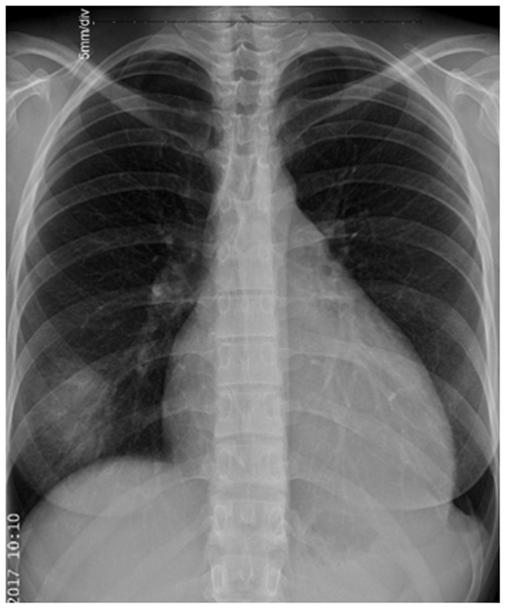
A chest X-ray showed enlargement of the cardiac shadow suggestive of pericardial effusion (Figure 1). Transthoracic echocardiography confirmed a large circumferential pericardial effusion and showed a rounded, well defined pericardial hyperechoic lesion attached to the right atrioventricular groove. There was no right ventricular dysfunction.
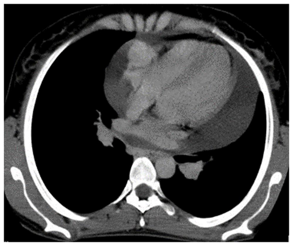
A thoracic computed tomography (CT) scan was performed, which showed a large pericardial effusion and confirmed a pericardial mass with homogenous contrast enhancement within the right atrioventricular groove (Figure 2).
Cardiac magnetic resonance imaging (MRI) confirmed the large pericardial effusion with a pedunculated ill-defined homogeneous hypointense mass on T1 and a hyperintense mass in the right atrioventricular groove with progressive enhancement after contrast administration on T2 (Figure 3).
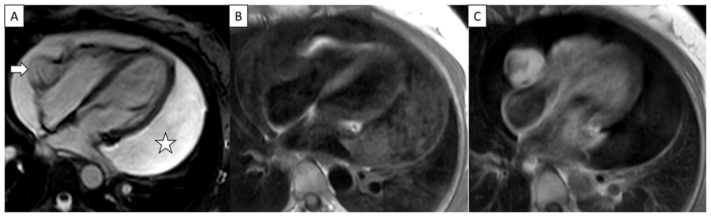
Cardiac MRI four-chamber view cine steady state free precession (A), black blood T1 weighted without (B) and after gadolinium administration (C): Rounded well defined homogenous hyperintense T2 hypointense T1 mass in right atrioventricular groove with homogenous enhancement after contrast administration (arrow). Note large pericardial effusion (star).
A coronary angiography was performed, which showed tumor blush.
The patient was referred to a cardiovascular surgery center to be operated on by an experienced cardiac surgeon. General anaesthesia was performed in supine position. Anaesthesia induction was performed by intravenous bolus of propofol (2mg/Kg), tracrium (0.5 mg/Kg) and fentanyl (2 mcg/Kg). Anaesthesia maintenance was performed by isoflurane 1.5% in oxygen and continuous intravenous infusion of tracrium (0.01 mg/Kg/min) and fentanyl (1 mcg/kg/hour). Surgery was initiated by a median sternotomy. Initial examination showed no extension of the mass into the cardiac chamber. A safety total excision of the mass was done using cutting diathermy. Vascular, pericardial and sternal sutures were performed by polypropylene, vicryl and wire, respectively. The anatomopathological examination of the mass revealed conjunctive tumor proliferation, vascular differentiated and concluded with a diagnosis of cavernous hemangioma. Post-procedural medication included antibiotic therapy with cefazolin (1 g intravenously, twice a day) for 48 hours, preventive anticoagulation by low molecular weight heparin (Enoxaparin 0.4 ml subcutaneously, once a day) and analgesic therapy by paracetamol (1 g intravenously, three times a day). Post-operative course was favorable and the patient was discharged after 72 hours.
Two months after surgery, the patient developed progressive dyspnea vomiting and precordial chest pain. CT scan found loculated left pleural effusion. Chest physiotherapy (one session a day) for two weeks and paracetamol (1 g orally, twice a day) for one week were prescribed with a favorable outcome. The patient remains well after two years of follow-up.
Cardiac hemangiomas are rare benign vascular tumors and constitute only 2.8% of primary cardiac tumors1. Pericardial localization is extremely rare2–4. Histopathologically, hemangiomas are characterized by benign proliferation of the endothelial cell lining of the blood vessel with increasing vascularization5.
Pericardial hemangioma is mostly asymptomatic. Clinical symptoms depend on location, size, and anatomic extension of the tumor5. The most frequents symptoms are dyspnea, cardiac arrhythmia, murmurs, and heart failure. Tamponade due to pericardial effusion can also occur. Imaging is very useful for the diagnosis, localization, and extension of the tumor. CT scans with contrast can show enhancing foci at the arterial phase with diffuse or heterogeneous enhancement at the delayed phase. Small calcifications might be seen also6. Cardiac MRI is a superior tool with a better contrast resolution5. Hemangiomas have an intermediate T1 signal with the same intensity as myocardium and a high T2 signal7. The dynamic postcontrast acquisition shows nodular enhancement with progressive fill-in on delayed images8. Feeding vessel, tumor blush, and flow voids might be seen also1. The tumors are usually ill-defined with no local invasion. Differential diagnoses can be made with solid pericardial masses such as mesothelioma, sarcoma, lymphoma, or paraganglioma4. Surgical total excision is the treatment of choice for resectable tumors9. The use of radiotherapy, corticosteroids, and beta blockers have been reported in some cases1.
Pericardial hemangiomas are extremely rare benign vascular tumors whose prognosis depends on their location and size. Surgical excision constitutes the treatment of choice. Our case demonstrates the importance of cardiovascular MRI as a tool to evaluate the resectability of the tumor.
All data underlying the results are available as part of the article and no additional source data are required.
Written informed consent for publication of their clinical details and clinical images was obtained from the patient.
| Views | Downloads | |
|---|---|---|
| F1000Research | - | - |
|
PubMed Central
Data from PMC are received and updated monthly.
|
- | - |
Provide sufficient details of any financial or non-financial competing interests to enable users to assess whether your comments might lead a reasonable person to question your impartiality. Consider the following examples, but note that this is not an exhaustive list:
Sign up for content alerts and receive a weekly or monthly email with all newly published articles
Already registered? Sign in
The email address should be the one you originally registered with F1000.
You registered with F1000 via Google, so we cannot reset your password.
To sign in, please click here.
If you still need help with your Google account password, please click here.
You registered with F1000 via Facebook, so we cannot reset your password.
To sign in, please click here.
If you still need help with your Facebook account password, please click here.
If your email address is registered with us, we will email you instructions to reset your password.
If you think you should have received this email but it has not arrived, please check your spam filters and/or contact for further assistance.
Comments on this article Comments (0)