Keywords
Acute leukemia, Immuno-pheno-typing, Acute lymphoblastic leukemia, Multi-parameter flowcytometry
Acute leukemia, Immuno-pheno-typing, Acute lymphoblastic leukemia, Multi-parameter flowcytometry
There are many characteristic categories of leukemia, which have well known prognoses and specific treatments. Differentiation between lymphoid and myeloid leukemia is performed using flow cytometry1. The analysis of acute leukemia immunophenotypic done by flow cytometry, this method recently become a tool for distinguishing between myeloid and lymphoid lineages. Several types of acute leukemias identified morphologically with or without enzyme cytochemical analysis, but immunophenotyping stay a vital for the right identification of myeloid lineages in minimally differentiated acute myeloid leukemia (AML-M0) and the differentiation of B or T-cell lineages in acute lymphoblastic leukemia (ALL)2. Flow cytometry (FC) is a newer technology that can be used to detect many CD markers in or on a single malignant cell, and also possesses multi-parametric capabilities for combining the study of physical characteristics, such as cell size and granularity, alongside multiple CD markers. The advantages of FC include simultaneous analysis of multiple antigens on the same cell, assessing small samples, and providing results to make an objective diagnosis within a few hours3. Immunophenotyping permits reproducible lineage detection and assignment of leukemia types. On the other hand, intra-permeable cytoplasmic markers, such as myeloperoxidase, cCD22, cCD3, and cCD79a, are more accurate and specific indicators4. Distinguishing AML-M0 from ALL remains a challenge in a minority of cases. The French–American–British (FAB) Cooperative Group recognized that many cases morphologically classified as types of ALL appeared to be of myeloid origin, as determined by monoclonal antibodies and ultra-structural cytochemistry5.
This study aimed to evaluate the immunophenotypic characterization and the expression of commonly used immuno-markers in both acute myeloid leukemia with minimal differentiation (AML-M0) and T-cell acute lymphoblastic leukemia (T-ALL), and to define the best use and the role of multi-parameter flowcytometry in the diagnosis and proper classification of these types of acute leukemia. Also, to evaluate the aberrant antigen expression in both AML-M0 and T-ALL and its correlation with clinical and prognostic criteria.
A retrospective cross-sectional study was conducted among 69 participants with newly diagnosed AL (M0 or T-ALL); there were 24 cases of AML-M0, 12 females and 12 males; and 45 cases of T-ALL, 40 males and five females. This study was conducted in the Pathology Department/Teaching Laboratories/Medical City/Iraq, and included all patients newly diagnosed with AL during the period from 5 January to 10 December 2018. Demographic characters of patients like age, and gender were collected from medical records of the participants. General clinical feature of each patients collected either when taking the patients history or during clinical examination included hepatosplenomegaly; lymphadenopathy (LAP); pallor; history of bleeding from gum, gingivae, and skin; fever; effusion of pleura; and presenting with a mediastinal mass.
1. Those presented have fever, abdominal fullness, and skin rash.
2. Those have pallor, petechiae (bleeding under the skin), sternal tenderness to palpation, lymphadenopathy, and hepatosplenomegaly on physical examination.
3. Those with history of bleeding from gum, and gingivae.
4. Those with elevated WBC count, normal RBC counts, decrease platelets counts in CBC.
5. Those with chest masses on chest radiograph.
6. Those with LAP and organomegaly on CT scans of the chest and abdomen/pelvis.
A sample size of 69 participants was calculated by the following formula:
where
z is the z score for 95% CI=1.96
ε is the margin of error=5%
n is sample size
p̂ is the population proportion=50%
This means 69 or more measurements/surveys are needed to have a confidence level of 95% that the real value is within ±5% of the measured/surveyed value. We the population proportion based on historical data from our center, whereby we found 50% of those attending were eligible for the study or in other words 50% were diagnosed with T-ALL or AML-M0.
Participants were recruited at their first visit to the center. Each patient attending our center meeting the inclusion criteria were invited to be including in our study after which written informed consent was obtained. Follow-up was performed at every subsequent visit. In EDTA anti-coagulated tubes (ATACO / China, Catalogue No.:443N980100E), the bone marrow aspirate (BMA) samples were collected (2–3 ml). The method of collection as follow: we marked and cleaned the area where the biopsy needle will be inserted. The bone marrow fluid (aspirate) and tissue sample (biopsy) were usually collected from the top ridge of the back of the hipbone (posterior iliac crest). A small incision was made, into which the needle was inserted into the bone and into the bone marrow. A sample of the liquid portion of the bone marrow was then drawn. Following sampling pressure was applied to the area of insertion to stop the bleeding and then a bandage will be placed on the site. Bone marrow was either aspirated and tested the same day or stored, if necessary, in a refrigerator (2–8°C), for no more than 48 hours. We pre-washed all the sample using at least 25 volumes excess 1X PBS (BD biosciences, Catalogue No.: 554781), mixed well. Then, pelleted cells by centrifugation and re-suspend in 1X PBS with 0.1% sodium azide to the original volume.
The panel used for diagnosis of both ALs included the following monoclonal antibodies (MoAbs): CD45, CD34, cMPO, CD13, CD33, CD14, CD15, CD64, CD117, cTdT, cCD79a, CD19, CD20, CD10, CD1a, CD2, sCD3, cCD3, CD4, CD5, CD7, CD8, and CD56, with an SSC/CD45 gating strategy, Table 1.
Immunophenotypic analysis was performed. Cells were stained using fluorescence-conjugated MoAbs and analyzed using a four-color Canto-2 BD flow cytometry device6. The device software was based on the FACS DIVA-8 operating system for multi-parametric data acquisition, display, analysis, and instrument control. The Canto-2 BD is a high performance, accurate, and cost-effective flow cytometry device that employs two lasers (red and blue), six optical parameters, and offers four colors with forward scatter (FSC) and side scatter (SSC) in combination with four fluorescence channels (FL1–FL4)7.
Each surface antibody (6 μl) was added to 100 μl of well-mixed EDTA (BD biosciences, Catalogue No.: 347689) anti-coagulated blood in labeled tubes, then incubated at room temperature in the dark for 15 minutes. Then, 2.5 ml 1X BD FACS lysis solution (BD biosciences, Catalogue No.: 349202) was added, and the samples were again incubated at room temperature in the dark for 15 minutes. The cells were washed three times by centrifugation at 1500 × g for 5 minutes before decanting the supernatant. FACS permeabilizing solution (BD biosciences, Catalogue No.: 347692) (0.5 ml) was added. Here, we vortex gently and incubated for 15 to 20 minutes in the dark at room temperature (20°C–25°C), and then the solution was re-suspended by vigorous shaking to mix. We then added 2 mL of CellWASH solution (BD biosciences, Catalogue No.: 349524) and centrifuged at 1200g for 5 minutes. Finally, we added 0.5 mL of CellFIX solution (BD biosciences, Catalogue No.: 340181) and mixed thoroughly. Then the samples were analyzed using the flow cytometer.
After adding surface markers, 0.5 ml of permeabilizing reagent was added to each tube. The tubes were vortexed then incubated for 15 minutes in the dark. Then, 6 μl of each cytoplasmic marker was added and the samples were incubated at room temperature in the dark for 30 minutes. Then, 0.5 ml of FACS solution was added, and the samples were re-suspended by vigorous mixing before being analyzed using the flow cytometer.
Permeabilizing Solution 2 (BD FACS™) is a premixed concentrate formulated specifically for use in flow cytometry. It is intended to permeabilize cell membranes prior to intracellular immunofluorescence staining with monoclonal antibodies.
Cell Lyse (BD biosciences, Catalogue No.: 559759), which is an erythrocyte lysing reagent kit for wash and no-wash procedures was used for cell lysis. Residual debris was removed by centrifugation and cell washing. Fixative reagent was used to fix and stabilize leukocytes. The fixed samples were stored for up to 24 hours at 2°C to 8°C before analysis.
1. BD FACS™ lysing solution (10X) (Catalog No. 349202)
2. BD CellWASH™ (Catalog No.349524)
3. BD CellFIX™ (Catalog No. 340181)
4. BD FACS™ permeabilizing solution 2 (Catalog No. 347692)
5. Wash buffer of PBS with 0.1% sodium azide (Catalog No. 554781)
6. Distilled water (BDH Ltd., Catalogue No.:DW005HH2).
1. Adjustable pipettes and their tips (Gelson/ France, Catalogue No.: 111-898-3).
2. Beakers (BD Biosciences, Catalogue No.: 33350410).
3. Flasks (BD Biosciences, Catalogue No.: 645392).
4. Centrifuge (Elite – Medichem/ (India), Catalogue No.: 153-33-IND).
5. Disposable slides (BD Biosciences, Catalogue No.: 335630).
6. EDTA tubes (ATACO / China, Catalogue No.:443N980100E).
7. Racks (BD Biosciences, Catalogue No.: 333489).
8. Refrigerator (HITACHI, Catalogue No.: HRPK0284A_E118-600L4D-STD-EX).
9. Stop watch (CTIZ / Japan, Catalogue No.:44448FHD).
10. Syringes (ATACO / China, Catalogue No.:CCD35560001).
11. Vortex mixer (Memmert/ Germany, Catalogue No.:CR076662088).
12. Wash bottle (BD Biosciences, Catalogue No.: 33635107).
13. Falcon® disposable 12 x 75-mm polystyrene test tubes (BD Biosciences, Catalogue No.: 343514).
The dominant population of leukemic blasts was identified using side scatter versus CD45 dot plots to isolate the blast population. A 20% cut-off value was used to indicate the positivity of surface markers, while the cut-off value for cytoplasmic markers was 10%.
Data acquisition and analysis was performed using FCS DIVA-8, made by the BD Software Company.
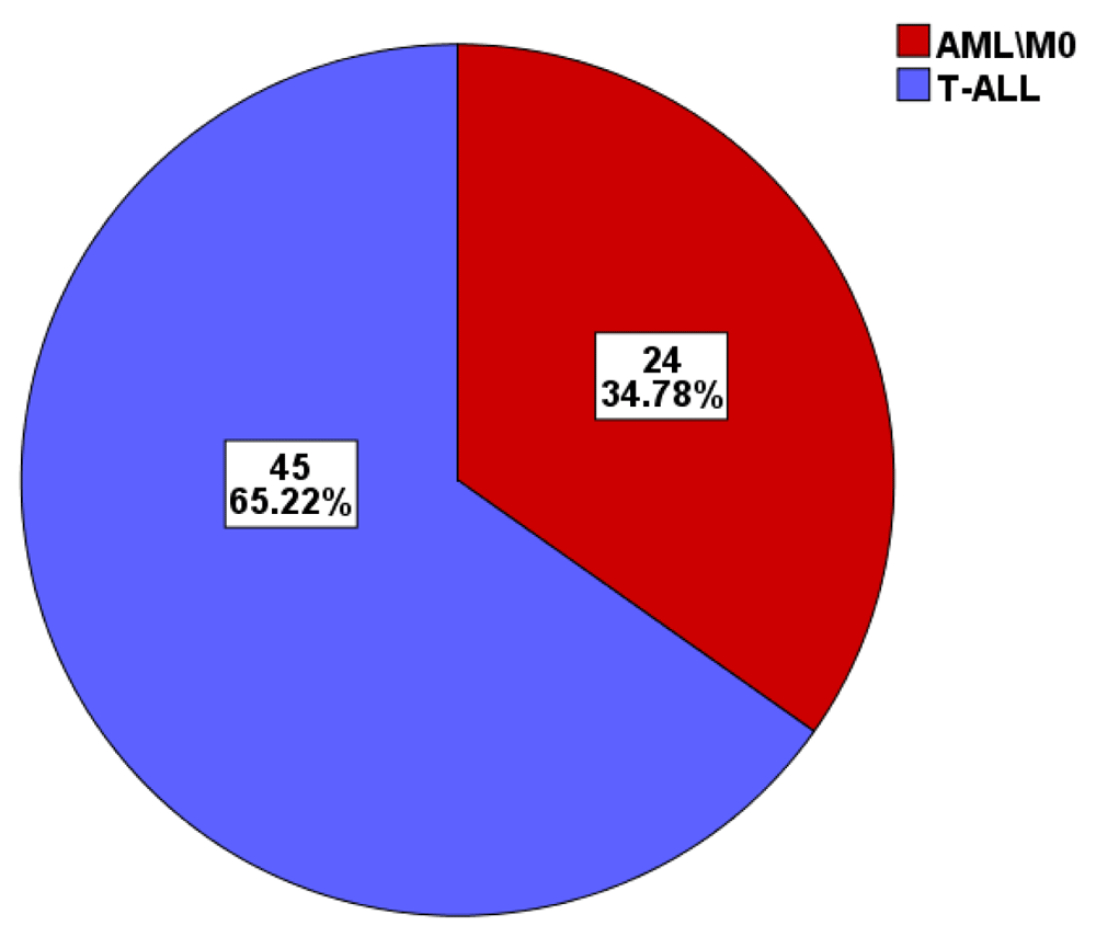
A total of 69 patients were newly diagnosed with AL; 24 patients with AML-M0, 12 females and 12 males; and 45 patients with T-ALL, 40 males and 5 females, as shown in Figure 1, the age and sex of patients is shown in Table 2 and Table 310.
| Gender | All patients N = 69 | AML\M0 N=24 | T ALL N=45 | |||
|---|---|---|---|---|---|---|
| N | % | N | % | N | % | |
| Male | 52 | 75.4 | 12 | 50 | 40 | 88.9 |
| Female | 17 | 24.6 | 12 | 50 | 5 | 11.1 |
| Age | N | Min-Max | Mean±SD | P value |
|---|---|---|---|---|
| AML\M0 | 24 | 7-76 | 43.73±18.62 | < 0.001 |
| T-ALL | 45 | 0.5-60 | 18.26±14.74 |
Regarding clinical findings, we listed in Table 410 as follow: hepatomegaly in 24.6%; splenomagaly in 36.2%; LAP in 44.9%; Pallor in 68.1%; bleeding in 71%; fever in 60.9%; pleural effusion in 1.4%; mediastinal mass in 5.8%.
This study identified 10 (41.6%) patients with AML-M0 who showed no aberrant antigen expression, while 33.3%, 16.6%, 8.3%, and 8.3% had aberrant CD7, CD56, CD2, and CD19 expression, respectively. In four (16.6%) cases of M0, there was more than one aberrant antigen expressed: two cases (8.3%) had both CD7 and CD56, one case (4.2%) had CD7 plus CD19, and one case expressed CD7 plus CD2. With regard to patients with T-ALL, we found the following: 7.0% of T-ALL patients had pro-T type, 58.0% had pre-T type, 13.0% had cortical type, and 22.0% had mature-T type, according to the European Group for the Immunological Classification of Leukemias (EGIL) criteria11. Our study also showed that 55.5% of all patients with T-ALL lacked aberrant antigen expression, while 13.3%, 13.3%, 11.1%, 6.7%, and 2.2% had aberrant CD13, CD117, CD33, CD19, and CD56 expression, respectively. Six cases (13.3%) showed expression of more than one aberrant antigen, mainly CD13 plus CD10, CD19, or CD56; CD13 plus CD117 in two cases; and one case with aberrant CD33 plus CD19, Table 5, Table 6 and Table 710.
| Frequency* | Percent 100% | |
|---|---|---|
| AML/M0 without expression | 10 | 41.6 |
| AML/M0 with aberrant CD7 expression. | 8 | 33.3 |
| AML/M0 with aberrant CD4 expression | 6 | 25 |
| AML/M0 with aberrant CD56 | 4 | 16.6 |
| AML/M0 with aberrant CD2 | 2 | 8.3 |
| CD19 | 2 | 8.3 |
Flow cytometry of leukemic cells plays an essential role in the identification of leukemia cell lines, maturation stage, and detection of residual disease. Immunophenotyping can improve both the accuracy and reproducibility of leukemia classification and is considered particularly useful for identifying AML with lymphoid marker expression and ALL with myeloid marker expression12. Some cases of AML associated with the expression of CD7 have a poor prognosis13, while other studies have reported CD2 and CD19 are better prognostic indicators in cases of AML14. In this study, 69 participants with de novo AL were enrolled, 45 of whom had T-ALL and 24 of whom had AML-M0. Most patients (88%) with T-ALL were male, which was in accordance with a study by Abid Salih and colleagues15 and also similar to another study that showed a predominance of males among patients with T-ALL16. The male:female ratio was 1:1 in patients with AML-M0 in our study, which was consistent with earlier work17.
In the present study, the most common features patients with T-ALL presented with were pallor and fever14,17–19, followed by hepato-splenomegaly and lymphadenopathy, which is in accordance with the findings of other studies14,17,18. The least common presenting features were CNS involvement, pleural effusion, and mediastinal mass19,20.
Regarding the clinical features of AML-M0, the current study found that pallor and fever were present in 16 (66.7%) of patients, consistent with the findings of Ghosh et al.17. Anemia is a common feature of all acute leukemias and is due to bone marrow infiltration; again, our results are comparable with other studies16. Pahloosye et al. reported pallor and fever were prominent symptoms20. The current study showed that bleeding occurred in 19 (79.2%) of patients with AML-M0. Such bleeding may be due to a number of causes, such as thrombocytopenia due to bone marrow suppression by infiltrating malignant leukemic cells, and by platelet dysfunction that accompanies leukemic involvement in the bone marrow16. Gaydos et al. were the first to report this in 1962, when they found that there was a linear relationship between bleeding and platelet count21. Qazi et al. correlated this feature with thrombocytopenia alone or disseminated intravascular coagulopathy (DIC)22. Extramedullary infiltration by leukemic cells may result in lymphadenopathy, splenomegaly, and/or hepatomegaly. In our patients, we observed hepatomegaly in 24.6% of them. We concluded that pleural effusion was absent in AML-M0 patients but present in one (2.2%) patient with T-ALL. Regarding mediastinal mass, this was absent in cases of AML-M0, and this agree with that noted by Ghosh et al.17.
The incidence of lymphoid antigen expression in AML-M0 cases was 58.4%, which was higher than that seen by El‑Sissy et al.12 and Khalidi et al.23, who found aberrant lymphoid antigen expression in AML to be 47% and 48.1%, respectively. The incidence of CD56 expression was 16.6% in this study, which was similar to rates recorded in previous studies24–28. Many studies have concluded that overall survival was significantly decreased in individuals who expressed CD5624–26. Regarding CD7, it was expressed in (33.3%) of patients with AML-M0, which was in agreement with other studies that found that CD7 to be aberrantly expressed in 10%–40% of blasts in AML patients26. Kita et al.13 showed that CD7 expression on cells is indicative of phenotype, is functional when immature, and is regarded as a prognostic risk factor.
Here, the most commonly expressed lymphoid marker was CD7 (33.3%), which is the same as recent reports by Bahia et al.29 and Zheng et al.30, although it is higher than the results reported by Venditti et al.31. CD4 was expressed in 25% of patients with AML-M0, which is comparable to one other study32 but higher than values reported by some other studies, e.g., 17.5%33 and 16%23.
In the current study, CD19 was expressed in 8.3% of patients, which is in accordance with the range of 8.6%–10% found in other studies like Khalidi et al.23, Bahia et al.29 and Venditti et al.31 but contrasts with other reports of 14%34 and even lower than 2.5%35. The current study showed that CD2 was expressed in 8.3% of cases of AML, similar to the proportion reported by Thalhammer-Scherrr et al.36, who found the expression of CD2 to be 3%, while Bahia et al.29 and Venditti et al.31 found higher values, of 11.4% and 14%, respectively.
The pattern of myeloid expression is correlated with some genetic features of blast cells. CD15, CD33, and CD65 are expressed in ALL cases with a rearranged MLL gene, and CD13 and CD33 are expressed in cases with the ETV6-RUNX1 gene37. The presence of these antigens lacks prognostic significance in contemporary treatment programs38.
The CD13 and CD33 were expressed in 6 (13.3%) and 5 (11.1%) of cases, respectively, which is consistent with another study39. Acute T-cell lymphoblastic leukemia with aberrant CD117 presented in 6 (13.3%) patients and is also comparable with the proportion seen in other studies40–43. Three (6.7%) patients with T-cell acute lymphoblastic leukemia expressed aberrant CD19, a lower value than other studies which found the expression of CD19 in cases of T-ALL to be 22%44. T-cell acute lymphoblastic leukemia expressing aberrant CD56 was only seen in one (2.2%) patient, and this is consistent with other studies45.
We concluded that aberrant expression of CD markers was present in some cases of acute leukemia. The incidence of aberrant antigen expression was comparable with other studies and represents an indicator of poor prognosis. CD13, CD33, and CD117 were the most frequent aberrant myeloid antigens expressed in T-ALL cases, while CD19 and CD56 were expressed in fewer cases. A large number of AML cases had aberrant lymphoid phenotypes, with T-cell markers more common than B‑cell markers. In addition, CD7 was a common aberrantly expressed lymphoid marker in AML. In the future, correlation studies of cytogenetic and lymphoid phenotypes should be performed to determine if there is any association between specific cytogenetic anomalies and the aberrant expression of certain lymphoid markers in patients with AML, or between specific cytogenetic anomalies and myeloid markers in patients with ALL.
Zenodo: Leukemia data. http://doi.org/10.5281/zenodo.402133410
This project contains the following underlying data:
- Pdf files for each participant including a immunophenotyping report with scatter plots
- Data.xlsx (Dataset containing immunophenotyping results for all participants)
Data are available under the terms of the Creative Commons Attribution 4.0 International license (CC-BY 4.0).
| Views | Downloads | |
|---|---|---|
| F1000Research | - | - |
|
PubMed Central
Data from PMC are received and updated monthly.
|
- | - |
Provide sufficient details of any financial or non-financial competing interests to enable users to assess whether your comments might lead a reasonable person to question your impartiality. Consider the following examples, but note that this is not an exhaustive list:
Sign up for content alerts and receive a weekly or monthly email with all newly published articles
Already registered? Sign in
The email address should be the one you originally registered with F1000.
You registered with F1000 via Google, so we cannot reset your password.
To sign in, please click here.
If you still need help with your Google account password, please click here.
You registered with F1000 via Facebook, so we cannot reset your password.
To sign in, please click here.
If you still need help with your Facebook account password, please click here.
If your email address is registered with us, we will email you instructions to reset your password.
If you think you should have received this email but it has not arrived, please check your spam filters and/or contact for further assistance.
Comments on this article Comments (0)