Keywords
Dens invaginatus, pulpectomy, apicoectomy
Dens invaginatus, pulpectomy, apicoectomy
Tooth development may produce various anomalies during differentiation of sensitive structures, such as the dental papilla. Genetic causes as well as local trauma and microbial encroachment upon immature dental tissues may result in anatomical structures that are difficult to negotiate when endodontic treatment may be required1. Reported prevalence of permanent teeth affected with dens invaginatus (DI) ranges from 0.3% to 10%; after routine examination of individuals, this anomaly was observed in 0.25% to 26.1%2. The most frequently affected tooth in permanent dentition appears among maxillary lateral incisor (47%)3. Upon eruption, remnants of the dental papilla or connective tissues from the periodontium may exist in the invagination. These elements might undergo necrosis and provide a nutrient-rich conditions for bacterial growth due to infiltration from the mouth flora2. The early diagnosis of DI is important as it helps in preventing the occurrence of complications from caries through to pulpal degeneration, and periodontal implications, requiring more advanced endodontic procedures later in life3. Among treatment options for DI, the operator might opt for a restoration of the defect, nonsurgical endodontic or surgical treatment, or extraction3. Morphological changes in the root canal may be associated with the invagination itself1,4. Based on radiographic observation, the depth of invagination and connection with periodontal ligament or periapical tissues, Oehlers classified DI into three categories. The three types are: Type I – small invagination, seen only on a radiograph and affecting the cervical third of the root exclusively; Type II – a more severe condition, advancing toward the pulp chamber and extending toward the middle third of the affected root; Type III – the most severe invagination compromising the remaining apical third of the root5. Endodontic treatment is complicated due to the unusual anatomical presentations of the canal spaces(s), which inhibits proper cleansing and shaping of these spaces6.
Microdontic teeth with DI are uncommon and present with a reduced labial to lingual and/or mesial to distal diameter. To date, examination of reported cases in literature, revealed the description of only two clinical situations, both affecting maxillary lateral incisors7. Our web search (Table 1), revealed more instances of case reports concerning microdontia combined with DI in peg-lateral maxillary incisors. One described a tooth with three patent canals to the apical foramen; in another case report, the peg-lateral had five canals with the same pathway to the foramen4,8. Treatment modalities varied according to clinical conditions: conventional endodontics alone, conventional endodontics followed by surgical treatment, and conventional endodontics with pulp revascularization. This case reports a Type II dens invaginatus with the formation of two distinct root canal spaces. Each canal was treated separately, and with different approaches: one portion of the canal was conventionally accessible through the crown and was treated non-surgically, while the other canal portion could only be reached surgically, with curettage of the periapical abscess, followed by root end cavity and filling.
MTA: Mineral Trioxide aggregate.
| Authors | Patient age/gender | Pulpal diagnosis | Radiographic signs | Number of canals | Nature of the procedure | Filling material | Outcome/ follow-up |
|---|---|---|---|---|---|---|---|
| Gharechahi, Ghodussi9 | 15/F | Necrotic | Large lesion | 1 | Conventional | MTA plug | Successful, 6 months |
| Kato8 | 16/M | Necrotic | Large lesion | 3 | Conventional | Sealapex/ softened gutta- carrier | Reported success/no documentation |
| Yang et al.10 | 12/M | Necrotic | Large lesion | 2 | 1st canal conventional, 2nd pulp revascularization | Guttaflow for conventional endo | Successful, 2 years recall |
| Jaikalash et al.4 | 17/M | Necrotic | Large lesion | 5 | Conventional | Gutta-percha | Successful, 1 year |
| Wayama et al.11 | 25/F | Necrotic | Large lesion | 2 | 1st canal conventional, 2nd canal surgical | Gutta-percha for both canals | Successful, 18 years |
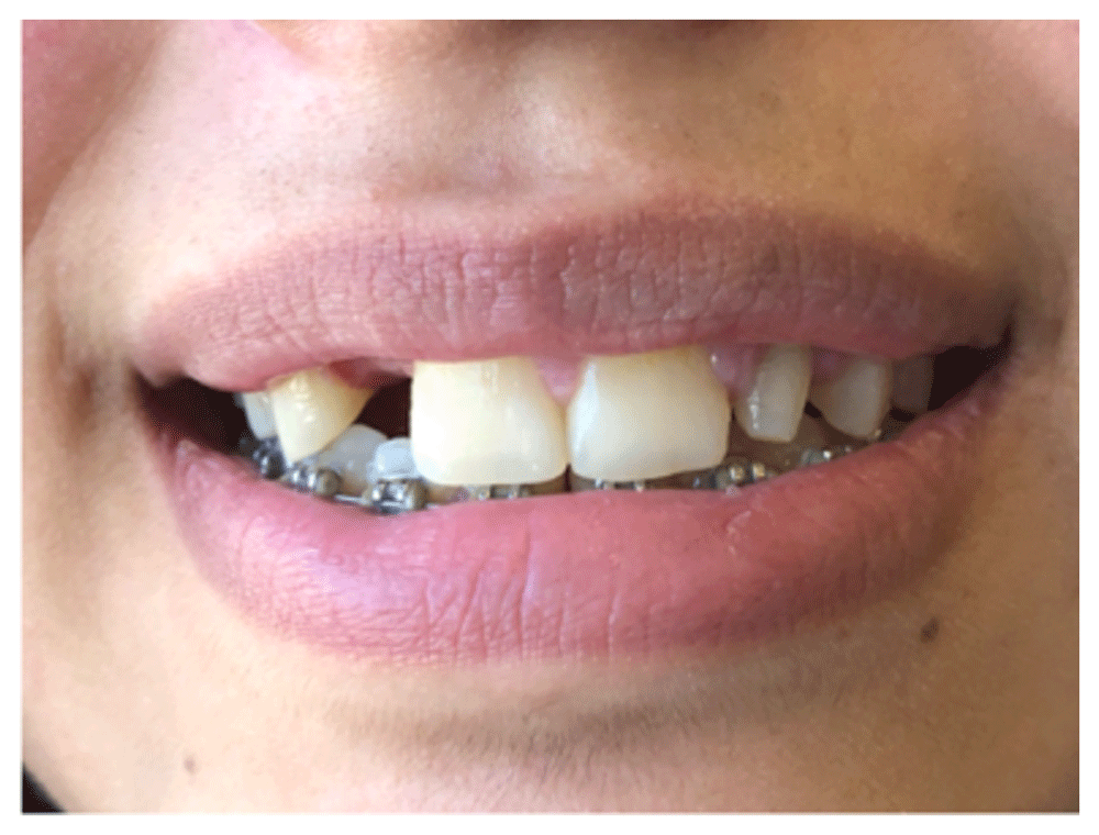
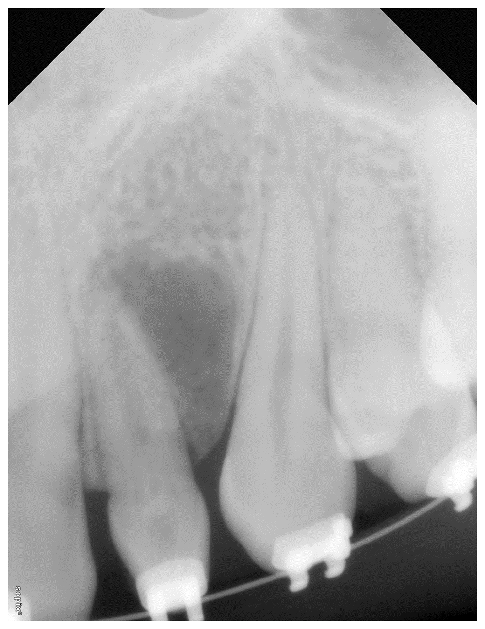
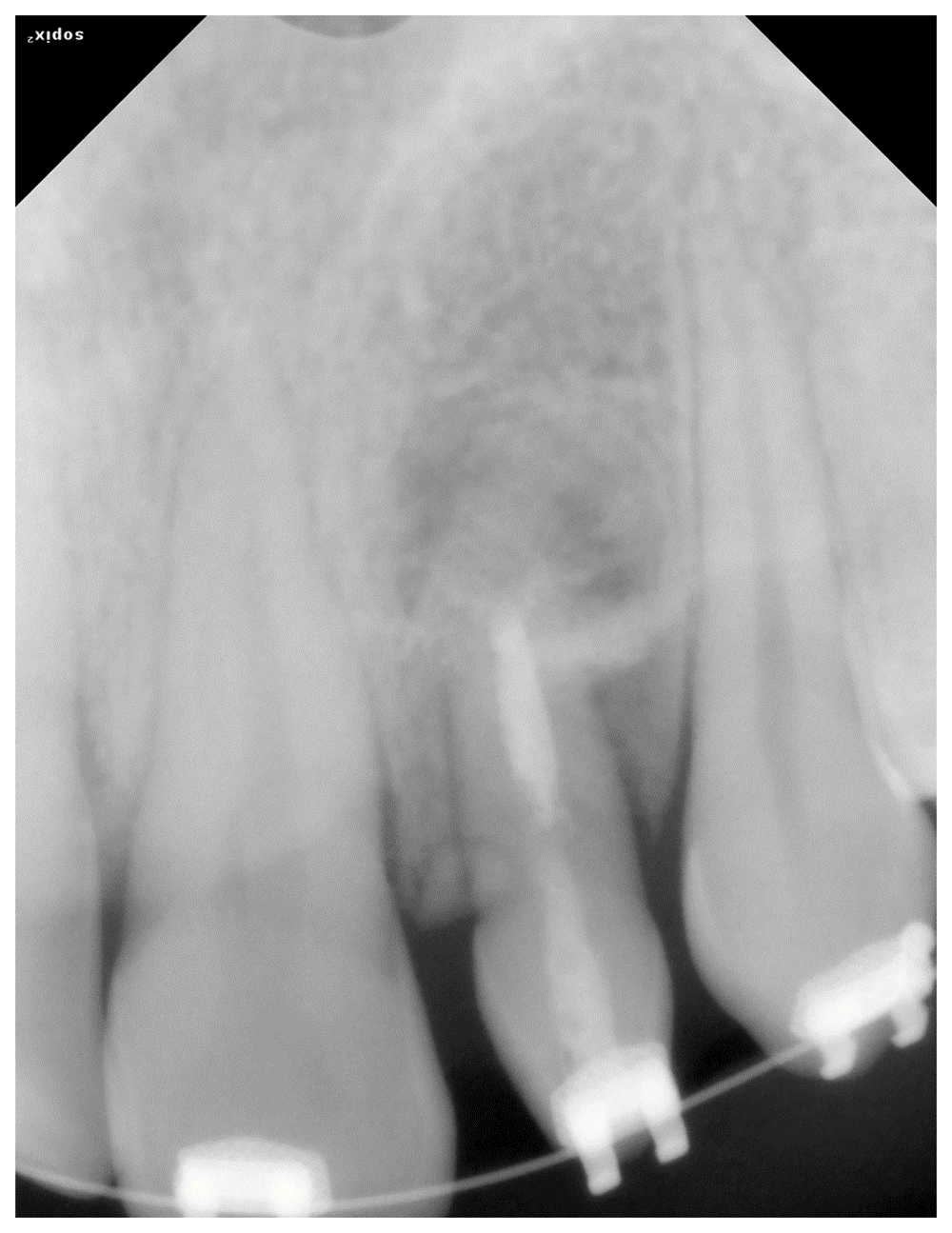
A 16-year-old caucasian female patient presented to our private practice in October 2017 for evaluation and eventual endodontic therapy of a maxillary right lateral incisor (Figure 1). The patient had no significant medical history, and the extra-oral exam revealed no unusual findings. The patient’s chief complaint was pain during mastication and swelling in the apical mucosa of the affected tooth. The patient was currently undergoing orthodontic treatment. The maxillary right lateral incisor exhibited a small crown volume with conical shape. Clinical tests on this tooth revealed tenderness to percussion and palpation, with grade 2 mobility. A negative response resulted from the thermal (cold) pulp test on this tooth. The periapical radiograph also revealed an extended type II invagination from the crown reaching the middle part of root, with no evidence of communication toward the main image of the canal. It also demonstrated an extended radiolucent image at the mesial aspect of the apex (Figure 2). Further radiographic assessment via CBCT was proposed, and subsequently declined by both the patient and her parents. The contralateral tooth (upper left lateral incisor) was extracted one year ago due to the same pathology of development but unsuccessfully treated by the referring doctor. The treatment plan was explained, and written informed consent was obtained to include documentation such as radiographs and clinical photographs for scientific publication. The proposed treatment plan was comprised of two parts: the first being the non-surgical cleaning, shaping and obturation of the conventionally accessible canal spaces, followed by a surgical procedure to access and treat the remaining canal space by root end preparation and filling.
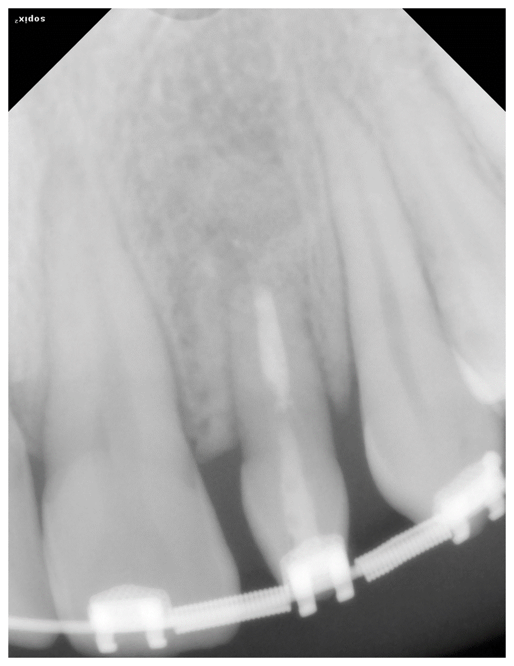
Clinical procedure part 1: The affected area was anesthetized by periapical infiltration of 1.8ml of 2% scandicaine Septodont® (Saint-Maur-des-fossés, France) and 1/100.000 vasoconstrictor, followed by isolation of the concerned tooth using rubber dam. The access cavity was achieved aided by a surgical microscope pico ZEISS OPMI® (Jena, Germany), and the high-speed Endo Access Kit® Dentsply-Sirona (Ballaigues, Switzerland), first using a diamond round bur to initiate pulpal exposure, followed by a tapered diamond bur (Figure 3) A Micro-Opener® Dentsply-Sirona (Ballaigues, Switzerland) was used to locate the canal orifice, but dystrophic deposits prevented deeper penetration apically. A # 10 K file Dentsply-Sirona (Ballaigues, Switzerland) was inserted to scout the canal, and working length (WL) was established based on radiographic estimation and clinical tactile feedback. This was in lieu of an electronic measurement; the apex locator failed to demonstrate any patency or continuity indications. Shaping of this part of the canal was completed with a set of S1, S2, F1 and F2 ProTaper® files, Universal Dentsply-Sirona (Ballaigues, Switzerland), in conjunction with copious irrigation with NaOCL. The canal was medicated with a calcium hydroxide temporary dressing AH Temp™ Dentsply-Sirona (Ballaigues, Switzerland). The access cavity was then temporary sealed with a cotton pellet and Cavit™ G (3M ESPE, St Paul, MN, USA). The patient received a prescription of amoxicillin (500 mg orally every 8 hours) and ibuprofen (200 mg every 6 hours). At the second visit, the patient’s symptoms had subsided partially; the patient was not experiencing pain to percussion or palpation; the swelling had disappeared. Removal of the AH Temp™ was accomplished using a K 25 ultrasonic file Irrisafe™ (Satelec, Halifax, Canada) and 2ml of 17% EDTA. After drying the canal space with adequate paper points, a #30 diameter master gutta-percha point from TotalFill® BC (La-Chaud-de-Fonds, Switzerland) was adjusted and sealed in the canal using TotalFill® BC sealer (La-Chaud-de-Fonds, Switzerland) in a single cone technique (Figure 4a).
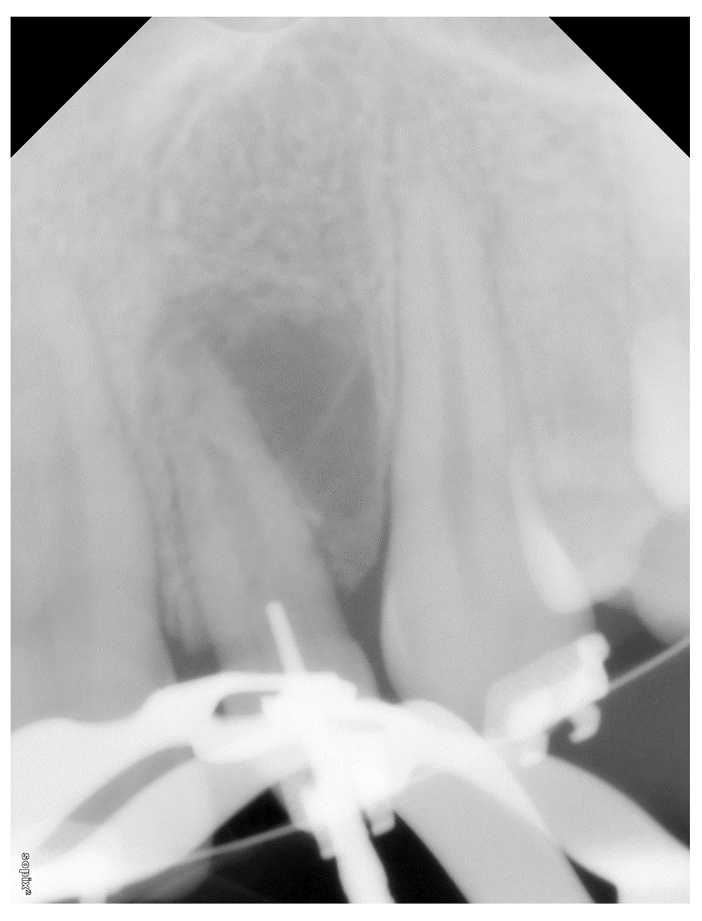
A tapered round-end high-speed bur placed in the access cavity and an X-ray exposed to make sure of the direction toward the pulp space.
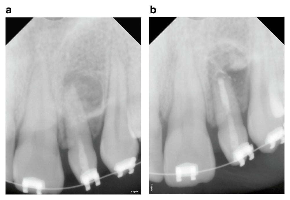
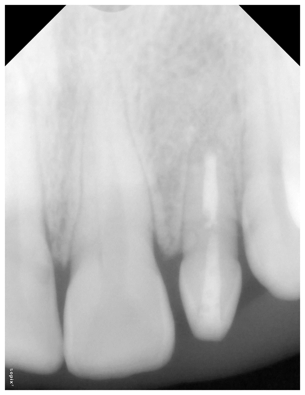
(a) post-operative X-ray after conventional endodontics (b) post-operative X-ray after surgical procedure.
The surgical procedure of the treatment was scheduled after 2 days. Administration of anesthesia (two cartridges of 1.8 ml of 2% lidocaine with 1: 50 000 epinephrine Dentsply Pharmaceutical, York, PA, USA) was accomplished and a muco-periosteal flap was raised extending from the mesial aspect of tooth #4 to the distal aspect of tooth #8. Under microscopic magnification the apical lesion was observed to be fenestrating the buccal cortical bone. After curettage of the granulation tissue, the refinement of osteotomy, and 1 mm thickness of the root apex resection was executed. Assessment at high magnification (X12) of the resected root surface was conducted to evaluate the extent and completeness of the resection. The root end preparation was created using a diamond coated 4 mm KiS-1D4 (Obtura Spartan, Fenton MO, USA). The prepared apical cavity was filled with Fast set putty TotalFill® BC (La-Chaud-de-Fonds, Switzerland) using the MAP® system (Vevey, Switzerland) for delivery of the apical filling material (Figure 4b). After repositioning of the flap suturing was conducted with 5-0 nylon sutures. The granulomatous soft tissue was harvested and sent for histopathological examination; the biopsy examination confirmed a pathological diagnosis of a periapical cyst. The patient presented 1 week later for control and suture removal with no postoperative pain and uneventful healing. Clinical and radiographic exam of the patient at 6-month intervals exhibited progressive healing of the lesion at recall appointments (Figure 5, Figure 6 and Figure 7).
Microdontia is a condition of one single tooth or more with a size smaller than normal. A peg-shape anomaly is characteristic of a tooth presenting with a crown width at incisal mesio-distal edge smaller than the cervical edge; this abnormal development usually affects upper lateral incisors. This situation may be the source of esthetic and orthodontic concern, and cause a problematic issue for dentists. Peg-lateral prevalence varies between 0.6% and 9.9% depending on ethnicity and sex, however, the overall prevalence reaches 1.8% which is equivalent to 1 in 55 people worldwide12. According to Kim et al. a slightly higher prevalence of peg-laterals and dental anomalies affects females (51.5%)6. In the same study, the authors also concluded that peg-laterals exhibit shorter root lengths compared to normal lateral incisors; they found also an incidence of 19.7% for dens invaginatus. Patients with peg-laterals often complain of esthetic issues, which need treatment orthodontic alignment and/or prosthetic restoration. Coexistence of both anomalies (peg-lateral and DI) would complicate this situation; an effective treatment plan would require endodontic therapy combined with the restorative treatment for esthetic concerns. The scarcity of case reports about microdontia combined with dens invaginatus is remarkable, with only two case reports of this anatomical feature described in the endodontic literature7. Jaikailash et al. reported a five canal peg-lateral treated in nonsurgical approach, with all canals patent from pulp chamber to foramen4. To our knowledge, one single case report has been published using a combined nonsurgical and surgical approach to treat DI affecting a mandibular incisor macrodontia13. If the success of endodontic treatment is dependent upon thorough cleaning of all irritants from canal space, then the complex configuration in DI leaves areas such as fins, communications and cul-de-sac unattainable by conventional instrumentation and disinfecting agents. The presence of a large apical lesion might compromise the conventional drying and sealing of the canal space, requiring a surgical approach to remove the apical lesion and staunch the flow of exudates. In their case report on DI type III with large apical lesion, Falcao et al. stated that, although the complexity of DI configuration could be controlled non-surgically, the authors had to eliminate the apical lesion surgically14. Fregnani et al. also stated that a surgical approach is only necessary in cases where conventional root canal therapy has failed, and in teeth with complex anatomic variations, impeding mechanical access and cleaning of all parts of the canal system. This occurs in some cases of dens invaginatus type III presenting with a periapical lesion15.
The case here described, had a large area of the canal that was inaccessible by nonsurgical means, thus the combination of non-surgical and surgical endodontics was indicated. The combination of both procedures has been reported to effectively treat cases DI13 with a long term success of 18 years follow-up11. The nature of the root end filling material has been evolving through the last two decades, with ProRooT MTA largely recommended for root end filling13. Recently, FKG (La-Chaud-de-Fonds, Switzerland) has developed a new bioceramic sealer with putty consistency and a fast setting time16. This material is ready-to-use, which eliminates the mishaps of incorrect ratios of powder-to-liquid: mixing errors, and delayed setting. Moreover, the consistency of this material enables easy transport and condensing in the root end preparation, enhancing the quality of the seal and optimizing the tooth prognosis.
Because of the unusual anatomy of DI, it is recommended to develop a thorough diagnosis and treatment plan, which contributes to the completion of the assessment of the configuration of the canal system16. CBCT exam is particularly helpful for diagnostic purposes and management of teeth presenting a particular anatomy10. However, in the present case report, the radiographic exam was limited to periapical radiographs, which prevented a more complete documentation and visualization of the canal anatomy. Patient and parents signed a written consent to justify their insistence not to proceed with a CBCT examination. Pulp vascularization is also a treatment modality of DI that could not be recommended for the present case17.
The challenges in treating this case, with a complex anatomy, and an unusual blockage of the canal system, resides in the decision-making; and the ability to perform a conventional approach for treating the accessible part of the canal, and surgically treat the residual portion of the canal. Additionally, the patient feared losing another tooth, providing a further challenge. The contralateral tooth had been extracted 2 years prior, most likely due to the tooth having a similar anatomy resulting in unsuccessful treatment.
To our knowledge, this is the first case report discussing the use of Fast set putty TotalFill® bioceramics as a retrograde filling material in the treatment of dens invaginatus with 2 years of follow-up. This material has two major advantages compared to MTA; first, the ease of manipulation as a ready mix paste to introduce into the retrograde cavity; second, the prevention of discoloration with the substitution of heavy metallic oxides and ferric oxide which cause discoloration of remaining tooth structure, and thus leads to esthetic failure9.
However, this study has a limitation in the radiographic analysis. It would have been more accurate to have a CBCT image as a diagnosis tool in order to elaborate a more adequate treatment plan, knowing the important role of CBCT as a potential aid in management of such cases, the present case has been unfortunately evaluated and negotiated with the aid of conventional radiographs, at the insistence of the patient’s parents.
All efforts should be aimed to save permanent teeth in adolescents. Knowledge of the treatment challenges caused by DI, and the use of operating microscope, which has proved to be an invaluable clinical asset, are of great help to control intricacies of complex cases, in both conventional and surgical procedures. The use of newly developed materials with improved qualities and ease of manipulation can enhance the outcomes in challenging cases, such as this case of dens invaginatus and microdontia. The importance of communication with the patient and the parents is of capital importance; it consolidates the relationship and encourages recall visits and long term follow-up.
Written informed consent for publication of their clinical details and clinical images was obtained from the patient and the patient’s parent.
| Views | Downloads | |
|---|---|---|
| F1000Research | - | - |
|
PubMed Central
Data from PMC are received and updated monthly.
|
- | - |
Provide sufficient details of any financial or non-financial competing interests to enable users to assess whether your comments might lead a reasonable person to question your impartiality. Consider the following examples, but note that this is not an exhaustive list:
Sign up for content alerts and receive a weekly or monthly email with all newly published articles
Already registered? Sign in
The email address should be the one you originally registered with F1000.
You registered with F1000 via Google, so we cannot reset your password.
To sign in, please click here.
If you still need help with your Google account password, please click here.
You registered with F1000 via Facebook, so we cannot reset your password.
To sign in, please click here.
If you still need help with your Facebook account password, please click here.
If your email address is registered with us, we will email you instructions to reset your password.
If you think you should have received this email but it has not arrived, please check your spam filters and/or contact for further assistance.
Comments on this article Comments (0)