Keywords
Prostatitis, Tuberculosis, Prostate Cancer, Genitourinary Tuberculosis, Prostate Enlargement
Prostatitis, Tuberculosis, Prostate Cancer, Genitourinary Tuberculosis, Prostate Enlargement
Tuberculosis (TB) is a chronic condition that is still endemic in Indonesia, with more than 800,000 new cases in 2018. Most cases are primary lung TB (88%); however, the genitourinary tract is one of the most common sites of hematogenous spread of extra-pulmonary TB. Genitourinary TB (GUTB), first introduced in 1937 by Wildbolz, is the second most common form of extra-pulmonary TB in endemic countries1. While male genital TB incidence are relatively low, in men, TB may affect the epididymis and the prostate (most common), followed by the seminal vesicles and the testicles1–3.
We present the case of a 56-year-old male who presented with recurrent gross hematuria and moderate lower urinary tract symptoms.
A 56-years-old male presented to the urology clinic with gross hematuria, which had been experienced for the past three months. Hematuria was intermittent and recurrent, with no history of urinary retention or previous trauma. The patient was also experiencing moderate lower urinary tract symptoms (LUTS) with daytime urinary frequency, poor urinary stream, nocturia up to three times a night, straining on bladder emptying and a sensation of incomplete emptying. His International Prostate Symptom Score was 18/35, indicating moderate symptoms, with 4/6 for the quality-of-life component, indicating mostly dissatisfied due to the symptoms. Physical examination was unremarkable, with no other comorbidities and no history of previous surgery. Digital rectal examination was performed, revealing a normal anal tone, normal perianal sensation, but an enlargement of the prostate with a firm consistency and smooth surface. Tenderness and palpable nodes were not found.
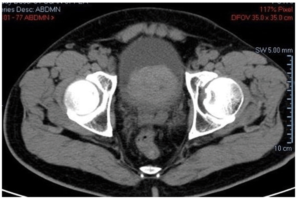
Workup was performed, revealing that blood markers were unremarkable with normal renal function, and a prostate specific antigen (PSA) value of 5.5 ng/dL (normal range, <4 ng/dL). Ultrasound and CT scan revealed prostate enlargement (Figure 1), with estimated volume of 95 gr. Cystoscopy and transurethral resection of the prostate (TUR-P) were performed for diagnosis as well as therapy to reduce prostate volume. During cystoscopy, the bladder was found to be unremarkable; the bladder wall was normal, with no mass or stone found. The prostate was found enlarged with protrusion to the bladder cavity, and a kissing lobe were found. Resection of the prostate was performed systematically and resected tissue was sent to pathology.
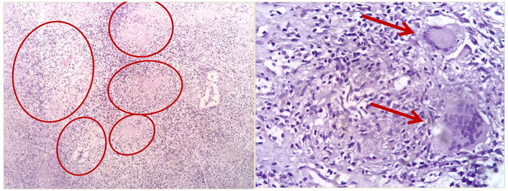
Pathology result showed a typical tuberculosis specimen with tubercles, consisting of epithelioid cells, central necrosis mass accompanied by chronic inflammatory lymphocyte cells, and presence of Langhans type of multinucleated giant cells (Figure 2). Interferon gamma release assay (IGRA) test was then performed to confirm TB diagnosis, which was positive.
The patient was given standard regimen treatment (combination of isoniazid, rifampicin, ethambutol and pyrazinamide for 2 months and isoniazid and rifampicin for the next 7 months) for TB for nine months, with evaluation of symptoms every three months. Evaluation and follow up consisted of history and physical examination, urinalysis and urine cytology and ultrasound of bladder and prostate. After 9 months of oral therapy, an additional CT scan was performed resulting normal prostate image.
TB is a long-term health issue in Indonesia. Current challenges with TB are increased incidence due to drug-resistant cases and HIV spread1. One of the most common sites of extra-pulmonary TB is the genitourinary tract, as it is the primary target of hematogenous infection. The kidney is the most common site of infection in the genitourinary tract. Prostatitis TB is a rare and uncommon presentation of extra-pulmonary tuberculosis. As a type of GUTB, diagnosis of this disease are challenging, as signs and symptoms, such as LUTS and hematuria, may mimic other disorders, such as urinary tract infection, benign prostate hyperplasia, and prostate cancer. Most of the time, diagnosis of prostate TB are incidental, for example made by the pathologist while performing examination of prostate tissue specimen taken from biopsy or prostate resection1,2,4. TB in the male genitalia is commonly found in the prostate or epididymal. Some of the most common symptoms are frequent urination, nocturia, dysuria, hematuria, urgency, and hematospermia. On physical examination indurations and nodules on the prostate can be found, which makes it difficult to distinguish from another malignancy; indeed it might be concurrent with other malignancies2,3. However, TB in the prostate is often asymptomatic, and has been shown to be found in about 10–14% of autopsy cases2.
Investigation for suspected prostate TB can be done by examining leukocytes on a three glass urine test, prostate massage secretions, ultrasound examination, and anatomical and cultural pathology examination. The gold standard is biopsy, and confirmation can be done by PCR test3. In our case, the patient had moderate LUTS symptoms and hematuria for the last three months. After examination, an enlargement of the prostate with high PSA value was found. After confirming the enlargement with ultrasound and CT scan, a TUR-P for biopsy was done which returned the result of TB. The positive IGRA test was used for confirmation. The IGRA test cannot be used alone because it will most likely always be positive in an endemic environment for TB, such as Indonesia. An alternative for TB confirmation is PCR.
Management of prostate TB is the same as other extra-pulmonary TB; administration of isoniazid, rifampicin, pyrazinamide and ethambutol or streptomycin every day for two or three months, followed by administration of isoniazid and rifampicin every day for six to seven months (for a total of nine months treatment). In our case, we used a nine month TB drug treatment, with follow-up of the symptoms every three months. Extra-pulmonary tuberculosis including genitourinary TB has a low mortality rate3.
Written informed consent for publication of their clinical details and clinical images was obtained from the patient.
| Views | Downloads | |
|---|---|---|
| F1000Research | - | - |
|
PubMed Central
Data from PMC are received and updated monthly.
|
- | - |
Provide sufficient details of any financial or non-financial competing interests to enable users to assess whether your comments might lead a reasonable person to question your impartiality. Consider the following examples, but note that this is not an exhaustive list:
Sign up for content alerts and receive a weekly or monthly email with all newly published articles
Already registered? Sign in
The email address should be the one you originally registered with F1000.
You registered with F1000 via Google, so we cannot reset your password.
To sign in, please click here.
If you still need help with your Google account password, please click here.
You registered with F1000 via Facebook, so we cannot reset your password.
To sign in, please click here.
If you still need help with your Facebook account password, please click here.
If your email address is registered with us, we will email you instructions to reset your password.
If you think you should have received this email but it has not arrived, please check your spam filters and/or contact for further assistance.
Comments on this article Comments (0)