Keywords
Giant hemangioma, Obstructive jaundice; gastric outlet obstruction; Acute spontaneous rupture
Giant hemangioma, Obstructive jaundice; gastric outlet obstruction; Acute spontaneous rupture
Hemangiomas are the most common primary benign liver tumours with an incidence estimated up to 20%1. Most of the lesions are less than 3 cm2. They are typically asymptomatic and are most often discovered incidentally during radiological investigations for other reasons. Although there is no general consensus on size, the most authors agree that hemangiomas >4 cm are described as giant3,4. Symptoms depend on the location and the size of the haemangioma. Concomitant gastric outlet obstruction (GOO) and obstructive jaundice is an extremely rare presentation of liver hemangiomas (LH)2.
We report the case of giant LH developing in the left lobe of the liver responsible for GOO and obstructive jaundice with fatal outcomes secondary to spontaneous rupture.
A 58-year-old male Tunisian patient was admitted to our department owing to a 3-week history of worsening postprandial vomiting, associated to abdominal pain in the right upper quadrant and jaundice. His past medical history was unremarkable.
The physical examination revealed a dehydrated patient with jaundice and moderately reduced general condition. His vital signs were stable, body temperature was 37.2°C, pulse rate was 72 beats/minute, and blood pressure was 120/75 mmHg. Abdominal palpation identified a large firm abdominal mass in the right upper quadrant extending to the epigastrum and the umbilicus.
Laboratory tests performed at admission showed hemoglobin levels of 12.6 g/dL, platelet count of 251,000/mL, international normalized ratio (INR) of prothrombin time of 1.12, albumin of 32 g/L (normal range: 36–48 g/L), aspartate aminotransferase of 45 UI/L (normal range up to 50 UI/L), alanine aminotransferase of 65 UI/L (normal range up to 40 UI/L), alkaline phosphatase of 360 U/L (normal range: 32–91 U/L), gamma glutamyl transpeptidase of 310 UI/L (normal range up to 50 UI/L) and notable hyper bilirubinemia (total bilirubin of 279 µmol/L (normal range up to 17 µmol/L) and direct bilirubin 90 of µmol/L (normal range up to 7 µmol/L). Renal function tests, electrolytes and c-reactive protein level were all within normal limits.
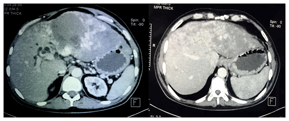
As the patient presented with symptoms related to gastric outlet obstruction, upper digestive endoscopy was performed one week after admission, following fluid resuscitation and nasogastric decompression. It showed that the stomach was full of fluid and food particles with evident extrinsic compression of the prepyloric region and the duodenum. The abdominal CT-scan revealed a 24 x 18 x 32 cm lobulated mass with a dense contrast uptake. It caused compression of the stomach, the duodenum, the common bile duct, and the hepatic pedicle (Figure 1). A diagnosis of compressive giant LH was made on the basis of typical radiological aspects.

A giant cavernous hemangioma, filling the left lobe of the liver, showing peripheral intermittent nodular contrast enhancement. with exophytic extension, compressing adjacent structures.
Although risky, surgery (partial liver resection) was decided upon because of the pressure being put on both the gastrointestinal system and the biliary tract. Unfortunately, before the intervention, the patient developed severe right upper quadrant abdominal pain, and became hemodynamically unstable. On palpation, the upper abdomen showed diffuse tenderness and fullness. Acute spontaneous rupture of the LH was strongly suspected and the patient died despite resuscitation attempts, three weeks after his admission.
Hemangioma is a frequent benign hepatic tumor with an estimated incidence of 0.4 to 7.3% in the general population5,6. Unfortunately, there are no national statistics for incidence in Tunisia. Regarding sex distribution, it seems that women are more frequently affected. The pathogenesis of this tumor is not yet completely understood. Female-specific hormones have been is involved and it has been suggested that they stimulate the enlargement of haemangiomas7.
A diagnosis of LH can be made easily through imaging techniques including ultrasonography, CT, magnetic resonance imaging, either alone or in combination, with a high sensitivity greater than 90%. Occasionally positron emission tomography (PET) or angiography can be used8. On ultrasound, small hemangioma appears as a uniform hyperechoic mass with relatively well- defined margins. Larger lesions, can appear hypoechoic or inhomogeneous, because of possible necrosis, hemorrhage or fibrosis9.
LH occurs more frequently in the right lobe of the liver10. Although there is no general agreement on size, LH are considered giant when they exceed 40 mm in diameter3. However, other sizes have been subsequently specified to categorize a LH as giant (4, 8 and 10 cm in diameter)11. Most lesions are less than 30 mm1, usually asymptomatic, and most often diagnosed incidentally on imaging tests for other reasons, laparotomy or autopsy.
Giant hemangiomas are symptomatic in 90% of cases, with various signs and findings depending on the location and size of the lesion12,13. The most frequently seen manifestation is abdominal pain resulting from the stretching of the Glisson capsule or necrosis of the lesion. In addition, giant LH may cause haematological abnormalities such as thrombocytopenia, anemia and leukopenia. Kasabach-Merritt syndrome is a life-threatening complication that may occur in 1.7% of patients with LH. It includes thrombocytopenia, consumptive coagulopathy and microangiopathic hemolytic anemia14. In rare cases, LH located in the left lobe, close to the hepatic hilum, may cause obstructive jaundice by biliary obstruction. GOO is also a rare manifestation of giant LH typically located in the left lobe of the liver, resulting from pressure on the stomach and the duodenum.
Spontaneous rupture of LH is an extremely rare and life-threatening event that is more likely to occur in lesions with a diameter ≥40 mm that are near the surface of the liver or with an exophytic growth15. It is associated with severe abdominal pain, peritonitis, and shock. The management of complications is challenging as the mortality rate ranges between 60 and 75%. Only a few cases have been reported16,17. Our patient had an extremely giant hemangioma exceeding 20 cm. To the best of our knowledge, this is the first case reported in literature to have LH with concomitant obstructive jaundice, GOO, and spontaneous rupture.
Treatment is only indicated when symptoms occur14,18.
As most liver hemangiomas do not increase in size over time, it has been suggested that asymptomatic patients with hemangiomas less than 5 cm require no intervention or observation. Additionally, giant LH with no or few symptoms requires no treatment since there is little risk of developing complications.
Symptomatic LH is conventionally treated surgically. Enucleation and resection are the most commonly used techniques and they are both safe. If feasible, enucleation is generally the preferred approach19. The choice of approach depends on the experience of the surgeon and the location and size of the tumor. Other options, such as hepatic artery ligation, transcatheter arterial embolization or in rare cases liver transplantation, have also been proposed. Surgical management of extremely giant LH >20 cm remains challenging18. In the case of our patient, we considered that hepatic resection was the most suitable therapeutic option. Unfortunately, the patient died before surgery because of the occurrence of a fatal spontaneous LH rupture.
To conclude, giant LH does not necessarily require any treatment unless symptoms appear. Lesions close to the hepatic hilum may cause compression of the bile duct and cause obstructive jaundice, those close to the stomach and duodenum may cause GOO. The exact risk of spontaneous rupture remains unknown. The association of several complications such as the case of our patient is a very uncommon clinical presentation that requires prompt surgical intervention.
All data underlying the results are available as part of the article and no additional source data are required.
Written informed consent for publication of their clinical details and clinical images was obtained from the family of the patient.
| Views | Downloads | |
|---|---|---|
| F1000Research | - | - |
|
PubMed Central
Data from PMC are received and updated monthly.
|
- | - |
Is the background of the case’s history and progression described in sufficient detail?
Yes
Are enough details provided of any physical examination and diagnostic tests, treatment given and outcomes?
Partly
Is sufficient discussion included of the importance of the findings and their relevance to future understanding of disease processes, diagnosis or treatment?
Partly
Is the case presented with sufficient detail to be useful for other practitioners?
Partly
Competing Interests: No competing interests were disclosed.
Reviewer Expertise: All aspects of General and Laparoscopic and Breast Surgery
Is the background of the case’s history and progression described in sufficient detail?
No
Are enough details provided of any physical examination and diagnostic tests, treatment given and outcomes?
Partly
Is sufficient discussion included of the importance of the findings and their relevance to future understanding of disease processes, diagnosis or treatment?
No
Is the case presented with sufficient detail to be useful for other practitioners?
No
References
1. Ferreira FG, Ribeiro MA, Abreu P, Ferreira R, et al.: Endoscopic Ultrasound-Guided Ethanol Injection Associated with Trans-arterial Embolization of a Giant Intra-abdominal Cavernous Hemangioma: Case Report and New Therapeutic Option.J Gastrointest Cancer. 2021; 52 (1): 381-385 PubMed Abstract | Publisher Full TextCompeting Interests: No competing interests were disclosed.
Reviewer Expertise: Abdominal Transplant Surgery, Liver and Pancreas Surgery
Alongside their report, reviewers assign a status to the article:
| Invited Reviewers | ||
|---|---|---|
| 1 | 2 | |
|
Version 1 20 Nov 20 |
read | read |
Provide sufficient details of any financial or non-financial competing interests to enable users to assess whether your comments might lead a reasonable person to question your impartiality. Consider the following examples, but note that this is not an exhaustive list:
Sign up for content alerts and receive a weekly or monthly email with all newly published articles
Already registered? Sign in
The email address should be the one you originally registered with F1000.
You registered with F1000 via Google, so we cannot reset your password.
To sign in, please click here.
If you still need help with your Google account password, please click here.
You registered with F1000 via Facebook, so we cannot reset your password.
To sign in, please click here.
If you still need help with your Facebook account password, please click here.
If your email address is registered with us, we will email you instructions to reset your password.
If you think you should have received this email but it has not arrived, please check your spam filters and/or contact for further assistance.
Comments on this article Comments (0)