Keywords
Thyroid nodules, Cytomorphological features, Fine Needle Aspiration, Sudan.
This article is included in the Neglected Tropical Diseases collection.
Thyroid nodules, Cytomorphological features, Fine Needle Aspiration, Sudan.
Fine-needle aspiration cytology (FNA) biopsy of the thyroid gland is an accurate and useful diagnostic tool in the initial evaluation of the nodular thyroid lesions1–4. Since ≤5% of thyroid nodules are malignant, most thyroid swellings are non-neoplastic and may not require surgical intercession5–7. Although surgical management of thyroid nodules is a feasible option, it is associated with many adverse effects, including development of hypoparathyroidism and an enduring need for thyroid hormone replacement therapy8, and also risk during surgical intercession such as injury of the recurrent nerves, bacterial infections of the surgery wound, and other related factors9. Therefore, to avoid unnecessary thyroid surgery, the standardized Bethesda scoring system and the ACR Thyroid Imaging Reporting and Data System (ACR TI-RADS) have been developed to empower clinicians to perform suitable therapeutic intercessions10,11. When an accurate preoperative diagnosis can be made, and no subsequent risk of malignant changes was suspected, surgery can be avoided12,13. FNA is one of the initial preoperative screening procedures for the diagnosis of nodular thyroid disease and considered as the most accurate diagnostic modality4,13–16, with a sensitivity ranging between 43 to 95%, and specificity of 47 to 100% for thyroid lesions16,17. This wide range of sensitivity and specificity is attributed to the testing of suspicious and rare cases and the inclusion of occult papillary carcinoma in the category of false negative diagnosis18,19. Also, FNA is a safe and useful procedure for the differential diagnosis of thyroid malignancies, such as medullary thyroid carcinoma20.
In Sudan, ultrasound-guided FNA is considered to be an expensive technique in order to be either provided by the hospital or requested by the physician managing the patient. Lacking the epidemiological data addressing the prevalence of thyroid malignancy in Sudan, besides, there is no recent descriptive study updating the status of thyroid malignancy through the past six years. Therefore, this study aimed at determining the cytomorphological patterns of thyroid lesions diagnosed by FNA among Sudanese patients attending a cytology clinic in Khartoum state, Sudan from January 2016 to December 2017.
A descriptive retrospective, clinic-based study in which we reviewed records of all patients referred to the Thyroid Nodule Center at the Total Lab Care Clinic in Khartoum state, Sudan for the evaluation of nodular thyroid disease from January 2016 to December 2017. Other than the presence of thyroid disease, no other eligibility criteria were used. The retrospective data, including type of disease, macroscopic appearance and malignancy status, were collected from the cytopathological records and sorted synonymously for analysis.
FNA was performed previously by two expert physicians using the palpation technique21. In cases of multinodular lesions and complex nodules consisting of solid and fluid matters, FNA was done multiple times to avoid sampling from single nodule or aspirating cystic fluid only. Uninodular lesions with solid composition were aspirated four times to avoid bias due to sampling from single area and to insure coverage of the whole nodule. Meanwhile, in case of small nodules which were difficult to palpate (<1 cm), cystic nodules and/or nodules located posteriorly in the thyroid gland, ultrasound guided FNA were done by experienced radiologist using a LOGIQ P5 Trackball device (Guangzhou Rongtao Medical Technology Co., Ltd, China). Four smears for each patient were taken from different regions of the thyroid nodule in order to prevent misdiagnosis. The four FNA smears of each patient were fixed in 95% ethanol according to procedures outlines by Safneck et al.22. Then, two smears were stained using Papanicolaou (Pap) stain and the other two were stained using May–Grünwald Giemsa (MGG) stain, as described previously23,24. Cytopathologic diagnosis was performed by two experienced cytopathologists using microscopy with a high-power magnification lenses (X40). Both cytopathologists’ results were compared with each other to confirm the diagnosis. Results of diagnosis of smears were categorized as non-diagnostic if insufficient cellular material was present and no evidence of cellular atypia was found (<6 groups of cells containing ≥15 cells each), and into negative for malignancy, positive for malignancy and indeterminate for malignancy according to the Bethesda System for Reporting Thyroid Cytopathology10.
A total of 1646 patients were identified and their smears were reviewed. The main cytomorphological features used for classification of thyroid nodules used included; cellularity of smears, cellular architectures (microfollicles and macrofollicles), cohesiveness, nuclear to cytoplasmic ratio, intactness of cell membrane, size of nuclei, pleomorphism, shape of nuclei, condensation of chromatin, alteration of nuclear contours, prominence of conspicuous nucleoli, and presence of pseudo-inclusions, nuclear grooves, and nuclear enlargement. Additionally, the presence or absence of Hurthle cells, amyloid, colloid, psammoma bodies, inflammatory cells, necrotic material, and tumor diathesis. The criteria of the Bethesda System for Reporting Thyroid Cytopathology were followed25.
Descriptive data were analyzed using the Statistical Package for the Social Sciences (SPSSv16). Pearson Chi Square Test was used to test the relationship between the different categorical variables. P value < 0.05 was considered a statistically significant.
Ethical approval was obtained from the ethics committee of the Faculty of Medical Laboratory Science, University of Khartoum-Sudan (UofK, FMLS 01/2017). Written informed consent for the procedure and for use of patient data for publication were obtained previously by the Thyroid Nodule Center at the Total Lab Care Clinic.
The study population consisted of 1385 (84.1%) females with mean age of 42.46±14.19, and 261 (15.9%) males with mean age of 48.29±16.84. The male to female ratio was 1:5.3. Age of patients was grouped based on interval of 10 years, age group with peak thyroid lesions was 31 to 40 years followed by 41 to 50 years.
Based on the Bethesda system, results of both cytopathologists were compared with each other to obtain the final diagnosis. Each smear was diagnosed by the first cytopathologist was matching to the diagnosis of the second cytopathologist. The retrospective results of the cytomorphological features of the FNA and data recorded about the size of swellings, site of swelling distributed based on age group were shown in Table 1. Size of swilling was significantly associated with malignancy (P=0.006), whereas site of swelling was not. Individual results for each patient are available as Underlying data26.
In total, 13/1646 patients only reported past medical history of thyroidectomy of one thyroid lobe, while the remaining 1633/1646 were not having any previous surgical removal of any of the thyroid lobes.
Family history of thyroid diseases were only reported among the negative and positive for malignancy, 77/1646 and 39/1646 respectively. Having family history of thyroid diseases was statistically significant (p = 0.0001).
Family history with thyroid diseases among patients diagnosed with malignancies was mostly noted among anaplastic carcinoma 11/39, followed by papillary thyroid cancer and follicular thyroid cancer (8/39 and 7/39, respectively). However, the remaining malignancies were also reported: 5/39 patients with metastatic cancer, 4/39 with medullary thyroid cancer and 3/39 patients with non-Hodgkin lymphoma.
Patients positive for malignancy were classified as follows: 11 (28.2%) had anaplastic thyroid cancer, 8 (20.5%) papillary thyroid cancer, 7 (17.9%) follicular thyroid cancer, 5 (12.8%) metastatic thyroid cancer, 4 (10.2%) medullary thyroid cancer, 3 (7.8%) non-Hodgkin lymphoma and 1 (2.6%) patient with thyroid follicular adenoma presenting malignant cells. Patients aged 61–70 years showed higher frequency of malignancy (Table 2).
In patients indeterminate for malignancy, the most frequent ages were 21 to 50 years, the peak age group was between the 20–39 years. The FNA diagnosis of the indeterminate for thyroid malignancy were grouped into two diagnostic verdicts, 24 (57.1%) as follicular neoplasm and 18 (43.9%) as Hurthle cell neoplasm. Figure 1 shows the different cytomorphological patterns of the thyroid lesions diagnosed in this study.
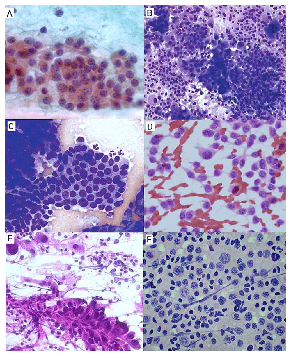
(A) Monolayer sheets of follicular cells from patient with goiter (Giemsa stain, X100). (B) Follicular neoplasm with Hurthle cells. (C) Papillary thyroid carcinoma with flat sheet that showed nuclear grooves (Giemsa stain, X40). (D) smear showed medullary thyroid carcinoma note the single cells with plasmacytoid and spindle cells differentiation with scant cytoplasm and coarsely granular chromatin (Papanicolaou, X40). (E) Anaplastic carcinoma showed bizarre and giant malignant cells with marked pleomorphic cells (Giemsa stain, X40). (F) Non-Hodgkin’s lymphoma (Giemsa stain, X40).
Thyroid nodules are a common clinical problem27. FNA of the thyroid is practiced worldwide and is reliable and cost-effective diagnostic procedure to diagnose thyroid lesions that may need surgical excision or conservative management20,28. The key factors to ensure informative thyroid FNA are having an adequate or representative cell sample and expertise of the healthcare professional performing the thyroid cytology4,29,30.
Thyroid cancer is the most common endocrine cancer (approximately 1.0%–1.5% of all new cancers diagnosed each year)31 and its incidence has continuously increased in the last three decades all over the world, including Sudan32–34. In this study, thyroid cancers were seen predominantly among those aged 30–59 years with the peak age in those aged 50–59 years. These results are in agreement with previous reports35,36. In this study, most of the diagnosed thyroid lesions were found to be benign in nature. However, in another study conducted by Caruso and his colleagues, they reported a lower percentage (74%) of benign thyroid lesions in more than 9000 patients37. Gharib et al.38, after reviewing 11,000 specimens, found 69% of examined thyroid lesions were benign lesions. Although benign nodules were reported to display morphologic changes over time, these were referred to as degenerative or mummified nodules30,39,40. This observation can lead to a non-diagnostic result on FNA, which could increase the incidence of unnecessary thyroid surgery39,41. Remarkably, the higher rate of colloid goiter among the benign lesions detected in this study can be related to the following reasons as discussed in the literature42,43: iodine deficiency, overgrowth of normal thyroid tissue, thyroid cyst, chronic inflammation of the thyroid (thyroiditis), multinodular goiter, thyroid cancer. In the current study, the relatively higher frequency of the colloid goiter from all retrieved thyroid lesions was in parity with previous reports conducted in Sudan30,44,45. Also, these results were in concordance with Nadira et al.46; which shows that colloid goiter represent about 77.2% of the thyroid lesions. Additionally, Hadi et al.47 revealed quite similar results of colloid goiter in 66.6% of the total cases.
Regarding thyroiditis, Hashimoto thyroiditis had the highest prevalence. Hashimoto thyroiditis is a common autoimmune disease characterized by marked lymphoid infiltrate destroying the thyroid follicles; it has a peak incidence between 40 and 60 years of age and a female predominance10,48. In the present study, Hashimoto thyroiditis were common among patients falling in the age between 21 and 50 years. But, Bhatia and his colleagues observed the commonest age group is those 20–39 years of age49. Also, female predominance has been observed in this study which agrees with previous published studies48,50–52.
With respect to malignant thyroid lesions, nearly 3% of the study cases were diagnosed as malignant and the majority of these were in females. Moreover, the malignant cases were reported to be in those between 30 and 59 years of age. Although anaplastic carcinoma occurs in approximately 2% of reported thyroid cancer cases53, this study depicted that anaplastic carcinoma accounted the highest proportion of thyroid malignancies followed by papillary thyroid carcinoma and follicular thyroid carcinoma in Sudan. This result is comparably different from other studies showed higher prevalence of thyroid malignancy34,54.
Our study has highlighted several potential benefits of FNA which is consistent with the previous studies addressing the FNA reimbursements4,38,55,56. The most important benefits are that FNA is a simple, safe and cost-effective first-line method to investigate thyroid lesions, particularly in low-resource settings such as Sudan, in which most of the patients suffering from thyroid lesions presented late to the clinics due to their low-income status. Furthermore, the diagnosis of benign lesions is 50-fold that of malignant ones; this can be interpreted as an increase in the community awareness about thyroid diseases as well as more clinicians recognizing the utility of FNA in evaluating thyroid nodule57,58. However, further studies considering this increase in benign lesions showed be well investigated in respect to all factors associated with developing benign thyroid lesions.
Most of the patients underwent surgical removal in different clinics. Therefore, follow-up data of the negative for malignancy lesions that showed non-neoplastic features were not correlated with the clinical presentation and ultrasonographic findings to conclude the final diagnosis. Follow up data of the repeated palpation, FNA and ultrasound at 6–18 months intervals to detect the appearance of significant growth or suspicious sonographic changes were not feasible to obtain from all the study participants and were excluded to avoid bias.
This study showed the usefulness of FNA in the evaluation of thyroid lesions and a high diagnostic performance for detection and differentiation of benign lesions from malignancy, with low rates of technical failures and complications with respect to patients’ economic status since ultrasound guided FNA were only used for unpalpable nodules. Also, this study addresses the increased predominance of benign thyroid lesions among young patients and thyroid malignancy among those aged 30–39.
Harvard Dataverse: Cytomorphological Patterns of Thyroid Lesions among 1646 Sudanese Patients: What we can learn from Fine Needle Aspiration Cytology Retrospective Analysis? https://doi.org/10.7910/DVN/AYEZVP (version 2)26.
This project contains the following underlying data:
Thyroid data (underlying data, including demographic details and information about malignancy, in SAV format).
Thyroid research (2) (underlying data in XLSX format).
Data are available under the terms of the Creative Commons Zero "No rights reserved" data waiver (CC0 1.0 Public domain dedication).
| Views | Downloads | |
|---|---|---|
| F1000Research | - | - |
|
PubMed Central
Data from PMC are received and updated monthly.
|
- | - |
Provide sufficient details of any financial or non-financial competing interests to enable users to assess whether your comments might lead a reasonable person to question your impartiality. Consider the following examples, but note that this is not an exhaustive list:
Sign up for content alerts and receive a weekly or monthly email with all newly published articles
Already registered? Sign in
The email address should be the one you originally registered with F1000.
You registered with F1000 via Google, so we cannot reset your password.
To sign in, please click here.
If you still need help with your Google account password, please click here.
You registered with F1000 via Facebook, so we cannot reset your password.
To sign in, please click here.
If you still need help with your Facebook account password, please click here.
If your email address is registered with us, we will email you instructions to reset your password.
If you think you should have received this email but it has not arrived, please check your spam filters and/or contact for further assistance.
Comments on this article Comments (0)